. 2000 Oct;11(10):3589-99.
doi: 10.1091/mbc.11.10.3589.

Rotational coupling of the transmembrane and kinase domains of the Neu receptor tyrosine kinase

Affiliations
Free PMC article

Rotational coupling of the transmembrane and kinase domains of the Neu receptor tyrosine kinase

C A Bell et al. Mol Biol Cell. 2000 Oct.
Free PMC article

Abstract

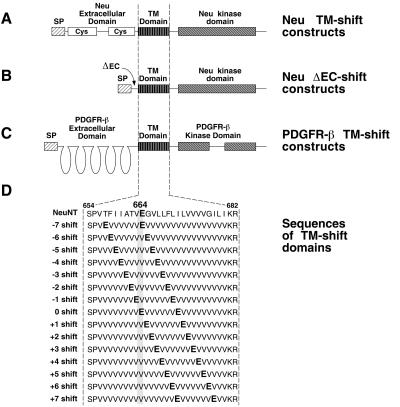
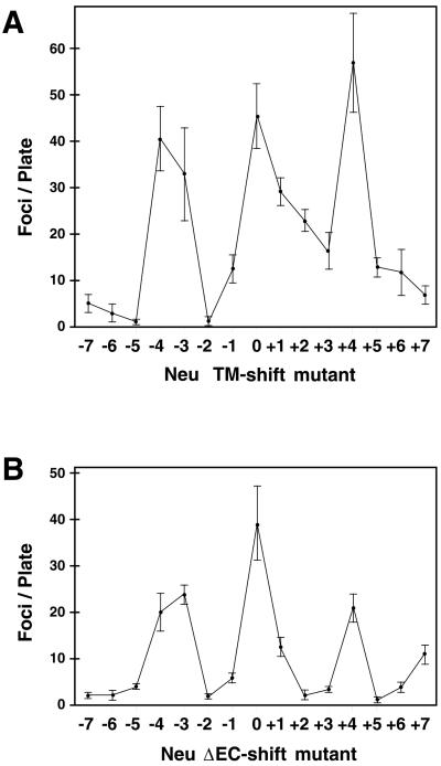
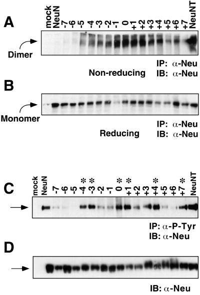
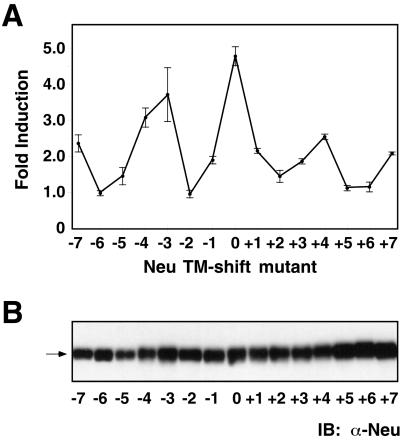
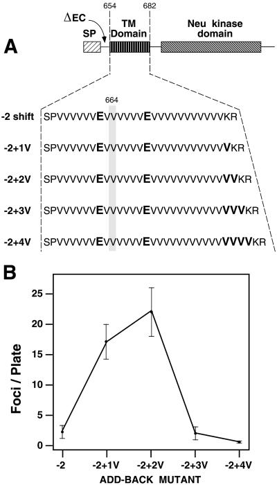
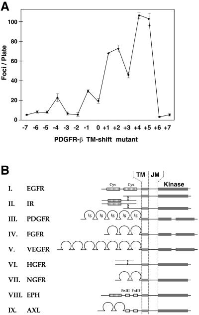
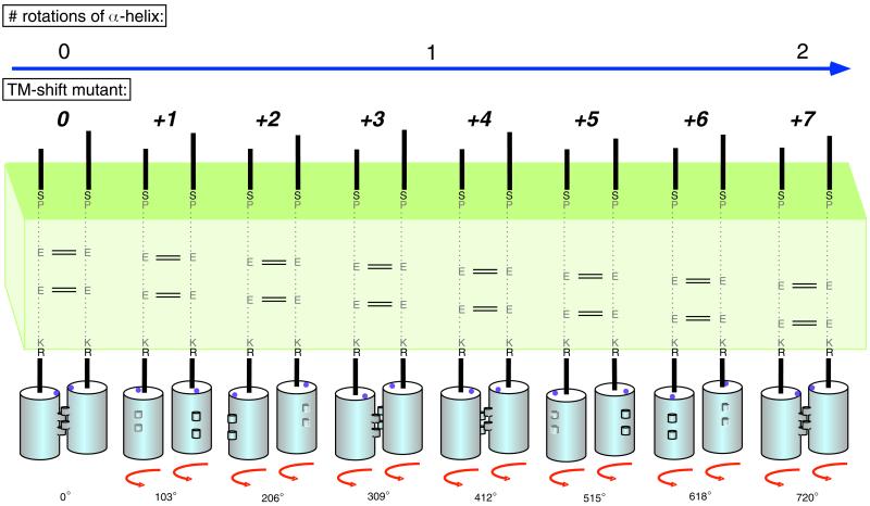
Ligand binding to receptor tyrosine kinases (RTKs) regulates receptor dimerization and activation of the kinase domain. To examine the role of the transmembrane domain in regulation of RTK activation, we have exploited a simplified transmembrane motif, [VVVEVVV](n), previously shown to activate the Neu receptor. Here we demonstrate rotational linkage of the transmembrane domain with the kinase domain, as evidenced by a periodic activation of Neu as the dimerization motif is shifted across the transmembrane domain. These results indicate that activation requires a specific orientation of the kinase domains with respect to each other. Results obtained with platelet-derived growth factor receptor-beta suggest that this rotational linkage of the transmembrane domain to the kinase domain may be a general feature of RTKs. These observations suggest that activating mutations in RTK transmembrane and juxtamembrane domains will be limited to those residues that position the kinase domains in an allowed rotational conformation.


Figures

Figure 1
Figure 1
Structure of the TM-shift mutants. (A) Neu TM-shift constructs containing simplified transmembrane domains with a dimerization motif. (B) Neu ΔEC-shift constructs containing simplified transmembrane domains with a dimerization motif. In these constructs, nearly the entire extracellular domain (amino acids 30–628) was deleted (ΔEC). (C) PDGFR-β constructs containing TM-shift domains with a dimerization motif. (D) The amino acid sequence is shown for each of the 15 transmembrane domains of the TM-shift mutants. The cysteine-rich regions (Cys) of the extracellular domain of Neu are shown. All constructs contain a signal peptide (SP) for membrane translocation and a mutated transmembrane (TM) domain that replaces residues 654–682 of Neu and residues 499–527 of murine PDGFR-β.
Figure 2
Figure 2
Transforming activity is presented for Neu TM-shift mutants (A) and ΔEC-shift mutants (B). The number of foci/10-cm plate/2.5 μg of DNA is presented. Four to ten plates were counted for each mutant. The SE of the mean is shown. The mock and the positive NeuNT controls are described in MATERIALS AND METHODS.
Figure 3
Figure 3
(A) Dimerization of Neu TM-shift mutants assayed by SDS-PAGE electrophoresis under nonreducing conditions, or reducing conditions (B). Receptors were immunoprecipitated with mAB 7.16.4, run on a 4–12% gradient gel, transferred to nitrocellulose, immunoblotted with C-18 Neu antisera and visualized by ECL. With the exception of the −7 shift, −6 shift, and +7 shift mutants, all other mutants exhibit a significant percentage of the receptor present as a dimer. Note that in the immunoblot shown, recovery of the −1 sample was anomalously low for unknown reasons. This was not observed for the −1 sample in other repeats of this experiment. (C) Tyrosine phosphorylation of Neu TM-shift mutants. RIPA lysates immunoprecipitated with 4G10 anti-phosphotyrosine mAb were electrophoresed on a 4–12% gradient SDS-PAGE gel, transferred to nitrocellulose, immunoblotted for Neu by using C-18 Neu antisera, and detected by ECL. ∗, mutants that exhibit an increase in P-tyr incorporation, which coincides with transformation. (D) Lysates from C were immunoblotted with C-18 Neu antisera to demonstrate approximately equivalent expression of Neu in all samples. Arrows in C and D indicate monomeric Neu receptors.
Figure 4
Figure 4
Fos-luciferase reporter assay. (A) Luciferase activity corresponds to transforming activity in cells transfected with Neu TM-shift mutants. The fold induction relative to mock transfected cells is set at 1. The positive control NeuNT exhibited a 5.4-fold induction. (B) Neu immunoblot of cell lysates demonstrates equivalent expression of the receptor in each sample. The arrow indicates the mobility of receptor monomers.
Figure 5
Figure 5
Restoration of activity to the inactive −2 ΔEC-shift mutant by insertion of additional Val residues in the add-back series. (A) The sequence of each transmembrane domain in this series of mutants is presented. (B) Transforming activity presented as the number of foci/10-cm plate/2.5 μg of DNA. Six plates from three independent experiments were counted. The SE of the mean is shown. The mock and positive NeuNT controls are described in MATERIALS AND METHODS.
Figure 6
Figure 6
(A) Transformation assay of PDGFR-β constructs containing the TM-shift domains. Transforming activity is presented as the number of foci/10-cm plate/1.25 μg of DNA. Four plates were counted for each mutant and the SE of the mean is shown. The negative and positive controls, PDGFR-β/neu and PDGFR-β/neu*, respectively, are described in MATERIALS AND METHODS. (B) Partial summary of RTK families is presented, indicating the transmembrane (TM) and juxtamembrane domains (JM). Separation between the transmembrane domain and the kinase domain varies from ∼35 to 75 residues, depending upon the specific RTK.
Figure 7
Figure 7
Model showing the role of rotational positioning in RTK activation. Moving the dimerization motif across the transmembrane domain rotates the Neu kinase domain monomers in the dimeric complex, leading to periodic activation of the kinase domains.

References

    1. Bargmann CI, Hung M-C, Weinberg RA. The neu oncogene encodes an epidermal growth factor receptor-related protein. Nature. 1986a;319:226–230. - PubMed
    1. Bargmann CI, Hung M-C, Weinberg RA. Multiple independent activations of the neu oncogene by a point mutation altering the transmembrane domain of p185. Cell. 1986b;45:649–657. - PubMed
    1. Bargmann CI, Weinberg RA. Increased tyrosine kinase activity associated with the protein encoded by the activated neu oncogene. Proc Natl Acad Sci USA. 1988a;85:5394–5398. - PMC - PubMed
    1. Bargmann CI, Weinberg RA. Oncogenic activation of the neu-encoded receptor protein by point mutation and deletion. EMBO J. 1988b;7:2043–2052. - PMC - PubMed
    1. Brandt-Rauf PW, Rackovsky S, Pincus MR. Correlation of the structure of the transmembrane domain of the neu oncogene-encoded p185 protein with its function. Proc Natl Acad Sci USA. 1990;87:8660–8664. - PMC - PubMed

Publication types

MeSH terms

Substances