. 2000 Oct;11(10):3601-15.
doi: 10.1091/mbc.11.10.3601.

Role for the silencing protein Dot1 in meiotic checkpoint control

Affiliations
Free PMC article

Role for the silencing protein Dot1 in meiotic checkpoint control

P A San-Segundo et al. Mol Biol Cell. 2000 Oct.
Free PMC article

Abstract

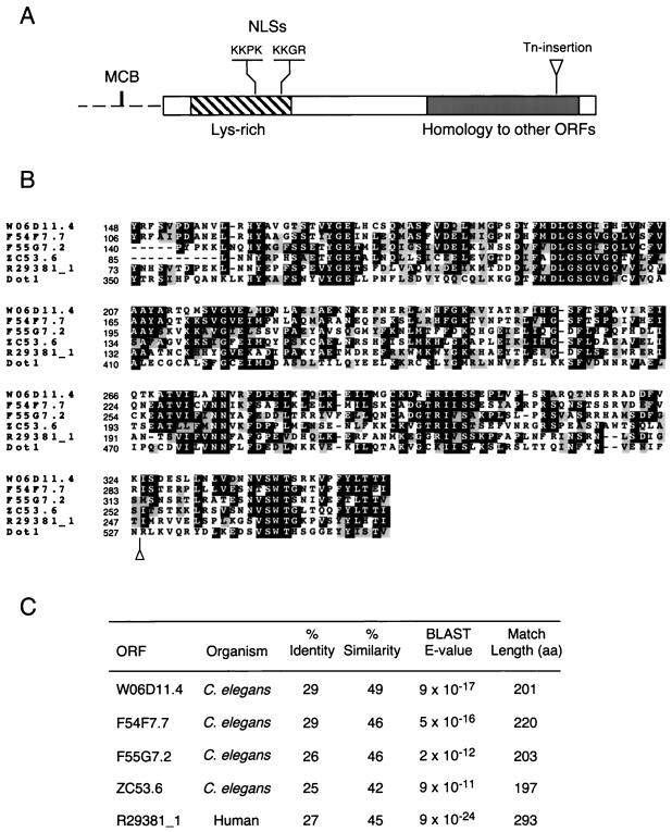
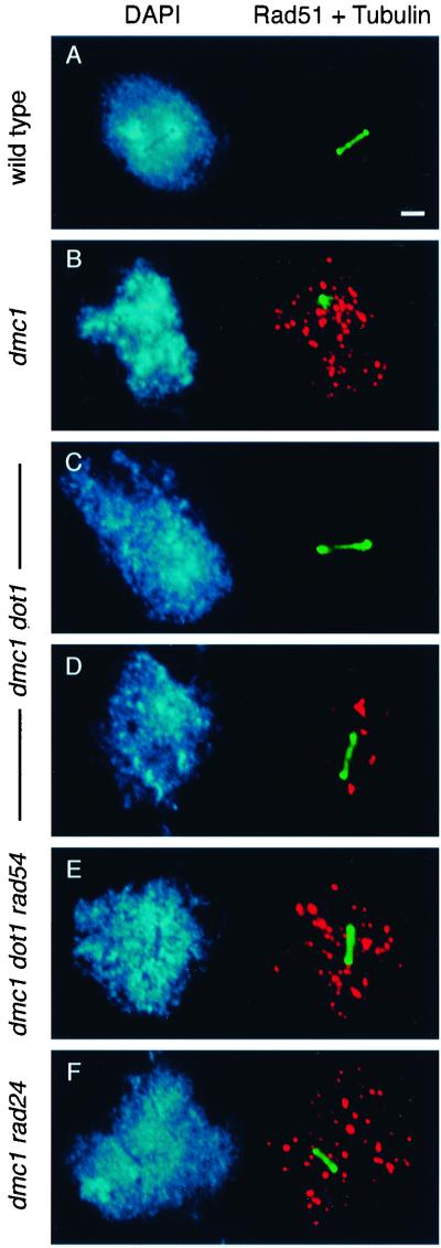
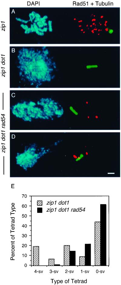
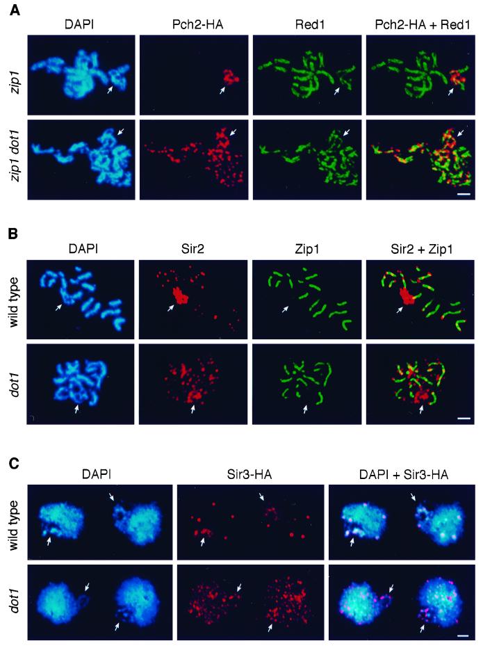
During the meiotic cell cycle, a surveillance mechanism called the "pachytene checkpoint" ensures proper chromosome segregation by preventing meiotic progression when recombination and chromosome synapsis are defective. The silencing protein Dot1 (also known as Pch1) is required for checkpoint-mediated pachytene arrest of the zip1 and dmc1 mutants of Saccharomyces cerevisiae. In the absence of DOT1, the zip1 and dmc1 mutants inappropriately progress through meiosis, generating inviable meiotic products. Other components of the pachytene checkpoint include the nucleolar protein Pch2 and the heterochromatin component Sir2. In dot1, disruption of the checkpoint correlates with the loss of concentration of Pch2 and Sir2 in the nucleolus. In addition to its checkpoint function, Dot1 blocks the repair of meiotic double-strand breaks by a Rad54-dependent pathway of recombination between sister chromatids. In vegetative cells, mutation of DOT1 results in delocalization of Sir3 from telomeres, accounting for the impaired telomeric silencing in dot1.


Figures

Figure 1
Figure 1
DOT1 sequence features. (A) Diagram of the Dot1 protein (rectangle) and DOT1 promoter (discontinuous line). Indicated are a lysine-rich domain (striped box; 21.5% of residues between amino acids 35 and 169 are lysines), two putative NLSs starting at positions 126 and 162, and the region of homology to human and C. elegans ORFs (gray area). The sequence ACGCGTCA at positions −172 to −165 in the DOT1 promoter corresponds to a consensus MCB element (MluI cell cycle box site is underlined). (B) Alignment of the regions of homology from S. cerevisiae Dot1, human ORF R29381_1 and C. elegans ORFs W06D11.4, F54F7.7, F55G7.2, and ZC53.6. Identities are highlighted in black and conservative substitutions in gray. Numbers at the left represent amino acid positions. Alignment and shading were performed with ClustalW 1.7 and Boxshade 3.21, respectively, at the BCM Search Launcher (http://dot.imgen.bcm.tmc.edu:9331/). In A and B, the position of the C-terminal transposon insertion recovered from the mutant screen is marked with a triangle. (C) Percentage identity and similarity of Dot1 to the human and nematode homologs over the indicated length. The significance of the homology is indicated by the BLAST E-values.
Figure 2
Figure 2
Dot1 is required for checkpoint-induced meiotic arrest (or delay) of zip1 and dmc1 mutants. (A) Mutation of DOT1 alleviates the meiotic arrest of zip1 in BR2495 isogenic strains. Time course of sporulation of strains BR2495 (wild type), MY63 (zip1), DP138 (dot1), and DP139 (zip1 dot1). (B) Mutation of DOT1 bypasses the meiotic delay of zip1 in the SK1 strain background. Dityrosine fluorescence, an indicator of sporulation, was examined after 1 and 2 d on sporulation plates. Strains are DP185 (wild type), DP246 (zip1), and DP371 (zip1 dot1). (C) Distribution of tetrad types. The percentages of tetrads with 4, 3, 2, 1 and 0 viable spores (4-sv, 3-sv, 2-sv, 1-sv, and 0-sv, respectively) are represented. Tetrads (389 and 513) were dissected from DP246 (zip1) and DP371 (zip1 dot1), respectively. (D) Bypass of dmc1 arrest by dot1 and effects of the rad54 mutation. Dityrosine fluorescence was examined after 2 d on sporulation plates. (%) MI + MII represents the fraction of cells that have undergone at least one meiotic nuclear division (i.e., containing two or more DAPI-stained bodies) after 24 h in liquid sporulation medium. (%) Spo. represents the fraction of cells that formed mature or immature asci. The asterisks indicate that most asci are immature. At least 300 cells were scored for each strain. Strains are DP338 (wild type), DP339 (dmc1), DP341 (dmc1 dot1), DP343 (dmc1 dot1 rad54), DP352 (dmc1 rad24), and DP342 (dmc1 rad54).
Figure 3
Figure 3
Cytological analysis of unrepaired DSBs and spindle elongation. Spread meiotic nuclei from strains DP338 (wild type; A), DP339 (dmc1; B), DP341 (dmc1 dot1; C and D), DP343 (dmc1 dot1 rad54; E), and DP352 (dmc1 rad24; F), stained with DAPI (left) and anti-Rad51 (red) and anti-tubulin (green) antibodies (right). Spreads were prepared after 6 h in sporulation medium. Bar, 2 μm.
Figure 4
Figure 4
Repair of DSBs in zip1 dot1 is largely independent of Rad54. (A–D) Cytological analysis of ongoing recombination and entry into MI, assessed by the presence of Rad51 foci and spindle elongation, respectively. Spread meiotic nuclei from strains MY152 (zip1; A), DP381 (zip1 dot1; B), and DP382 (zip1 dot1 rad54; C and D), stained with DAPI (left) and anti-Rad51 (red) and anti-tubulin (green) antibodies (right). Spread nuclei were prepared after 16 h in sporulation medium. Bar, 2 μm. (E) Distribution of tetrad types. The percentages of tetrads with 4, 3, 2, 1, and 0 viable spores (4-sv, 3-sv, 2-sv, 1-sv, and 0-sv, respectively) are represented. Tetrads (88 and 87) were dissected from strains DP381 (zip1 dot1) and DP382 (zip1 dot1 rad54), respectively.
Figure 5
Figure 5
Nuclear localization of Dot1. (A and B) Cells from strain DP138 (dot1) transformed with pSS64 (2 μ DOT1-GFP) growing vegetatively (A) or at different times in sporulation medium (B) were fixed, stained with DAPI, and observed at the fluorescence microscope. The merged images of DAPI (red) and Dot1-GFP (green) are shown in the bottom panels. Overlap appears yellow. The corresponding differential interference contrast images are also presented (top). The approximate cell-cycle stage was estimated based on bud size and nuclear morphology and position. In B, only the large mother cells have entered meiosis; the small buds should not be considered. (C) Spread nuclei from strain DP138 (dot1) transformed with pSS63 (2 μ DOT1-HA) growing vegetatively, stained with DAPI (blue) and anti-HA antibodies (red). (D) Spread leptotene/zygotene (top row) and pachytene (bottom row) nuclei from strain DP138 (dot1) containing pSS63 (2 μ DOT1-HA) stained with DAPI (blue) and anti-Zip1 (green) and anti-HA (red) antibodies. Bar, 2 μm.
Figure 6
Figure 6
Pch2, Sir2, and Sir3 are mislocalized in the absence of DOT1. (A) Spread meiotic nuclei from DP201 (zip1 PCH2-HA, top) and DP285 (zip1 dot1 PCH2-HA, bottom) stained with DAPI (blue) and anti-HA (red) and anti-Red1 (green) antibodies. (B) Spread pachytene nuclei from BR2495 (wild type, top) and DP138 (dot1, bottom) stained with DAPI (blue) and anti-Sir2 (red) and anti-Zip1 (green) antibodies. (C) Spread mitotic nuclei from DP365 (SIR3-HA, top) and DP366 (dot1 SIR3-HA, bottom) stained with DAPI (blue) and anti-HA (red) antibodies. In all panels, arrows point to the nucleolar region. Bar, 2 μm.

References

    1. Ahmed S, Hodgkin J. MRT-2 checkpoint protein is required for germline immortality and telomere replication in C. elegans. Nature. 2000;403:159–164. - PubMed
    1. Arbel A, Zenvirth D, Simchen G. Sister chromatid-based DNA repair is mediated by RAD54, not by DMC1 or. TID1. EMBO J. 1999;18:2648–2658. - PMC - PubMed
    1. Bailis JM, Roeder GS. Synaptonemal complex morphogenesis and sister-chromatid cohesion require Mek1-dependent phosphorylation of a meiotic chromosomal protein. Genes Dev. 1998;12:3551–3563. - PMC - PubMed
    1. Bailis JM, Roeder GS. The pachytene checkpoint. Trends Genet. 2000a;16:395–403. - PubMed
    1. Bailis JM, Roeder GS. Pachytene exit controlled by reversal of Mek1-dependent phosphorylation. Cell. 2000b;101:211–221. - PubMed

Publication types

MeSH terms