. 2000 Oct;11(10):3629-43.
doi: 10.1091/mbc.11.10.3629.

Yeast exocytic v-SNAREs confer endocytosis

Affiliations
Free PMC article

Yeast exocytic v-SNAREs confer endocytosis

S Gurunathan et al. Mol Biol Cell. 2000 Oct.
Free PMC article

Abstract

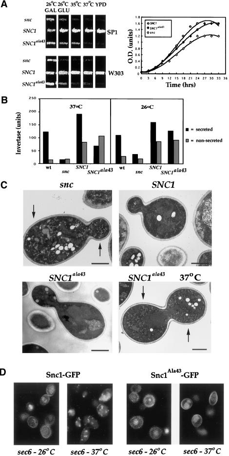
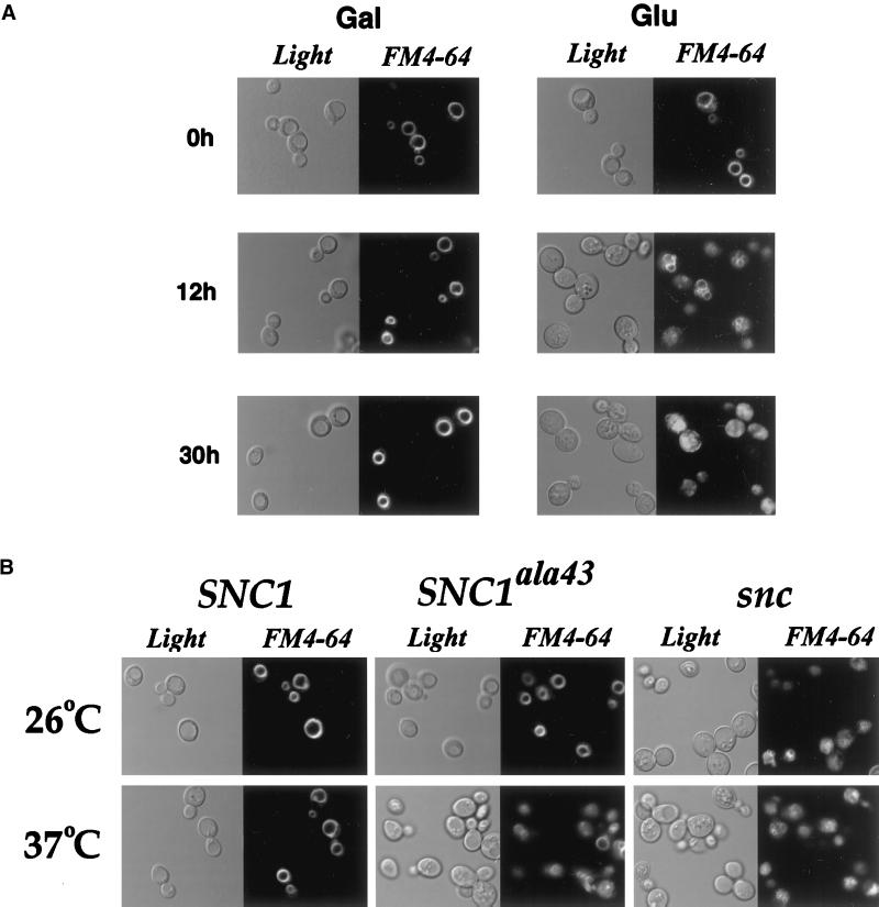
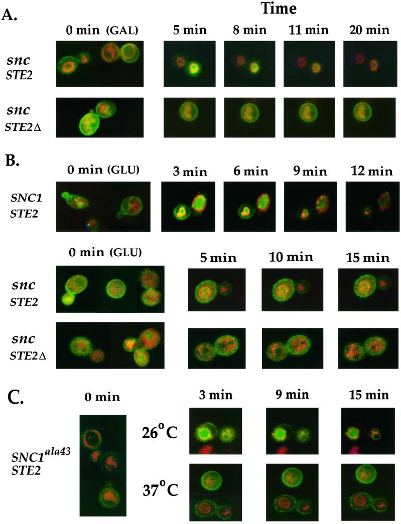
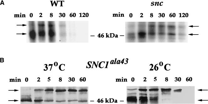
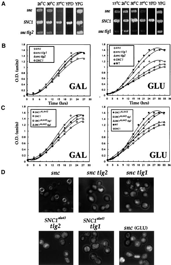
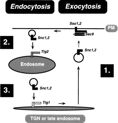
In yeast, homologues of the synaptobrevin/VAMP family of v-SNAREs (Snc1 and Snc2) confer the docking and fusion of secretory vesicles at the cell surface. As no v-SNARE has been shown to confer endocytosis, we examined whether yeast lacking the SNC genes, or possessing a temperature-sensitive allele of SNC1 (SNC1(ala43)), are deficient in the endocytic uptake of components from the cell surface. We found that both SNC and temperature-shifted SNC1(ala43) yeast are deficient in their ability to deliver the soluble dye FM4-64 to the vacuole. Under conditions in which vesicles accumulate, FM4-64 stained primarily the cytoplasm as well as fragmented vacuoles. In addition, alpha-factor-stimulated endocytosis of the alpha-factor receptor, Ste2, was fully blocked, as evidenced using a Ste2-green fluorescent protein fusion protein as well as metabolic labeling studies. This suggests a direct role for Snc v-SNAREs in the retrieval of membrane proteins from the cell surface. Moreover, this idea is supported by genetic and physical data that demonstrate functional interactions with t-SNAREs that confer endosomal transport (e.g., Tlg1,2). Notably, Snc1(ala43) was found to be nonfunctional in cells lacking Tlg1 or Tlg2. Thus, we propose that synaptobrevin/VAMP family members are engaged in anterograde and retrograde protein sorting steps between the Golgi and the plasma membrane.


Figures

Figure 1
Figure 1
Mutation of methionine-43 to alanine in Snc1 yields a thermosensitive exocytic v-SNARE. (A) SNC1ala43 encodes a thermosensitive v-SNARE. Left panel: snc cells (SP1 background–JG8 and W303 background–JG10) transformed with a control vector (snc) or plasmids constitutively expressing SNC1 or SNC1ala43 were patched onto synthetic complete plates containing galactose. Cells were grown for 3 d at 26°C, before replica plating onto medium containing glucose to deplete SNC1. After 24 h, patches were replica plated onto the following: galactose- (GAL) and glucose-containing (GLU) plates at 26°C; prewarmed glucose-containing plates at 35°C and 37°C; and amino acid-rich medium (YPD). Plates were incubated for 30 h. Right panel: snc cells (JG8) transformed with a control vector (snc), or plasmids expressing SNC1 or SNC1ala43, were grown to log phase on galactose-containing medium and then were shifted to glucose-containing medium for 24 h to deplete SNC1. Cells were seeded in fresh glucose-containing medium at 26°C, and optical density (measured at 600 nm) was monitored as a function of time. (B) Invertase secretion is blocked in SNC1ala43 cells after a shift to restrictive temperatures. Wild-type (SP1), snc (JG8), and snc cells (JG8) transformed with plasmids expressing SNC1 or SNC1ala43 were grown to log phase on galactose-containing medium, shifted to glucose medium for 24 h at 26°C and then derepressed for 2 h on low glucose (0.05%) medium at either 26°C or 37°C. Both secreted (black) and nonsecreted (gray) invertase activities were measured. (C) SNC1ala43 cells accumulate secretory vesicles at restrictive temperatures. snc cells (JG8) expressing a control vector (snc), SNC1, or SNC1ala43 were grown to log phase on glucose-containing medium. Cells were maintained at 26°C, or were shifted for 2 h to 37°C (indicated as 37°C), and were processed for electron microscopy. Bar indicates 1 μm. (D) Localization of Snc1-GFP and Snc1ala43-GFP in sec6 cells. sec6–4 cells transformed with plasmids expressing Snc1-GFP or Snc1ala43-GFP were maintained at 26°C or were shifted for 2 h to 37°C, and then were processed for confocal microscopy.
Figure 2
Figure 2
Delivery of FM4–64 to the vacuole is impaired in snc cells and SNC1ala43 cells shifted to restrictive temperatures. (A) Delivery of FM4–64 to the vacuole is impaired in snc cells. snc cells (JG8) maintained on galactose-containing medium (GAL), or shifted to glucose-containing medium (GLU) for various times at 26°C, were incubated with FM4–64 (see MATERIALS AND METHODS) and were processed for microscopy. Cells were visualized using light (Nomarski optics) and fluorescence (rhodamine channel) microscopy. (B) Delivery of FM4–64 to the vacuole is impaired in SNC1ala43cells shifted to restrictive temperatures. snc cells (JG8) expressing a control vector (snc), SNC1, or SNC1ala43 were incubated with FM4–64 at 26°C or were shifted for 30 min to 37°C before labeling with FM4–64 at the restrictive temperature (see MATERIALS AND METHODS).
Figure 3
Figure 3
Ligand-mediated delivery of Ste2 to the vacuole is abolished in snc cells and SNC1ala43 cells shifted to restrictive temperatures. (A) Delivery of Ste2-GFP, but not Ste2Δ-GFP, to the vacuole occurs in snc cells grown on galactose. snc cells (JG8) expressing either Ste2-GFP (snc STE2) or a C-terminal truncated form of Ste2-GFP (snc STE2Δ), which does not undergo endocytosis, were grown to log phase at 26°C before labeling with FM4–64 (see MATERIALS AND METHODS). Cultures were split, whereby half were mounted in soft agar containing no additions, while the other half were mounted in agar containing α-factor (1 μM) and were processed immediately for microscopy. A composite image of untreated cells is representative of the zero time (0 min), while specific α-factor–treated cells were monitored as a function of time. (B) Delivery of Ste2-GFP to the vacuole is abolished in snc cells grown on glucose. snc cells (JG8) expressing SNC1 constitutively or bearing a control plasmid were transformed with plasmids expressing Ste2-GFP to give SNC1 STE2 and snc STE2 strains, respectively. In addition, snc cells expressing the truncated form of Ste2-GFP (snc STE2Δ) also were used. Cells were grown to log phase at 26°C, before labeling with FM4–64. Next, samples of cells were mounted in agar containing no additions, while an equal amount was mounted in agar containing α-factor (1 μM) before visualization, as described above. (C) Delivery of Ste2-GFP to the vacuole is abolished in SNC1ala43 cells shifted to the restrictive temperature. snc cells (JG8) expressing SNC1ala43 were grown to log phase and either incubated with FM4–64 at 26°C or shifted for 30 min to 37°C before labeling with FM4–64 at the restrictive temperature. Cells that were treated with α-factor (1 μM) were monitored over time by microscopy.
Figure 4
Figure 4
Vacuolar degradation of Ste2 is severely delayed or blocked in snc cells and temperature-shifted SNC1ala43 cells. (A) Vacuolar degradation of Ste2 is delayed in snc cells. Wild-type (WT) and snc cells grown on glucose-containing medium at 26°C for 30 h were pulse-labeled with [35S]-methionine and chased for 30 min in medium containing 5 mM methionine/cysteine, before treatment with α-factor (1 μM). At various times after α-factor treatment, cell extracts were prepared and Ste2HA was immunoprecipitated using anti-HA antibodies. Ste2HA was resolved by SDS-PAGE and was detected by autoradiography. The lower and upper arrows indicate the respective 46–47-kDa and 54-kDa forms of Ste2HA observed before and after treatment with α-factor. (B) Vacuolar degradation of Ste2 is blocked in temperature-shifted SNC1ala43 cells. snc cells expressing SNC1ala43 were grown on glucose-containing medium at 26°C for 30 h before pulse-chase labeling and exposure to α-factor (1 μM) for different amounts of time. Cells either were maintained at the permissive temperature (26°C) during labeling and α-factor treatment or were shifted to the restrictive temperature (37°C) during the chase and thereafter. Ste2HA was resolved on gels and detected as described above.
Figure 5
Figure 5
Genetic interactions between TLG and SNC genes in yeast. (A) Disruption of TLG1 or TLG2 in snc cells (JG8) slows cell growth and results in conditional lethality. Left panel: snc cells (JG8) transformed with a control plasmid (snc), a plasmid expressing SNC1 constitutively (SNC1), or snc tlg2 cells. Right panel: snc cells (JG8) transformed with a control plasmid (snc), a plasmid expressing SNC1 constitutively (SNC1), or snc tlg1 cells. All cells first were grown at 26°C on medium containing galactose, before replica plating onto medium containing glucose to deplete Snc1. After 24 h, patches were replica-plated onto the following: glucose-containing plates preequilibrated to 15°C, 26°C, 30°C, and 37°C; rich medium (YPD); and rich medium containing galactose (YPG). Plates were incubated for 30 h. (B) Growth curves of snc tlg cells on galactose- and glucose-containing medium. Left panel: snc (JG8), snc cells expressing SNC1, snc tlg1, and snc tlg2 cells were grown to log phase at 26°C on galactose-containing medium (to induce SNC1). Equal amounts of cells then were seeded into freshgalactose-containing medium (GAL), and optical density (measured at 600 nm) at 26°C was monitored as a function of time. Right panel: Wild-type (WT), snc (JG8), snc cells expressing SNC1, snc tlg1, and snc tlg2 cells were grown to log phase at 26°C on galactose-containing medium before transfer to glucose-containing medium for 24 h to deplete Snc1. Equal amounts of cells then were seeded into glucose-containing medium (GLU) and optical density (measured at 600 nm) at 26°C was monitored as a function of time. (C) snc tlg cells expressing SNC1ala43 are not rescued for growth defects. Left panel: snc (JG8), snc cells expressing SNC1, snc tlg1, and snc tlg2 cells were grown to log phase at 26°C on galactose-containing medium (to induce SNC1). Equal amounts of cells then were seeded into fresh galactose-containing medium (GAL) and optical density (at 600 nm) at 26°C was monitored as a function of time. Right panel: WT, snc (JG8), snc cells expressing SNC1, SNC1ala43 tlg1, and SNC1ala43 tlg2 cells were grown to log phase at 26°C on galactose-containing medium before transfer to glucose-containing medium for 24 h to deplete Snc1. Equal amounts of cells then were seeded into fresh glucose-containing medium (GLU) and optical density (at 600 nm) at 26°C was monitored. (D) FM4–64 labeling of vacuoles is impaired in snc tlg and SNC1ala43 tlg cells. snc (JG8), snc tlg1, and snc tlg2 cells were grown continually on galactose-containing medium at 26°C before labeling with FM4–64 (top three panels). snc (JG8), SNC1ala43 tlg1, and SNC1ala43 tlg2 cells were grown continually on glucose-containing medium at 26°C before labeling with FM4–64 (bottom three panels).
Figure 6
Figure 6
A model for Snc v-SNARE functioning in exocytosis and endocytosis. (1) Secretory vesicles derived from the trans Golgi (TGN) or, perhaps, a late endosomal sorting compartment undergo docking and fusion at the PM. Snc v-SNAREs confer the docking and fusion of two classes of secretory vesicles by forming functional SNARE complexes in trans with partner t-SNAREs from the PM, Sso1,2, and Sec9. (2) After membrane fusion and subsequent disassembly of the cis SNARE complexes (catalyzed by Sec17/18 [not illustrated]), Snc proteins are recruited to newly forming endocytic vesicles. Next, they interact with Tlg2 at an early endosomal compartment and mediate fusion of the endocytic vesicles, as suggested from this and other studies (see DISCUSSION). (3) Finally, Snc proteins are retrieved back to the Golgi by entering an endosome-derived vesicle and subsequently conferring its ability to fuse with the TGN. This last step is probably mediated by Tlg1, which has been shown to localize to that compartment. Given the long half-life of Snc proteins (approximately 8 h; Couve et al., 1995), it is likely that these v-SNAREs participate in many rounds of exocytic and endocytic vesicle trafficking.

Similar articles

Cited by

References

    1. Abeliovich H, Darsow T, Emr SD. Cytoplasm to vacuole trafficking of aminopeptidase I requires a t-SNARE-Sec1p complex composed of Tlg2p and Vps45p. EMBO J. 1999;18:6005–6016. - PMC - PubMed
    1. Abeliovich H, Grote E, Novick P, Ferro-Novick S. Tlg2p, a yeast syntaxin homolog that resides on the Golgi and endocytic structures. J Biol Chem. 1998;273:11719–11727. - PubMed
    1. Banfield DK, Lewis MJ, Rabouille C, Warren G, Pelham HR. Localization of Sed5, a putative vesicle targeting molecule, to the cis-Golgi network involves both its transmembrane and cytoplasmic domains. J Cell Biol. 1994;127:357–371. - PMC - PubMed
    1. Brennwald P, Kearns B, Champion K, Keranen S, Bankaitis V, Novick P. Sec9 is a SNAP-25-like component of a yeast SNARE complex that may be the effector of Sec4 function in exocytosis. Cell. 1994;79:245–258. - PubMed
    1. Bryant NJ, Stevens TH. Vacuole biogenesis in Saccharomyces cerevisiae: protein transport pathways to the yeast vacuole. Microbiol Mol Biol Rev. 1998;62:230–247. - PMC - PubMed

Publication types

MeSH terms