. 2000 Oct;11(10):3645-60.
doi: 10.1091/mbc.11.10.3645.

Wpkci, encoding an altered form of PKCI, is conserved widely on the avian W chromosome and expressed in early female embryos: implication of its role in female sex determination

Affiliations
Free PMC article

Wpkci, encoding an altered form of PKCI, is conserved widely on the avian W chromosome and expressed in early female embryos: implication of its role in female sex determination

T Hori et al. Mol Biol Cell. 2000 Oct.
Free PMC article

Abstract

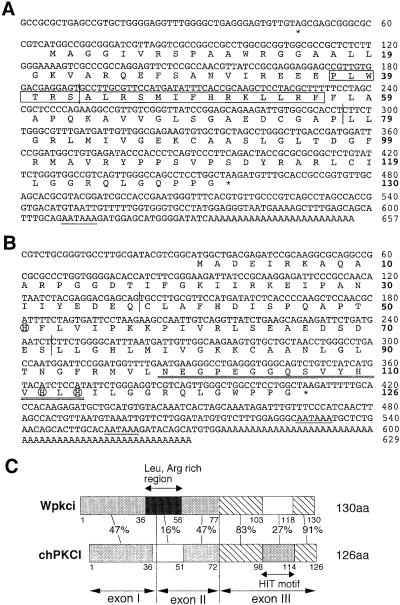
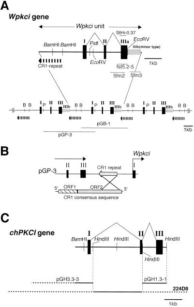
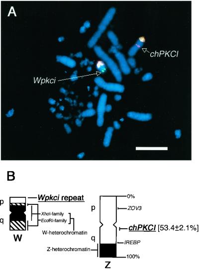
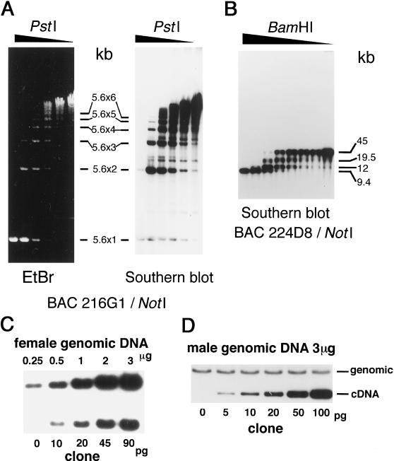
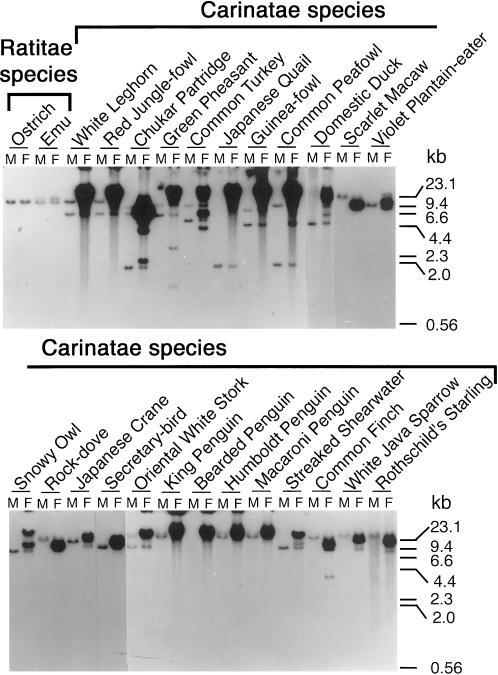
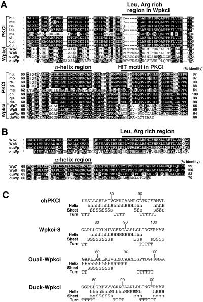
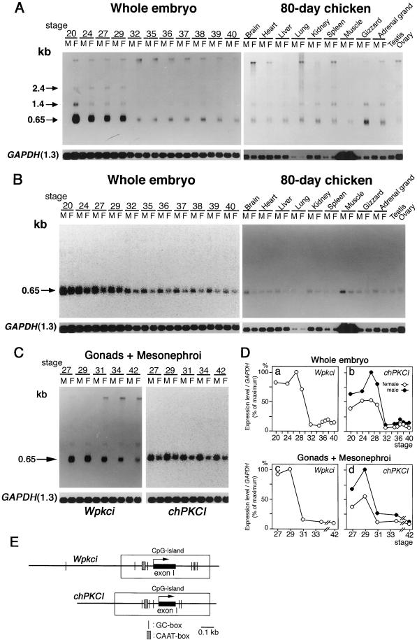
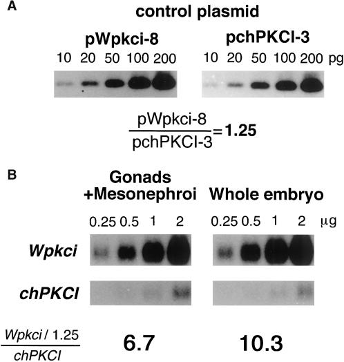
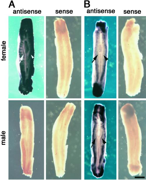
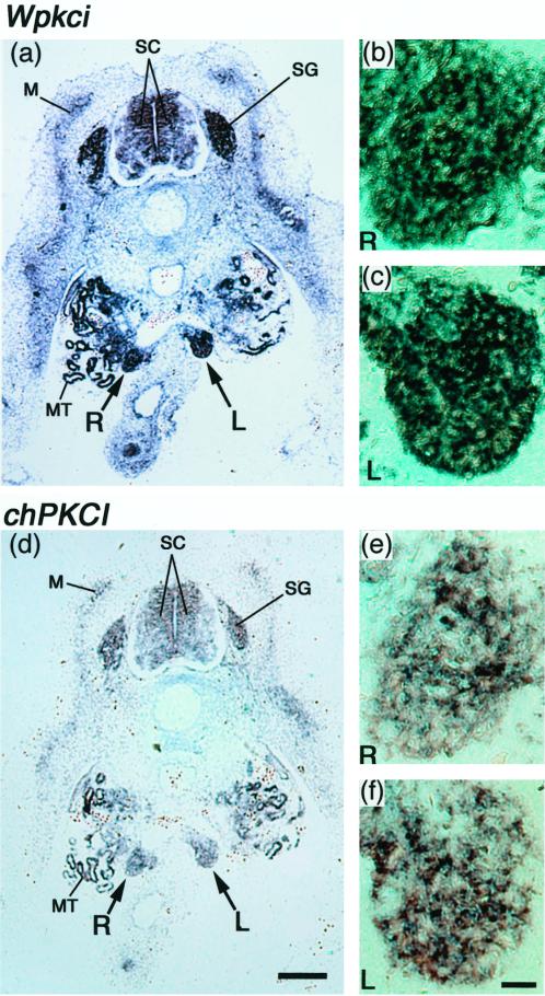
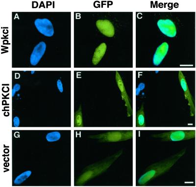
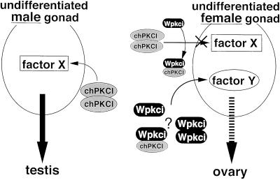
Two W chromosome-linked cDNA clones, p5fm2 and p5fm3, were obtained from a subtracted (female minus male) cDNA library prepared from a mixture of undifferentiated gonads and mesonephroi of male or female 5-d (stages 26-28) chicken embryos. These two clones were demonstrated to be derived from the mRNA encoding an altered form of PKC inhibitor/interacting protein (PKCI), and its gene was named Wpkci. The Wpkci gene reiterated approximately 40 times tandemly and located at the nonheterochromatic end of the chicken W chromosome. The W linkage and the moderate reiteration of Wpkci were conserved widely in Carinatae birds. The chicken PKCI gene, chPKCI, was shown to be a single-copy gene located near the centromere on the long arm of the Z chromosome. Deduced amino acid sequences of Wpkci and chPKCI showed approximately 65% identity. In the deduced sequence of Wpkci, the HIT motif, which is essential for PKCI function, was absent, but the alpha-helix region, which was conserved among the PKCI family, and a unique Leu- and Arg-rich region, were present. Transcripts from both Wpkci and chPKCI genes were present at significantly higher levels in 3- to 6-d (stages 20-29) embryos. These transcripts were detected in several embryonic tissues, including undifferentiated left and right gonads. When the green fluorescent protein-fused form of Wpkci was expressed in male chicken embryonic fibroblast, it was located almost exclusively in the nucleus. A model is presented suggesting that Wpkci may be involved in triggering the differentiation of ovary by interfering with PKCI function or by exhibiting its unique function in the nuclei of early female embryos.


Figures

Figure 1
Figure 1
The cDNA and deduced amino acid sequences of Wpkci (A) and chPKCI (B) and comparison of the levels of sequence identity of their protein subregions (C). The cDNA sequences of pWpkci-8 (A) and pchPKCI-3 (B) were determined. The N-terminal Met residue in A was assigned because of its location nearest to the termination codon in the 5′ untranslated region, and that in B was assigned by comparison with the mammalian PKCI sequences. In A and B, termination codons (asterisks), polyadenylation signals (underlines), and boundaries of exons (vertical lines) are indicated. The Leu- and Arg-rich region of Wpkci is boxed in A. The HIT motif containing the conserved His triad (HVHLH) is double-underlined, and the three His residues involved in the binding of zinc are circled in B. In C, the residue numbers for the N terminus, the C terminus, and the last residue of each region, correspondence to exons I–III, and levels (%) of identity of the deduced sequences between corresponding regions of Wpkci and chPKCI are indicated. The sequences of the inserts of pWpkci-8 and pchPKCI-3 are deposited in the DDBJ, EMBL, and GenBank nucleotide sequence databases with accession numbers AB026677 and AB026675, respectively.
Figure 2
Figure 2
Genomic structure of the tandemly reiterated Wpkci (A and B) and the nonreiterated chPKCI (C) genes determined by restriction mapping and sequencing of BAC clones (216G1 for Wpkci and 224D8 for chPKCI) and their subclones. Exons are indicated with closed rectangular boxes. The location of the partial sequence of CR1 between Wpkci genes is indicated in A and B. A minor 1.4-kb transcript of Wpkci contains an additional region to exon III, indicated as IIIb in A. Positions of cDNA clones (5fm2, 5fm3, and fst5.2-5) and genomic subclones (StHi-0.37, pGP-3, pGB-1, pGH3.3-3, and pGH1.3-1) are indicated. Accession numbers for the DDBJ, EMBL, and GenBank nucleotide sequence databases are AB026678 for pGB-1 and AB026679 for pGP-3.
Figure 3
Figure 3
Chromosomal locations of Wpkci and chPKCI demonstrated by FISH with the mixed probes. (A) A metaphase set from the female chicken embryonic fibroblast was hybridized with biotinylated pGP-3 (for Wpkci), DIG-labeled BAC clone 224D8 (for chPKCI), a 1:1 mixture of biotinylated and DIG-labeled pUGD1201 (for the EcoRI family on the W chromosome), and a 1:1 mixture of biotinylated and DIG-labeled pCZTH5–8 (for the pFN-1 macrosatellite on the Z chromosome). The biotinylated probes were detected with FITC–avidin, and the DIG-labeled probes were detected with sheep anti-DIG Fab fragment followed by rhodamine-labeled anti-sheep Fab. Locations of Wpkci on the nonheterochromatic end of the W chromosome and chPKCI near the centromere on the long arm of the Z chromosome are illustrated in B. The chPKCI locus on the Z chromosome was assigned by measuring 122 FISH figures.
Figure 4
Figure 4
Determination of reiteration frequencies of Wpkci and chPKCI in the chicken genome. (A) The 140-kb insert containing Wpkci genes was obtained from the BAC clone 216G1 by digestion with NotI and further digested with PstI at six different concentrations. The digests were separated by agarose gel electrophoresis, stained with ethidium bromide (left panel), and subjected to Southern blot hybridization with the 32P-labeled insert of the cDNA clone fst5.2-5 (right panel; see Figure 2A), which showed signals corresponding to multiples of the 5.6-kb repeating unit. (B) The insert of BAC clone 224D8 containing the chPKCI gene sequence was obtained by NotI digestion, further digested with BamHI at eight different concentrations, and subjected to Southern blot hybridization with the 32P-labeled cDNA fragment (nucleotide positions 30–410) of chPKCI. (C) Different amounts of EcoRV-digested genomic DNA of the female chicken (upper panel) and EcoRV-digested genomic DNA of the male chicken (3 μg each) mixed with different amounts of the linearized cDNA clone p5fm2 (3.83 kb) (lower panel; see also Figure 2A) were subjected to agarose gel electrophoresis and Southern blot hybridization with the 32P-labeled insert of p5fm2. Comparing the slopes of signal intensities for the former and the latter samples, 1 μg of the female genomic DNA and 72 pg of p5fm2 gave the same signal intensity. (D) The BamHI-digested genomic DNA of the male chicken (3 μg each) and different amounts of the HindIII-digested, linearized cDNA clone pchPKCI-3 were subjected to electrophoresis and Southern blot hybridization with the 32P-labeled subfragment (nucleotide positions 30–410) of pchPKCI-3. Comparing the mean signal intensity of the former and the slope of signal intensities of the latter samples, 3 μg of the male genomic DNA and 12 pg of pchPKCI gave the same signal intensity.
Figure 5
Figure 5
Presence of the reiterated Wpkci gene in the female genomes of Carinatae birds. EcoRI-digested genomic DNA preparations from male (M) and female (F) species of birds as indicated were subjected to agarose gel electrophoresis and Southern blot hybridization with the 32P-labeled genomic fragment StHi-0.37 (see Figure 2A).
Figure 6
Figure 6
Comparison of deduced amino acid sequences of mammalian, plant, and avian PKCI and avian Wpkci, and prediction of the α-helix for the fourth region of avian PKCI and Wpkci. (A) Deduced sequences of PKCI from human (hu.), mouse (mo.), rabbit (ra.), bovine (bo.), maize (ma.), chicken (ch.), Japanese quail (qu.), and domestic duck (du.) and of Wpkci from chicken (Wp7 and Wp8), Japanese quail, and domestic duck, abbreviated as above, were aligned, and three characteristic regions or a motif are indicated. Identical residues are shown with white letters on a black background and similar residues are shown on a stippled background. The overall level (%) of identity relative to the sequence of chicken PKCI is indicated at the end of each deduced sequence. Only a single residue is different between Wpkci-7 (Wp7) (H-81, minor type) and Wpkci-8 (Wp8) (R-81, major type) of chicken. (B) Alignment and comparison of the deduced sequences of Wpkci from chicken (Wp7 and Wp8), Japanese quail (qu), and domestic duck (du). Wpkci of domestic duck lacks the last 14 residues. (C) Prediction of α-helix (H or h) formation for the fourth region (see Figure 1C) of chPKCI and Wpkci of chicken (Wpkci-8), Japanese quail, and domestic duck. The cDNA sequences for Japanese quail PKCI (AB033882), domestic duck PKCI (AB033884), Wpkci-7 (AB033880), Japanese quail Wpkci (AB033881), and domestic duck Wpkci (AB033883) are deposited in the DDBJ, EMBL, and GenBank nucleotide sequence databases with the accession numbers shown in parentheses.
Figure 7
Figure 7
Transcripts of both Wpkci and chPKCI genes are present at higher levels in early embryonic stages. (A and B) Poly(A)+ RNA preparations from 3- to 14.5-d (stages 20–40) male (M) and female (F) chicken whole embryos or from different tissues of 80-d chickens, as indicated, were subjected to Northern blot hybridization with the 32P-labeled cDNA probe specific for Wpkci (63-bp probe) (A) or chPKCI (72-bp probe) (B) and with 32P-labeled GAPDH cDNA probe (A and B), followed by autoradiography (A and the right panel of B for the 80-d chicken) or fluorescence image analysis (the left panel of B for the whole embryo). (C) Poly(A)+ RNA preparations from undifferentiated gonads plus mesonephroi of 5- to 16-d (stages 27–42) male (M) or female (F) chicken embryos, as indicated, were subjected to Northern blot hybridization with the 32P-labeled Wpkci-specific, chPKCI-specific, or GAPDH cDNA probe, followed by autoradiography (the left panel for Wpkci) or fluorescence image analysis (the right panel for chPKCI). (D) Relative values of the signal intensity of Wpkci mRNA or chPKCI mRNA to GAPDH mRNA, determined from the same Northern blot analyses, were plotted for female (○) and male (●) embryos at different stages. (E) Some common transcriptional elements around exon I of Wpkci and chPKCI predicted from the genomic sequences determined for pGP-3 and pGH3.3-3 (see Figure 2).
Figure 8
Figure 8
Determination of the molar ratio of mRNAs for Wpkci and chPKCI. Different amounts of the linearized cDNA clone, pWpkci-8 or pchPKCI-3 (A), or different amounts of poly(A)+ RNA from gonads plus mesonephroi of 6-d (stage 29) female embryos or 6-d female whole embryos (B) were electrophoresed, blotted onto nylon membranes, and hybridized together with 32P-labeled cDNA probe specific for Wpkci, chPKCI, or GAPDH, respectively, and subjected to fluorescence image analysis. The ratio of signal intensity in A was obtained by comparing slopes of signal intensity for each cDNA clone. In B, the signal intensity for Wpkci mRNA or chPKCI mRNA was first corrected with that for GAPDH mRNA (the intensity of Wpkci mRNA was multiplied by 1.003 for the sample from whole embryo and by 1.38 for the sample from gonads plus mesonephroi), the slope of signal intensity for Wpkci mRNA was corrected by the ratio obtained in A, and the molar ratio of mRNAs for Wpkci and chPKCI was calculated.
Figure 9
Figure 9
Whole mount in situ hybridization to the part of a 4.5-d (stage 25) female or male chicken embryo containing mesonephroi and undifferentiated left (arrowhead) and right (arrow) gonads, with DIG-labeled antisense or sense riboprobe for Wpkci (A) or chPKCI (B). Wpkci transcripts were detected only in the female embryo, and the chPKCI transcripts were detected in both female and male embryos with the antisense probe, followed by the reaction with anti-DIG antibody–coupled alkaline phosphatase. Bar, 1 mm.
Figure 10
Figure 10
In situ detection of Wpkci and chPKCI transcripts in the tissue section of 5-d (stages 26 to 27) female chicken embryo. In situ hybridization was carried out as in Figure 9 to the 4-μm-thick paraffin sections containing left (L) and right (R) undifferentiated gonads with the antisense riboprobe for Wpkci (a–c) or chPKCI (d–f). Significant signals of hybridization were detected on both sides of gonads (a and d, and enlarged in b, c, e, and f), mesonephric tubule (MT), spinal cord (SC), spinal ganglion (SG), and myotome (M). Bar in d, 500 μm; bar in f, 50 μm.
Figure 11
Figure 11
Intracellular distribution of the GFP-fused form of Wpkci or chPKCI in male chicken embryonic fibroblasts. DAPI-stained nuclei (left panels), GFP fluorescence of the protein expressed (middle panels), and merged images of DAPI staining and GFP fluorescence (right panels) are shown for cells transfected with the expression vector for GFP-Wpkci (A–C), GFP-chPKCI (D–F), or GFP alone (G–I). Bars, 10 μm.
Figure 12
Figure 12
A model suggesting the involvement of chPKCI and Wpkci in triggering gonadal differentiation in chicken embryos.

References

    1. Asakawa S, Abe I, Kudoh Y, Kishi N, Wang Y, Kubota R, Kudoh J, Kawasaki K, Minoshima S, Shimizu N. Human BAC library: construction and rapid screening. Gene. 1997;20:69–79. - PubMed
    1. Barber DL, Sanders EJ, Aebersold R, Schneider WJ. The receptor for yolk lipoprotein deposition in the chicken oocyte. J Biol Chem. 1991;266:18761–18770. - PubMed
    1. Bennett CP, Docherty Z, Robb SA, Ramani P, Hawkins JR, Grant D. Deletion 9p and sex reversal. J Med Genet. 1993;30:518–520. - PMC - PubMed
    1. Brenner C, Garrison P, Gilmour J, Peisach D, Ringe D, Petsko GA, Lowenstein JM. Crystal structures of HINT demonstrate that histidine triad proteins are GalT-related nucleotide-binding proteins. Nat Struct Biol. 1997;4:231–238. - PMC - PubMed
    1. Brzoska PM, Chen H, Levin NA, Kuo WL, Collins C, Fu KK, Gray JW, Christman MF. Cloning, mapping, and in vivo localization of a human member of the PKCI-1 protein family (PRKCNH1) Genomics. 1996;36:151–156. - PubMed

Publication types

MeSH terms