. 2000 Nov;279(5):C1656-64.
doi: 10.1152/ajpcell.2000.279.5.C1656.

Smooth muscle myosin light chain kinase expression in cardiac and skeletal muscle

Affiliations

Smooth muscle myosin light chain kinase expression in cardiac and skeletal muscle

B P Herring et al. Am J Physiol Cell Physiol. 2000 Nov.

Abstract

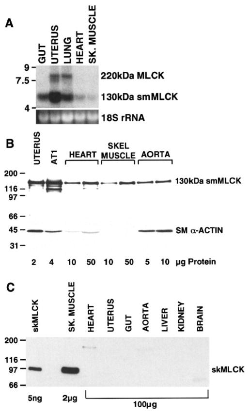
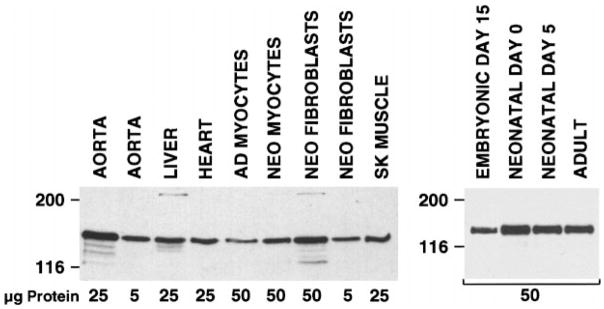
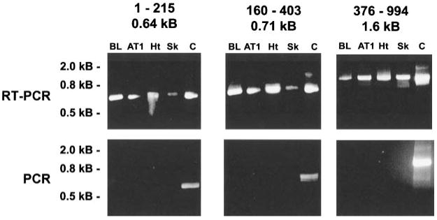
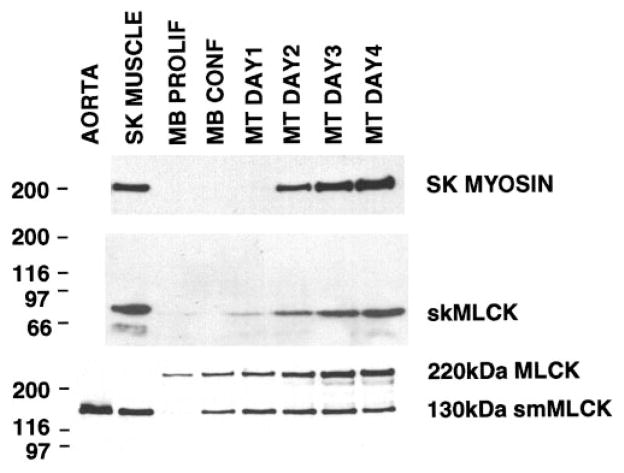
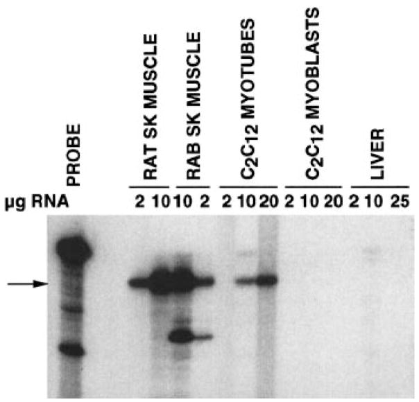
The purpose of this study was to characterize myosin light chain kinase (MLCK) expression in cardiac and skeletal muscle. The only classic MLCK detected in cardiac tissue, purified cardiac myocytes, and in a cardiac myocyte cell line (AT1) was identical to the 130-kDa smooth muscle MLCK (smMLCK). A complex pattern of MLCK expression was observed during differentiation of skeletal muscle in which the 220-kDa-long or "nonmuscle" form of MLCK is expressed in undifferentiated myoblasts. Subsequently, during myoblast differentiation, expression of the 220-kDa MLCK declines and expression of this form is replaced by the 130-kDa smMLCK and a skeletal muscle-specific isoform, skMLCK in adult skeletal muscle. These results demonstrate that the skMLCK is the only tissue-specific MLCK, being expressed in adult skeletal muscle but not in cardiac, smooth, or nonmuscle tissues. In contrast, the 130-kDa smMLCK is ubiquitous in all adult tissues, including skeletal and cardiac muscle, demonstrating that, although the 130-kDa smMLCK is expressed at highest levels in smooth muscle tissues, it is not a smooth muscle-specific protein.


Figures

Fig. 1
Fig. 1
Northern and Western blot analysis of myosin light chain kinase (MLCK) expression in mouse tissues. A: Northern blot in which 15 μg of total RNA, isolated from the indicated mouse tissues, were separated on a 1.4% agarose gel. A 5.8-kb mRNA corresponding to the 130-kDa smooth muscle MLCK (smMLCK) and an 8-kb mRNA corresponding to the 220-kDa MLCK were detected using an anti-sense cRNA probe to smMLCK as described in METHODS (top). Bottom: ethidium bromide stain of 18S rRNA. B: Western blot analysis of extracts from various mouse tissues and AT1 cardiac myocytes, reacted with a monoclonal antibody to smMLCK (top) or to smooth muscle α-actin (bottom). The amount of each extract analyzed is indicated in μg below the blots. C: Western blot analysis of mouse tissue extracts reacted with a polyclonal antibody specific for skeletal muscle MLCK (skMLCK). Lane 1, 5 ng of purified rabbit skMLCK. The amount of each extract analyzed is indicated in μg below the blot. Note that 50-fold less extract from skeletal muscle was analyzed compared with the other tissues. The cross-reacting band at 180–200 kDa in heart extract likely represents nonspecific binding to cardiac myosin.
Fig. 2
Fig. 2
smMLCK expression in cardiac myocytes. Immunoblot analysis of various rat tissue and cell extracts (left) and mouse heart extracts (right) as indicated. Amounts of extract analyzed are indicated under the blot. Adult heart cells were isolated by retrograde perfusion of collagenase through an adult rat heart, and unfractionated cells were analyzed. Neonatal myocytes and fibroblasts were prepared by digestion of hearts from newborn rats. Myocytes and fibroblasts were then separated on a Percoll gradient as described previously (16). This procedure results in a myocyte fraction that is >95% cardiac myocytes. Mouse hearts were harvested from embryonic, neonatal, and adult mice at the developmental stages indicated.
Fig. 3
Fig. 3
Immunolocalization of smMLCK expression in AT1 cardiac myocytes. AT1 cardiac myocytes were prepared for analysis as described in METHODS. A monoclonal antibody to sarcomeric α-actinin was obtained from Sigma (clone EA-53), and polyclonal antisera to T-antigen was a generous gift from Dr. Loren Field. Top right: a polyclonal antibody (C-T) that binds to the carboxy-terminal “telokin” region of the smMLCK (6) was used. Middle: an anti-peptide antibody (REP) directed against the 15-amino acid repeated motif in the amino terminus of the smMLCK (7) was used. Bottom: a monoclonal antibody (N-T) that binds to the amino terminus of smMLCK (Sigma, clone K56) (9) was used. Monoclonal antibodies were visualized using rhodamine-conjugated anti-mouse IgG and polyclonal antibodies using fluorescein-conjugated anti-rabbit IgG. Cells are shown at ×1,000 original magnification.
Fig. 4
Fig. 4
Alignment of the protein sequence of the mouse cardiac 130-kDa MLCK to mammalian smMLCKs. The deduced amino acid sequence of the mouse 130-kDa smMLCK, obtained from AT2 cardiac myocytes, is shown aligned to the sequences reported for the bovine (18), human (27), and rabbit (7) smMLCKs. The symbols below the alignment refer to residues that are identical (★) in all 4 of the sequences. The positions of the 3 sets of oligonucleotide primer pairs used for PCR are shown above the sequence, and the position of the catalytic core is bracketed. Sequences of all fragments isolated by RT-PCR from mouse bladder and AT1 cells were identical to the sequence of the cDNA clone obtained from AT2 cells.
Fig. 5
Fig. 5
RT-PCR analysis of MLCK expression. Top: cDNA was generated by reverse transcription from RNA isolated from the indicated tissues and cell lines. MLCK cDNAs were amplified using oligonucleotide primers based on the sequence of the 130-kDa mouse smMLCK. Three sets of primers were used to generate three fragments spanning the coding region. The fragments from these reactions were compared by agarose gel electrophoresis and compared with the size of a positive control that used PCR and each primer set to amplify the corresponding fragment from the AT2-derived cDNA (Fig. 4). Bottom: negative control reactions incubated in the absence of RT. The numbers above each gel indicate the first and last amino acid residues of the mouse smMLCK that correspond to each fragment and the size of the PCR fragment. Molecular mass markers are indicated at left. PCR fragments from bladder and AT1 cells were subcloned and sequenced. BL, bladder; AT1, AT1 mouse cardiac myocytes; Ht, heart; Sk, skeletal muscle; C, positive control MLCK plasmid.
Fig. 6
Fig. 6
MLCK expression during C2C12 cell differentiation. Western blot analysis of skeletal, 130-kDa smooth, and 220-kDa MLCK and skeletal muscle myosin expression during in vitro differentiation of C2C12 skeletal muscle cells. Extracts were prepared from adult mouse aorta and skeletal (SK) muscle, from C2C12 myoblasts grown to low density (MB Prolif) or high density (MB Conf) in the presence of 20% FCS, or from confluent myoblasts that were switched to media containing 2% horse serum for 1–4 days as indicated. Twenty-five micrograms of cell extracts were analyzed in each lane with the exception of the skeletal muscle extract (top), and the aorta extract (bottom), which represent only 5 μg of extract. The following antibodies were used for the analysis; polyclonal anti-rabbit skMLCK (middle), monoclonal anti-fast skeletal myosin (Sigma clone MY32; top), monoclonal anti-smMLCK (Sigma clone K57, bottom). The positions of molecular mass markers (in kDa) are indicated at left.
Fig. 7
Fig. 7
RNase protection analysis of skMLCK mRNA in C2C12 cells. A 32P-labeled 335-bp antisense RNA probe derived from the catalytic core of the rat skMLCK cDNA (14) was hybridized in solution to the indicated amounts of total RNA isolated from adult rat and rabbit (RAB) skeletal muscle, C2C12 myoblasts or C2C12 myotubes (differentiated for 4 days) as described previously (14). After hybridization (42°C, 80% formamide), unprotected probe was digested with RNase as described in METHODS. Protected fragments were separated by electrophoresis on denaturing polyacrylamide gels and visualized by autoradiography. Undigested probe is shown in the left lane. Arrow, position of the 335-bp fragment fully protected from digestion by the skMLCK mRNA. The smaller fragment seen in the rabbit lanes corresponds to a partially protected fragment that results from species differences in the nucleotide sequence between rat and rabbit skMLCK.

Similar articles

Cited by

References

    1. Amano M, Ito M, Kimura K, Fukata Y, Chihara K, Nakano T, Matsuura Y, Kaibuchi K. Phosphorylation and activation of myosin by Rho-associated kinase (Rho-kinase) J Biol Chem. 1996;271:20246–20249. - PubMed
    1. Cavadore JC, Molla A, Harricane MC, Gabrion J, Benyamin Y, Demaille JG. Subcellular localization of myosin light chain kinase in skeletal, cardiac, and smooth muscles. Proc Natl Acad Sci USA. 1982;79:3475–3479. - PMC - PubMed
    1. Chew TL, Masaracchia RA, Goeckeler ZM, Wysolmerski RB. Phosphorylation of nonmuscle myosin II regulatory light chain by p21-activated kinase (gamma-PAK) J Muscle Res Cell Motil. 1998;19:839–854. - PubMed
    1. Field LJ. Atrial natriuretic factor-SV40 T antigen transgenes produce cardiac tumors and cardiac arrhythmias in mice. Science. 1988;239:1029–1033. - PubMed
    1. Fisher SA, Ikebe M. Developmental and tissue distribution of expression of nonmuscle and smooth muscle isoforms of myosin light chain kinase. Biochem Biophys Res Commun. 1995;217:696–703. - PubMed

Publication types

Substances