. 2000 Nov;182(21):5948-53.
doi: 10.1128/JB.182.21.5948-5953.2000.

Regulation of ferritin-mediated cytoplasmic iron storage by the ferric uptake regulator homolog (Fur) of Helicobacter pylori

Affiliations

Regulation of ferritin-mediated cytoplasmic iron storage by the ferric uptake regulator homolog (Fur) of Helicobacter pylori

S Bereswill et al. J Bacteriol. 2000 Nov.

Abstract

Homologs of the ferric uptake regulator Fur and the iron storage protein ferritin play a central role in maintaining iron homeostasis in bacteria. The gastric pathogen Helicobacter pylori contains an iron-induced prokaryotic ferritin (Pfr) which has been shown to be involved in protection against metal toxicity and a Fur homolog which has not been functionally characterized in H. pylori. Analysis of an isogenic fur-negative mutant revealed that H. pylori Fur is required for metal-dependent regulation of ferritin. Iron starvation, as well as medium supplementation with nickel, zinc, copper, and manganese at nontoxic concentrations, repressed synthesis of ferritin in the wild-type strain but not in the H. pylori fur mutant. Fur-mediated regulation of ferritin synthesis occurs at the mRNA level. With respect to the regulation of ferritin expression, Fur behaves like a global metal-dependent repressor which is activated under iron-restricted conditions but also responds to different metals. Downregulation of ferritin expression by Fur might secure the availability of free iron in the cytoplasm, especially if iron is scarce or titrated out by other metals.


Figures

FIG. 1
FIG. 1
Transcriptional organization of the H. pylori fur (A) and pfr (B) genes. The genes are numbered according to the annotated genome sequence of H. pylori strain 26695 (28). The insertion sites and orientations of promoterless cat cassettes inserted in the fur and pfr mutants of strains NCTC11638 and G27, respectively, are marked by circles (cat). The transcriptional direction of the cat gene is indicated by a short arrow.
FIG. 2
FIG. 2
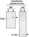
Analysis of the mutagenized fur gene by Southern hybridization. HindIII- and Sau3A-digested DNAs from the strain NCTC11638 (lanes 1) and the fur mutant NCTC11638-FUR (lanes 2) were hybridized with probes specific for fur and cat, respectively. The arrows indicate hybridizing DNA fragments of the predicted sizes, demonstrating correct replacement of the wild-type fur gene with the interrupted version. The sizes of individual marker DNA fragments are indicated on the left.
FIG. 3
FIG. 3
Growth characteristics of the H. pylori fur mutant. The H. pylori strain NCTC11638 (solid bars) and the fur mutant (shaded bars) were grown in BBF medium with iron and nickel at various concentrations. For high-iron, low-iron, and high-nickel conditions, the BBF medium was supplemented with 1 mM FeCl3, 20 μM desferal, or 1 mM nickel, respectively. Bacterial growth was determined after 48 h by measuring the optical density (OD) at 600 nm. The values represent the means of three independent determinations. The standard deviation is indicated above each bar.
FIG. 4
FIG. 4
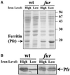
Influence of Fur on ferritin synthesis in response to iron starvation. (A) Bacteria of strain NCTC11638 (wild type [wt]) and the isogenic mutant (fur) were grown for 48 h under iron-rich conditions (BBF liquid medium [High]) and under iron-restricted conditions generated with desferal at a concentration of 20 μM (Low). Proteins were analyzed by SDS-PAGE (15% gel) and stained with Coomassie blue. The ferritin protein is marked by the arrow on the left. The sizes of individual marker proteins are indicated on the right. (B) Western blot analysis of ferritin production in strain NCTC11638 (wt) and the isogenic mutant (fur) grown under iron-rich and iron-restricted conditions as described for panel A with the ferritin-specific antiserum AK198. The Pfr protein is marked by the arrow on the right.
FIG. 5
FIG. 5
Influence of the fur mutation on transcription of the pfr gene. Total RNAs (10 μg) isolated from H. pylori strains NCTC11638 wild type (wt) and NCTC11638-FUR (fur) grown in BBF medium (high) and in BBF medium with the iron chelator desferal (20 μM) (low) were hybridized with DIG-labeled pfr antisense mRNA (upper gel). The bands corresponding to pfr mRNA are indicated on the right. The lower gel shows a methylene blue stain of transferred RNA prior to hybridization. The positions of 16S and 23S rRNAs are indicated on the right.
FIG. 6
FIG. 6
Role of Fur in repression of ferritin synthesis by different metals. Strain NCTC11638 (wild type [wt]) and the fur mutant (fur) were grown in BBF liquid medium in the presence of zinc, copper, manganese, and nickel at concentrations of 100 and 500 μM. Proteins were analyzed by SDS-PAGE (15% gel) and stained with Coomassie blue. The ferritin protein (Pfr) is indicated by the arrows on the right.
FIG. 7
FIG. 7
Expression of the pfr gene in response to iron starvation and nickel supplementation determined with a pfr::cat fusion. The isogenic pfr mutant strain, in which the promoterless cat gene is fused to the pfr gene, was grown in the presence of nickel at increasing concentrations and under conditions of iron starvation generated with the iron chelator desferal (20 μM). The production of the Cat protein resembling transcriptional activity of the pfr gene was monitored by Cat-specific ELISA. The amount of Cat produced under iron-replete conditions in BBF medium was set at 100%. The values represent the means of three independent determinations. The error bars indicate standard deviations.
FIG. 8
FIG. 8
Schematic representation of iron metabolism in H. pylori under iron-rich, iron-restricted, and metal-rich conditions. The putative regulatory roles of Fur are indicated. Iron flux in the cytoplasm is indicated by the arrows. Free iron is indicated by the solid circles. The possible competitive inhibitory interactions between free iron and other metals (shaded circles) are indicated by a double arrow. The protein shell of ferritin is shown by the open circle. Repression of ferritin synthesis under conditions of iron starvation or metal overload, as well as release of ferritin-bound iron, is indicated by the dashed circle. Thick and thin arrows at the membrane indicate high and low transport, respectively. Arrows with two bars indicate inhibition of ferritin synthesis.

Similar articles

Cited by

References

    1. Abdul-Tehrani H, Hudson A J, Chang Y S, Timms A R, Hawkins C, Williams J M, Harrison P, Guest J R, Andrews S C. Ferritin mutants of Escherichia coli are iron deficient and growth impaired, and fur mutants are iron deficient. J Bacteriol. 1999;181:1415–1428. - PMC - PubMed
    1. Alm R A, Ling L S, Moir D T, King B L, Brown E D, Doig P C, Smith D R, Noonan B, Guild B C, deJonge B L, Carmel G, Tummino P J, Caruso A, Uria-Nickelsen M, Mills D M, Ives C, Gibson R, Merberg D, Mills S D, Jiang Q, Taylor D E, Vovis G F, Trust T J. Genomic-sequence comparison of two unrelated isolates of the human gastric pathogen Helicobacter pylori. Nature. 1999;397:176–180. - PubMed
    1. Althaus E W, Outten C E, Olson K E, Cao H, O'Halloran T V. The Ferric Uptake Regulation (Fur) repressor is a zinc metalloprotein. Biochemistry. 1999;38:6559–6569. - PubMed
    1. Andrews S C. Iron storage in bacteria. Adv Microb Physiol. 1998;40:281–351. - PubMed
    1. Ausubel F M, Brent R, Kingston R E, Moore D D, Seidman J G, Smith J A, Struhl K. Short protocols in molecular biology. New York, N.Y: John Wiley & Sons; 1992.

Publication types

LinkOut - more resources