Comparative Study
. 2000 Nov;182(21):5997-6004.
doi: 10.1128/JB.182.21.5997-6004.2000.

Characterization of the hydrogen-deuterium exchange activities of the energy-transducing HupSL hydrogenase and H(2)-signaling HupUV hydrogenase in Rhodobacter capsulatus

Affiliations
Comparative Study

Characterization of the hydrogen-deuterium exchange activities of the energy-transducing HupSL hydrogenase and H(2)-signaling HupUV hydrogenase in Rhodobacter capsulatus

P M Vignais et al. J Bacteriol. 2000 Nov.

Abstract

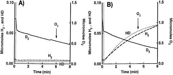
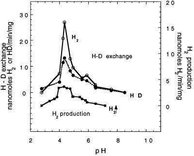
Rhodobacter capsulatus synthesizes two homologous protein complexes capable of activating molecular H(2), a membrane-bound [NiFe] hydrogenase (HupSL) linked to the respiratory chain, and an H(2) sensor encoded by the hupUV genes. The activities of hydrogen-deuterium (H-D) exchange catalyzed by the hupSL-encoded and the hupUV-encoded enzymes in the presence of D(2) and H(2)O were studied comparatively. Whereas HupSL is in the membranes, HupUV activity was localized in the soluble cytoplasmic fraction. Since the hydrogenase gene cluster of R. capsulatus contains a gene homologous to hoxH, which encodes the large subunit of NAD-linked tetrameric soluble hydrogenases, the chromosomal hoxH gene was inactivated and hoxH mutants were used to demonstrate the H-D exchange activity of the cytoplasmic HupUV protein complex. The H-D exchange reaction catalyzed by HupSL hydrogenase was maximal at pH 4. 5 and inhibited by acetylene and oxygen, whereas the H-D exchange catalyzed by the HupUV protein complex was insensitive to acetylene and oxygen and did not vary significantly between pH 4 and pH 11. Based on these properties, the product of the accessory hypD gene was shown to be necessary for the synthesis of active HupUV enzyme. The kinetics of HD and H(2) formed in exchange with D(2) by HupUV point to a restricted access of protons and gasses to the active site. Measurement of concentration changes in D(2), HD, and H(2) by mass spectrometry showed that, besides the H-D exchange reaction, HupUV oxidized H(2) with benzyl viologen, produced H(2) with reduced methyl viologen, and demonstrated true hydrogenase activity. Therefore, not only with respect to its H(2) signaling function in the cell, but also to its catalytic properties, the HupUV enzyme represents a distinct class of hydrogenases.


Figures

FIG. 1
FIG. 1
Gene organization at the hup locus of the chromosome of R. capsulatus. The coding region at the hup locus of R. capsulatus chromosome comprises 21 ORFs, all contiguous and transcribed from the same strand. At the 5′ end, it is separated by around 500 nt from the mcpA and mcpB genes transcribed in the opposite direction (28). The positions of known promoters and plasmid inserts are shown. B, BamHI; H, HindIII; P, PstI; S, SalI.
FIG. 2
FIG. 2
Nitrogenase-mediated H2 and HD production, in the presence of D2 and H2O, by the RCC44 mutant. The RCC44 mutant (lacking the hoxH, hupTUV, and the hypF genes) was grown photoheterotrophically in either MN medium (A) or MG medium (B). (A) The MN culture (1.5 ml, 0.7 mg of protein) was sparged with D2, and the reaction vessel was closed at the time indicated by the vertical dotted line. The curves (not corrected for the consumption of gasses of the apparatus) exhibit the real changes with time in D2 (▄), H2 (–––), and HD (•••) concentrations in the reaction chamber. (B) The MG culture (1.5 ml, 0.6 mg of protein) in the reaction chamber was sparged with D2. At the time indicated by the vertical dotted line, the cell was closed and the H-D exchange reaction in whole cells was measured under light (light on) or in darkness (light off) and after addition of 10 mM ammonium sulfate, as indicated by arrows. The curves have been corrected for gas consumption by the mass spectrometer.
FIG. 3
FIG. 3
Time course of H2 and HD production in the D2-H2O system by the HupUV protein complex in JBC13 cells. Cells were grown anaerobically in the light in MN medium. The H-D exchange reaction in whole cells of JBC13 (hupSL hoxH double mutant) (3.7 mg of protein) was measured in 1.5 ml of 50 mM Tris-HCl buffer (pH 8.0). After gassing the cell suspension with D2, the reaction chamber was closed (vertical grey lines), and the H-D exchange reaction was allowed to proceed. At 18 min, the reaction chamber was regassed with D2; at 22 min, the light was turned off and the vessel was closed; at 27 min, H2O2 (5 μl, 0.3%) was added and O2 was liberated by decomposition (the lower trace shows O2 concentrations measured at a mass of 32 Da); at 30 min, the chamber was regassed with D2; at 35 min, the light was turned off, the vessel was closed, and there was new addition of H2O2 (10 μl, 0.3%). The figure shows the real gas concentrations of H2 (▄), HD (••••), and O2 (▄ ▄ ▄) in the reaction chamber.
FIG. 4
FIG. 4
Time course of H2 and HD production and D2 consumption in the D2-H2O system by the HupSL (A) and HupUV (B) protein complexes. Cells were grown anaerobically in the light in MN medium. The H-D exchange reaction in whole cells of BSE16, a ΔhupUV mutant (2.5 mg of protein) (A), and in whole cells of JP91(pAC206), a hupSL mutant, with the hupTUV operon-containing plasmid pAC206 (5.2 mg of protein) (B), was measured in 50 mM citrate-phosphate buffer (pH 7.0). At the time indicated by the vertical dotted line, the reaction vessel was closed, and the concentrations of D2 (▄), H2 (----), and HD (····) were recorded. The arrows indicate the time of O2 appearance in the medium after H2O2 addition (2 μl of 0.3% H2O2) and the time of ferricyanide addition (10 mM). The changes in O2 concentration were monitored at a mass of 32 Da (data not shown). The figure shows the real concentrations of the hydrogen species present in the vessel.
FIG. 5
FIG. 5
Effect of acetylene on the H-D exchange reaction catalyzed by the hupSL-encoded hydrogenase (A) and by the hupUV-encoded hydrogenase (B). The conditions were the same as those in Fig. 4, with cells grown overnight anaerobically in the light in MN medium. H2 (▄ ▄ ▄) and HD (····) production in exchange with D2 (––––) uptake catalyzed by whole cells of BSE16 (2.5 mg of protein) (A) and JP91(pAC206) (5.2 mg of protein) (B) was measured at pH 7 after the cells had been incubated for 1 h at room temperature under a gas phase of C2H2-Ar (1:1). The figure shows the real concentrations of the hydrogen species in the vessel.
FIG. 6
FIG. 6
HupUV hydrogenase activities in the soluble cytosolic fraction of JP91(pAC206) cells as a function of pH. JP91(pAC206) cells grown photoheterotrophically in MN medium were broken by passage through a French pressure cell. The soluble cytoplasmic fraction obtained by centrifugation at 100,000 × g for 70 min was used to determine HupUV hydrogenase activities. (A) H2 production linked to MV oxidation at pH 4.0. The phosphate-citrate buffer (1.25 ml, final concentration of 100 mM) in the reaction chamber was first sparged with argon to remove O2, and then the reaction vessel was closed, and 0.25 ml of soluble cytosolic fraction (0.9 mg of protein) was added. Two minutes later, MV+ (50 μl, final concentration of 120 mM) was injected into the reaction vessel. (B) pH dependence of MV+-mediated H2 production and H2 and HD formation in exchange with D2. Initial rates determined for the first minute of H2 (○) and HD (●) production (in 1.5 ml, 0.8 mg of protein) are plotted versus pH. To measure the H-D exchange, the reaction vessel was sparged first with D2. H2 (■) was formed by proton reduction with MV+. The buffers used (final concentration of 100 mM) were phosphate-citrate (pH 2.9 to 7.0), phosphate-Tris (pH 6.6 to 8.5), phosphate-glycine-NaOH (pH 7.5 to 10), and glycine-NaOH (pH 9.0 to 12.8).
FIG. 7
FIG. 7
pH dependence of the H-D exchange reaction and H2 production catalyzed by the hupSL-encoded hydrogenase in BSE16 membranes. The experimental conditions were the same as those in Fig. 6. H2 (○) and HD (●) were produced by the H-D exchange reaction and 0.4 mg of protein. Production of H2 (■) was measured with MV+ and 0.8 mg of protein.

Similar articles

Cited by

References

    1. Appel J, Schulz R. Hydrogen metabolism in organisms with oxygenic photosynthesis: hydrogenases as important regulatory devices for a proper redox poising? J Photochem Photobiol. 1998;47:1–11.
    1. Black L K, Fu C, Maier R J. Sequences and characterization of hupU and hupV genes of Bradyrhizobium japonicum encoding a possible nickel-sensing complex involved in hydrogenase expression. J Bacteriol. 1994;176:7102–7106. - PMC - PubMed
    1. Cauvin B, Colbeau A, Vignais P M. The hydrogenase structural operon in Rhodobacter capsulatus contains a third gene, hupM, necessary for the formation of a physiologically competent hydrogenase. Mol Microbiol. 1991;5:2519–2527. - PubMed
    1. Colbeau A, Elsen S, Tomiyama M, Zorin N A, Dimon B, Vignais P M. Rhodobacter capsulatus HypF is involved in regulation of hydrogenase synthesis through the HupUV proteins. Eur J Biochem. 1998;251:65–71. - PubMed
    1. Colbeau A, Godfroy A, Vignais P M. Cloning of DNA fragments carrying hydrogenase genes of Rhodopseudomonas capsulata. Biochimie. 1986;68:147–155. - PubMed

Publication types

LinkOut - more resources