. 2000 Oct;131(4):695-704.
doi: 10.1038/sj.bjp.0703621.

Bicuculline, pentobarbital and diazepam modulate spontaneous GABA(A) channels in rat hippocampal neurons

Affiliations

Bicuculline, pentobarbital and diazepam modulate spontaneous GABA(A) channels in rat hippocampal neurons

B Birnir et al. Br J Pharmacol. 2000 Oct.

Abstract

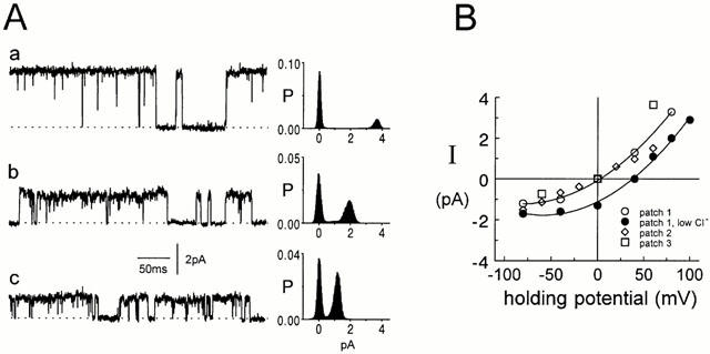
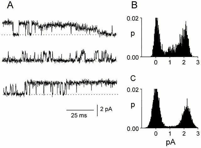
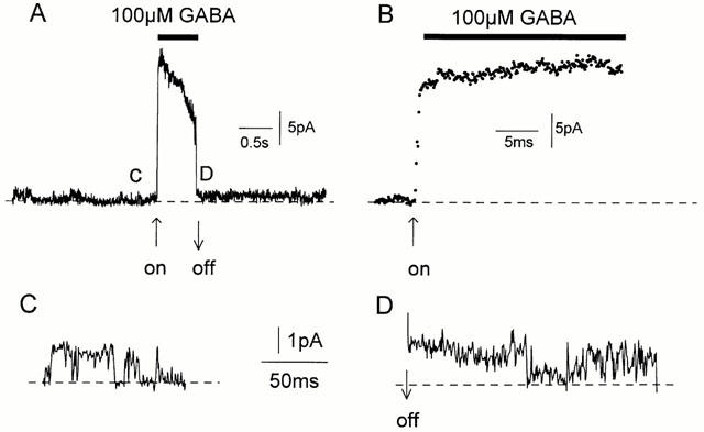
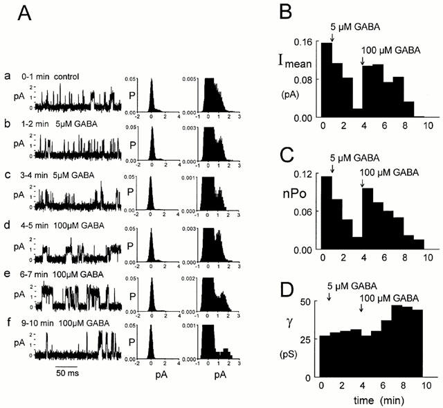
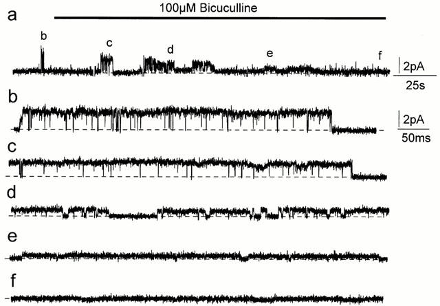
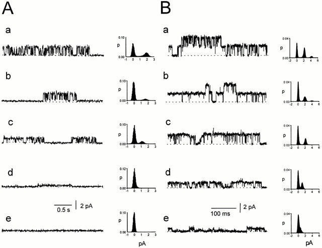
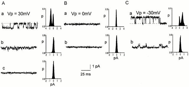
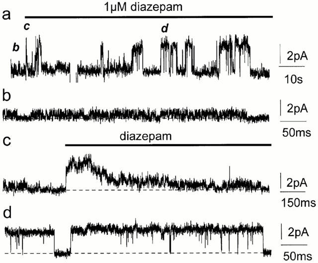
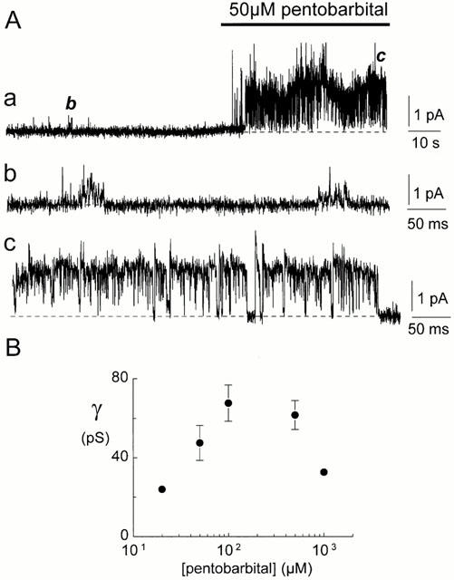
Spontaneously opening, chloride-selective channels that showed outward rectification were recorded in ripped-off patches from rat cultured hippocampal neurons and in cell-attached patches from rat hippocampal CA1 pyramidal neurons in slices. In both preparations, channels had multiple conductance states and the most common single-channel conductance varied. In the outside-out patches it ranged from 12 to 70 pS (Vp=40 mV) whereas in the cell-attached patches it ranged from 56 to 85 pS (-Vp=80 mV). Application of GABA to a patch showing spontaneous channel activity evoked a rapid, synchronous activation of channels. During prolonged exposure to either 5 or 100 microM GABA, the open probability of channels decreased. Application of GABA appeared to have no immediate effect on single-channel conductance. Exposure of the patches to 100 microM bicuculline caused a gradual decrease on the single-channel conductance of the spontaneous channels. The time for complete inhibition to take place was slower in the outside-out than in the cell-attached patches. Application of 100 microM pentobarbital or 1 microM diazepam caused 2 - 4 fold increase in the maximum channel conductance of low conductance (<40 pS) spontaneously active channels. The observation of spontaneously opening GABA(A) channels in cell-attached patches on neurons in slices suggests that they may have a role in neurons in vivo and could be an important site of action for some drugs such as benzodiazepines, barbiturates and general anaesthetics.


Figures

Figure 1
Figure 1
Spontaneous channels in outside-out patches. (A) Variable conductance. Current records from three different outside-out patches from cultured hippocampal neurons (a–c) at a potential of +60 mV. The maximum conductance of the channels was 66 (Aa), 42 (Ab) and 25 pS (Ac). Currents were digitally filtered at 2 kHz. The corresponding all-points histograms on the right of each trace are from 10 s current records. The dotted lines represent the level of the baseline current. (B) Chloride selectivity. Single-channel current amplitudes are plotted against the pipette potential for three different outside-out patches (open circle, diamond and square). All currents reversed at 0 mV. In one of the patch currents were first recorded in symmetrical Cl solutions (146 mM, open circles) and then currents were recorded again when the bath Cl concentration had been lowered to 30 mM (iso-osmolality was maintained with gluconate, filled circles). The reversal potential of the currents was shifted to +38 mV.
Figure 2
Figure 2
Conductance states of channels in CA1 pyramidal neurons. The channels can adopt various conductance states but subconductance states are not as frequent as the main, largest conducting state. (A) The current traces are from a cell-attached patch at a pipette potential of −40 mV and are continuous. (B) All-points histograms from the 300 ms of current trace shown in (A). (C) All-points histogram from the same patch as in (B) but from a longer (3 s) current record.
Figure 3
Figure 3
Rapid activation of channels by GABA. (A) 100 μM GABA was rapidly applied (horizontal bar) to an outside-out patch (Vp=+40 mV) containing spontaneously active 37 pS channels (see C). The ‘peak' current response was 21 pA (525 pS). (B) The rising phase of the current shown in (A) at a faster time scale. The data points are at 100 μs intervals. (C). Spontaneous single-channel currents before GABA application. (D) Spontaneous single-channel currents immediately after GABA application was stopped.
Figure 4
Figure 4
GABA modulation of spontaneous channels recorded in an outside-out patch. (A) Traces (200 ms) of single-channel currents under the conditions shown above each trace (a–f). One-minute all-points histograms are shown on two vertical scales alongside each trace. The vertical scale on the histograms to the right was chosen to emphasise the peak representing the open channel currents. The dotted lines represent the level of the baseline current. Currents were filtered at 5 kHz. (B) Mean current (Imean). The mean current was determined in successive 1 min periods after patch formation (Vp=+40 mV). Five and 100 μM GABA were applied after 1 and 4 min, respectively. (C) Channel open probability (nPo). Results were obtained from the same current record as used in (B) and the open probability was determined in 1 min consecutive current segments. (D) Channel conductance as a function of time. The conductance was determined from the peak in consecutive 1 min all-points current amplitude histograms.
Figure 5
Figure 5
Bicuculline inhibition of spontaneously active channels. (A) 100 μM bicuculline inhibited spontaneous channel currents recorded in an outside-out patch from cultured hippocampal neurons (Vp=+60 mV). (a) A compressed 3.8 min current trace. Initially the maximum channel conductance was 42 pS (b) but after exposure to bicuculline (c–f) it decreased gradually. The conductances were 30 (c), 15 (d), 8 (e) and 0 pS (f) at 0.5, 1.1, 2.1 and 3.1 min after bicuculline application started, respectively. The horizontal bar denotes the bicuculline application and the breaks in the current record are due to brief changes in the holding potential. Currents are filtered at 5 kHz. Calibration bars for trace a are shown beside the trace. Calibrations for traces (b–f) are beside trace (b). The dotted lines represent the level of the baseline current.
Figure 6
Figure 6
Effects of bicuculline on spontaneous channels in CA1 pyramidal neurons. Spontaneously opening channels were recorded in a cell-attached patch on CA1 hippocampal neurons in the slice preparation at a pipette potential of −40 mV. (A) 100 μM bicuculline was then injected into the tip of the patch pipette. Initially the maximum channel conductance was 50 pS (a) but after exposure to bicuculline (b–e) it decreased gradually. The conductances were 40 (b), 29 (c), 9 (d) and 0 pS (e) at 5, 15, 25 and 40 s after bicuculline application, respectively. The all-points histograms were each obtained from 10 s current records and are in time sequence after bicuculline injection. The dotted lines represent the level of the baseline current. (B) Effect of 100 μM bicuculline plus GABA. (a) The all-points histogram is from 16 s of current record before bicuculline and GABA were injected into the pipette tip. (b–e). Each histogram is from 2 min of current record after bicuculline plus GABA had been injected into the pipette tip. The representative current traces shown were recorded 0.3 min (b), 3.3 min (c), 5.6 min (d) and 7.9 min (e) after injection of bicuculline plus GABA.
Figure 7
Figure 7
No effect of bicuculline on the reversal potential of channels in CA1 pyramidal neurons. Spontaneously opening channels were recorded in a cell-attached patch before (a) and after (b, c) injection of 100 μM bicuculline into the pipette tip. Representative current traces are shown at pipette potentials of: A. 30 mV, B. 0 mV and C. −30 mV. Histograms from 10 s current records are shown alongside each trace. The dotted lines represent the level of the baseline current.
Figure 8
Figure 8
Diazepam modulation of spontaneously active channels. Diazepam enhanced spontaneous channel currents in outside-out patches from cultured hippocampal neurons (Vp=+40 mV). (a) A compressed 1.7 min current trace, the letters (b–d) refer to the traces that follow and show the corresponding section of the experiment at a faster time scale. Initially the maximum spontaneous channel conductance was about 22 pS (a,b). The channel conductance increased rapidly to 77 pS when exposed to 1 μM diazepam but then decreased (a,c). 77 pS channels were later maintained in the presence of 1 μM diazepam (a,d). The horizontal bar in (a,c) denote the diazepam application and breaks in the current record are due to brief changes in the holding potential. The dotted lines represent the level of the baseline current. Currents are filtered at 5 kHz. Calibrations for each trace are shown on the right.
Figure 9
Figure 9
Pentobarbital modulation of spontaneously active channels. (A) Pentobarbital enhanced spontaneous channel currents in an inside-out patch from cultured hippocampal neurons (Vp=−40 mV). (a) A compressed 2.2 min current trace where the horizontal bar denotes the pentobarbital application. Initially the average spontaneous channel conductance was about 16 pS (a,b). The average channel conductance increased when the patch was exposed to 50 μM pentobarbital (a,c, 43 pS). The dotted lines represent the level of the baseline current. The current trace in (a) is filtered at 2 kHz, the traces in (b,c) at 5 kHz. Calibrations for each trace are shown on the right. (B) The effect of pentobarbital concentration on the average single-channel conductance of the spontaneously active channels. The data were obtained from ten inside-out and four outside-out patches. Data points represent the mean conductance±s.e.mean if larger than the symbol. The average conductance of the spontaneously active channels before exposure to pentobarbital was 15±2 pS.

Similar articles

Cited by

References

    1. BARKER J.L., HARRISON N.L., LANGE G.D., OWEN D.G. Potentiation of γ-aminobutyric-acid-activated chloride conductance by a steroid anaesthetic in cultured rat spinal neurons. J. Physiol. 1987;386:485–501. - PMC - PubMed
    1. BARNARD E.A., SKOLNICK P., OLSEN R.W., MOHLER H., SIEGHART W., BIGGIO G., BRAESTRUP C., BATESON A.N., LANGER S.Z. International Union of Pharmacology. XV. Subtypes of γ-aminobutyric acidA receptors: classification on the basis of subunit structure and receptor function. Pharmacol. Rev. 1998;50:291–313. - PubMed
    1. BIRNIR B., EGHBALI M., EVERITT A.B., COX G.B., GAGE P.W. Non-synaptic GABA-gated receptors exhibit great plasticity in channel conductance. Biophys. J. 1999;76:A338.
    1. BIRNIR B., EVERITT A.B., GAGE P.W. Characteristics of GABAA channels in rat dentate gyrus. J. Membrane. Biol. 1994;142:93–102. - PubMed
    1. BIRNIR B., EVERITT A.B., LIM M.S.F., GAGE P.W. Spontaneously opening GABAA channels in CA1 pyramidal neurones of rat hippocampus. J. Membrane Biol. 2000;174:21–29. - PubMed

LinkOut - more resources