. 2000 Oct 16;19(20):5269-80.
doi: 10.1093/emboj/19.20.5269.

X-ray crystal structure of rabbit N-acetylglucosaminyltransferase I: catalytic mechanism and a new protein superfamily

Affiliations

X-ray crystal structure of rabbit N-acetylglucosaminyltransferase I: catalytic mechanism and a new protein superfamily

U M Unligil et al. EMBO J. .

Abstract

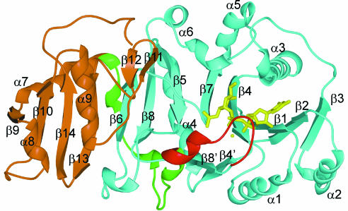
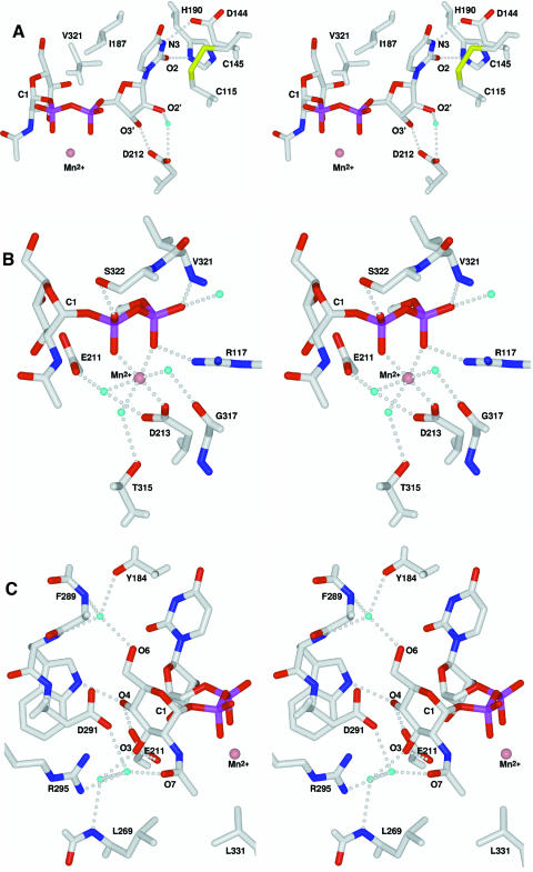
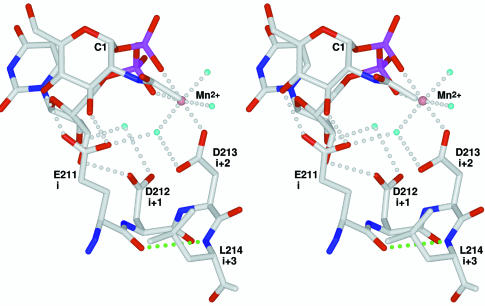
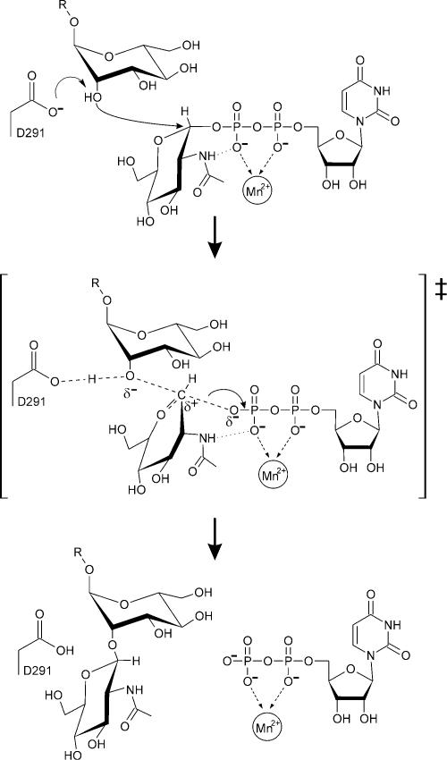
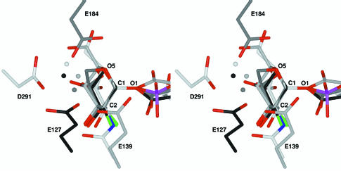
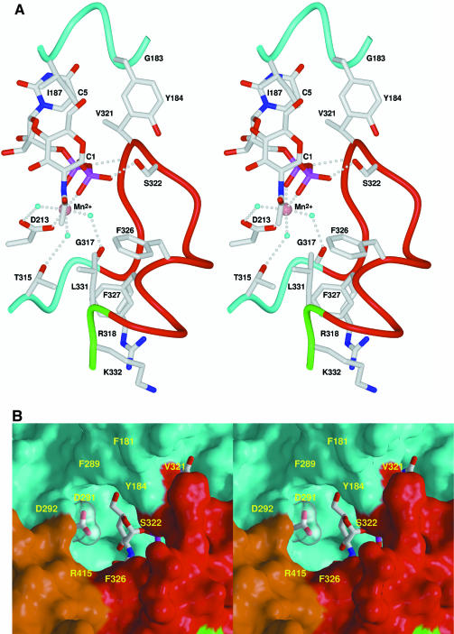
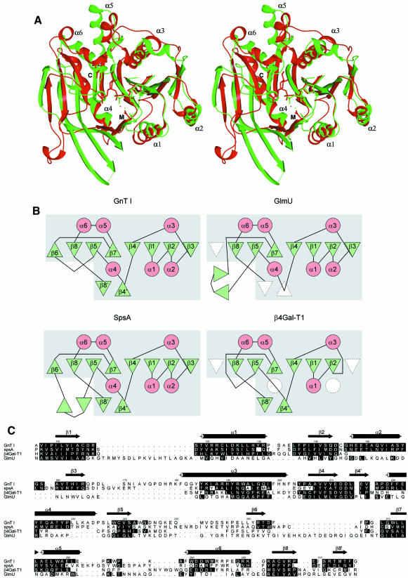
N:-acetylglucosaminyltransferase I (GnT I) serves as the gateway from oligomannose to hybrid and complex N:-glycans and plays a critical role in mammalian development and possibly all metazoans. We have determined the X-ray crystal structure of the catalytic fragment of GnT I in the absence and presence of bound UDP-GlcNAc/Mn(2+) at 1.5 and 1.8 A resolution, respectively. The structures identify residues critical for substrate binding and catalysis and provide evidence for similarity, at the mechanistic level, to the deglycosylation step of retaining beta-glycosidases. The structuring of a 13 residue loop, resulting from UDP-GlcNAc/Mn(2+) binding, provides an explanation for the ordered sequential 'Bi Bi' kinetics shown by GnT I. Analysis reveals a domain shared with Bacillus subtilis glycosyltransferase SpsA, bovine beta-1,4-galactosyl transferase 1 and Escherichia coli N:-acetylglucosamine-1-phosphate uridyltransferase. The low sequence identity, conserved fold and related functional features shown by this domain define a superfamily whose members probably share a common ancestor. Sequence analysis and protein threading show that the domain is represented in proteins from several glycosyltransferase families.


Figures

None
Fig. 1. GnT I ribbon diagram. Domain 1 is shown in cyan, the loop structured upon UDP-GlcNAc binding in red, the linker connecting domains 1 and 2 in green, domain 2 in brown and UDP-GlcNAc and Mn2+ ion in yellow. All molecular images were prepared using SPOCK (Christopher, 1998) and rendered using Raster3D (Bacon and Anderson, 1988; Merritt and Murphy, 1994).
None
Fig. 2. Stereo view of the UDP-GlcNAc/Mn2+ binding site. Carbon, oxygen, nitrogen, sulfur and phosphorus are coloured white, red, blue, yellow and purple, respectively; water molecules are cyan and the Mn2+ ion is salmon. Hydrogen bonds are shown as dotted lines. The C1 of the N-acetylglucos amine moiety is labelled for reference. (A) Uracil and ribose interactions; (B) Mn2+ and phosphate interactions; (CN-acetylglucosamine interactions.
None
Fig. 3. The DxD motif. Atom colours and labels are as in Figure 2. ‘i’, ‘i+1’, ‘i+2’ and ‘i+3’ correspond to the residues of the type I β-turn. The hydrogen bond characteristic of this turn type is shown in green.
None
Fig. 4. Schematic representation of the GnT I reaction mechanism. The oxocarbenium-ion-like transition state is enclosed in large square brackets.
None
Fig. 5. Stereo diagram of an overlay of the GnT I active site with three glycosyl-enzyme glycosidase intermediates. Carbon atoms are coloured white in GnT I (D291), light grey in Bacillus agaradhaerens Cel5A [E139, Protein Data Bank (PDB) entry code 6A3H] (Davies et al., 1998b), dark grey in B.agaradhaerens Xyl11 (E184, PDB code 1QH6) (Sabini et al., 1999) and black in Cellulomonas fimi Cex (E127, PDB code 2XYL) (Notenboom et al., 1998). Water molecules are shown as spheres in the same colour as the carbons of the protein to which they are bound. Oxygen (except for waters), nitrogen, fluorine and phosphorus atoms are coloured in red, blue, green and purple, respectively. The structures were overlaid using the positions of the carbohydrate atoms C1, O1, C2 and O5. In all cases the water molecules are positioned for nucleophilic attack at the donor sugar C1; in GnT I the water molecule will be replaced by the attacking 2-hydroxyl of the 3-arm mannose of the Man5GlcNAc2 acceptor. The structures of the glycosyl-enzyme intermediates of the glycosidases Streptomyces lividans CelB2 (PDB code 2NLR) (Sulzenbacher et al., 1999) and Bacillus circulans BCX (PDB code 1BVV) (Sidhu et al., 1999) can be overlaid with GnT I in the same way and yield a very similar picture.
None
Fig. 6. Stereo diagram of the structured loop and the acceptor-binding pocket. Atom colours and labels are as in Figure 2. Backbone tubes and molecular surfaces are colour-coded as follows: red, structured loop; green, linker region; cyan, domain 1; brown, domain 2. (A) Structured loop and UDP-GlcNAc/Mn2+ interactions; (B) Surface representation of the acceptor binding pocket. The side chain of the catalytic base (D291) and the N-acetylglucosamine moiety of the UDP-GlcNAc are seen at the base of the pocket.
None
Fig. 7. The SGC domain. (A) Stereo ribbon overlay of the SGC domains of GnT I (red) and SpsA (green). For clarity, only the α-helices are labelled. UDP (SpsA) and UDP-GlcNAc (GnT I) are shown in stick representation. M and C label the side chains of the metal binding and catalytic aspartic acid residues, respectively, which are also shown in stick representation. (B) Topology diagrams of the SGC domains of GnT I, SpsA, GlmU and β4Gal-T1. β-strands are shown as green triangles and α-helices as red circles, with missing elements shown in white. The secondary structural elements are labelled as in GnT I. The boxed grey region corresponds to the SGC domain. (C) A structure-based sequence alignment of GnT I, SpsA, β4Gal-T1 and GlmU, produced by STAMP (Russell and Barton, 1992) and modified slightly by manual intervention. Shown are residue numbers and secondary structure for GnT I. Residues with their α-carbon atoms <2.5 Å from the structurally equivalent GnT I α-carbon atom are shaded in black (as is the GnT I residue itself). The alignment was rendered using ALSCRIPT (Barton, 1993).

References

    1. Altschul S.F., Madden,T.L., Schäffer,A.A., Zhang,J., Zhang,Z., Miller,W. and Lipman,D.J. (1997) Gapped BLAST and PSI-BLAST: a new generation of protein database search programs. Nucleic Acids Res., 25, 3389–3402. - PMC - PubMed
    1. Artymiuk P.J., Rice,D.W., Poirrette,A.R. and Willett,P. (1995) β-Glucosyltransferase and phosphorylase reveal their common theme. Nature Struct. Biol., 2, 117–120. - PubMed
    1. Bacon D.J. and Anderson,W.F. (1988) A fast algorithm for rendering space-filling molecule pictures. J. Mol. Graph., 6, 219–220.
    1. Barton G.J. (1993) ALSCRIPT a tool to format multiple sequence alignments. Protein Eng., 6, 37–40. - PubMed
    1. Black C.B., Huang,H.-W. and Cowan,J.A. (1994) Biological coordination chemistry of magnesium, sodium, and potassium ions. Protein and nucleotide binding sites. Coord. Chem. Rev., 135/136, 165–202.

Publication types

MeSH terms

Substances

LinkOut - more resources