. 2000 Oct 16;19(20):5281-7.
doi: 10.1093/emboj/19.20.5281.

Crystal structure of Streptococcus pneumoniae acyl carrier protein synthase: an essential enzyme in bacterial fatty acid biosynthesis

Affiliations

Crystal structure of Streptococcus pneumoniae acyl carrier protein synthase: an essential enzyme in bacterial fatty acid biosynthesis

N Y Chirgadze et al. EMBO J. .

Abstract

Acyl carrier protein synthase (AcpS) catalyzes the formation of holo-ACP, which mediates the essential transfer of acyl fatty acid intermediates during the biosynthesis of fatty acids and lipids in the cell. Thus, AcpS plays an important role in bacterial fatty acid and lipid biosynthesis, making it an attractive target for therapeutic intervention. We have determined, for the first time, the crystal structure of the Streptococcus pneumoniae AcpS and AcpS complexed with 3'5'-ADP, a product of AcpS, at 2.0 and 1.9 A resolution, respectively. The crystal structure reveals an alpha/beta fold and shows that AcpS assembles as a tightly packed functional trimer, with a non-crystallographic pseudo-symmetric 3-fold axis, which contains three active sites at the interface between protomers. Only two active sites are occupied by the ligand molecules. Although there is virtually no sequence similarity between the S.pneumoniae AcpS and the Bacillus subtilis Sfp transferase, a striking structural similarity between both enzymes was observed. These data provide a starting point for structure-based drug design efforts towards the identification of AcpS inhibitors with potent antibacterial activity.


Figures
None
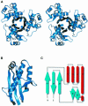
Fig. 1. (A) Stereoview showing a ribbon diagram of the AcpS homotrimer, viewed along a non-crystallographic 3-fold axis. (B) Ribbon diagram of the Cα backbone of one S.pneumoniae AcpS monomer structure. (C) A topology (Richardson) diagram of AcpS. β-strands are represented as arrows, while α helices are rectangles. The secondary structure elements are defined as follows: β1, Ile4–Glu13; α1, Leu14–Arg23; α2, Phe27–Val31; α3, Ala34–Ser42; α4, Gly45–Met66; α5, Ile70–Leu73; β2, Glu79–Asn82; β3, Pro88–Gln92; β4, Lys98–His105; β5, Phe109–Glu117.
None
Fig. 2. (A) Sequence alignment of bacterial AcpS genes from different species. The most conserved regions are shown in gray. Secondary structural elements observed in the S.pneumoniae AcpS crystal structure are indicated above the sequence pile-up. (B) Structural alignment of two S.pneumoniae AcpS monomer molecules with two domains of B.subtilis Sfp. The N-terminal half of Sfp from Met1 to Pro103 corresponds to one protomer of the AcpS trimer (shown in blue) and has a 22% sequence identity. The C-terminal half of Sfp from Ile104 to Pro209 corresponds to a second AcpS protomer (shown in green) and has 25% sequence identity. The remaining C-terminal portion from Asp210 to Leu224 has no counterpart in the AcpS structure. The three amino acid residues involved in Mg2+ binding are marked by a star. The regions involved in CoA binding are marked by plus signs.
None
Fig. 3. View of the 3′5′-ADP fragment of CoA bound to the active site of S.pneumoniae AcpS. The omitted electron density map corresponding to the ligand was contoured at the 1 σ level at 1.9 Å resolution.
None
Fig. 4. A superposition of apo-AcpS from S.pneumoniae (shown in blue) on the surfactin synthetase activating enzyme Sfp (4′-phosphopantetheinyl transferase) from B.subtilis complexed with CoA (shown in red). One protomer of AcpS is superimposed on the N-terminal domain of Sfp and a second AcpS protomer is superimposed on the C-terminal domain of Sfp. The third protomer of the AcpS trimer does not have a counterpart in Sfp structure. A sulfate ion was found in the AcpS binding site that corresponds to the position of the α-phosphate of CoA in the Sfp molecule.

References

    1. Ahamed J., Gangopadhyay,J. and Sinha,A.K. (1999) Mechanisms of quinolone resistance in clinical isolates of Shigella dysenteriae. Antimicrob. Agents Chemother., 43, 2333–2334. - PMC - PubMed
    1. Badger J., Kumar,R.A., Yip,P. and Szalma,S. (1999) New features and enhancements in the X-PLOR computer program. Proteins, 35, 25–33. - PubMed
    1. Brünger A.T. (1992) The free R value: a novel statistical quantity for assessing the accuracy of crystal structures. Nature, 355, 472–474. - PubMed
    1. Carreras C.W., Gehring,A.M., Walsh,C.T. and Khosla,C. (1997) Utilization of enzymatically phosphopantetheinylated acyl carrier proteins and acetyl-acyl carrier proteins by the actinorhodin polyketide synthase. Biochemistry, 36, 11757–11761. - PubMed
    1. Cohen M.L. (1992) Epidemiology of drug resistance: implications for a postantimicrobial era. Science, 257, 1050–1055. - PubMed

MeSH terms