. 2000 Oct 16;19(20):5300-7.
doi: 10.1093/emboj/19.20.5300.

Crystal structure of the cell division protein FtsA from Thermotoga maritima

Affiliations

Crystal structure of the cell division protein FtsA from Thermotoga maritima

F van den Ent et al. EMBO J. .

Abstract

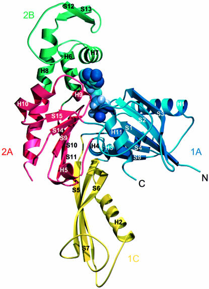
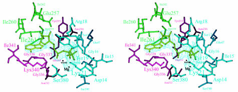
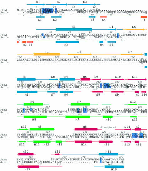
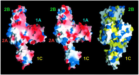
Bacterial cell division requires formation of a septal ring. A key step in septum formation is polymerization of FtsZ. FtsA directly interacts with FtsZ and probably targets other proteins to the septum. We have solved the crystal structure of FtsA from Thermotoga maritima in the apo and ATP-bound form. FtsA consists of two domains with the nucleotide-binding site in the interdomain cleft. Both domains have a common core that is also found in the actin family of proteins. Structurally, FtsA is most homologous to actin and heat-shock cognate protein (Hsc70). An important difference between FtsA and the actin family of proteins is the insertion of a subdomain in FtsA. Movement of this subdomain partially encloses a groove, which could bind the C-terminus of FtsZ. FtsZ is the bacterial homologue of tubulin, and the FtsZ ring is functionally similar to the contractile ring in dividing eukaryotic cells. Elucidation of the crystal structure of FtsA shows that another bacterial protein involved in cytokinesis is structurally related to a eukaryotic cytoskeletal protein involved in cytokinesis.


Figures

None
Fig. 1. Stereo drawing of the 2FoFc electron-density map of crystal form 1 at 1.9 Å resolution. Residues 208–222 and 230–242 are shown. The figure was prepared with the program MAIN (Turk, 1992).
None
Fig. 2. Ribbon plot of the crystal structure of FtsA. The structure is divided into four domains, in analogy to the actin family of proteins, which are designated 1A (blue), the FtsA-specific domain 1C (yellow), subdomain 2A (red) and subdomain 2B. Secondary structure elements are labelled according to their order of appearance in the primary sequence of FtsA (Figure 4). Mg-ATP is depicted in purple. The figure was prepared using the program MOLSCRIPT (Kraulis, 1991).
None
Fig. 3. Stereo view of the active site of FtsA. Residues within hydrogen-bonding distance of Mg-ATP are labelled. Colours of the residues correspond to those of the subdomains as defined in Figure 2. The nucleotide is depicted in light green, within the 2FoFc electron-density map. The figure was prepared using MOLSCRIPT (Kraulis, 1991).
None
Fig. 4. Structure-based sequence alignment between FtsA and actin (Protein Data Bank entry 1YAG-A) [using the DALI web server (Holm and Sander, 1993)]. Boundaries of the secondary structure elements were defined using the program DSSP (Kabsch and Sander, 1983). The colours of the secondary structure elements correspond to those of the domains shown in Figure 2. Active site residues are coloured in purple. Conserved residues are blocked. The figure was prepared using the program ALSCRIPT (Barton, 1993).
None
Fig. 5. Plot of the solvent-accessible surface of FtsA. Left and middle: electrostatic potential [–6 (red) to +6 kT (blue)] plotted on to the surface of FtsA in a slightly tilted orientation around the y-axis compared with Figure 2. The left picture shows the structure derived from crystal form 2 (with Mg-ATP bound); the middle picture shows the structure based on crystal form 1 (with an empty active site). The putative peptide-binding groove is located perpendicular to the plane between subdomains 1A and 1C (left and middle). The right-hand hydrophobicity plot is rotated 90° around the y-axis, with respect to the middle figure, to show the depth of the putative peptide-binding groove across the molecule. The colour code is as follows: yellow for hydrophobic residues (Ala, Val, Leu, Ile, Met, Phe, Pro), blue for hydrophilic residues (Lys, Arg, Glu, Asp) and white for Ser, Thr, Tyr, His, Cys, Asn, Gln, Trp and Gly. Domains are labelled as in Figure 2. This figure was prepared using GRASP (Nicholls, 1993).

References

    1. Abrahams J.P. and Leslie,A.G.W. (1996) Methods used in the structure determination of bovine mitochondrial F1-ATPase. Acta Crystallogr. D, 52, 30–42. - PubMed
    1. Addinall S.G., Bi,E. and Lutkenhaus,J. (1996) FtsZ ring formation in fts mutants. J. Bacteriol., 178, 3877–3884. - PMC - PubMed
    1. Addinall S.G., Cao,C. and Lutkenhaus,J. (1997) FtsN, a late recruit to the septum in Escherichia coli. Mol. Microbiol., 25, 303–309. - PubMed
    1. Anderson C.M., McDonald,R.C. and Steitz,T.A. (1978) Sequencing a protein by X-ray crystallography. Interpretation of yeast hexokinase B at 2.5 Å resolution by model building. J. Mol. Biol., 123, 1–13. - PubMed
    1. Ayala J.A., Garrido,T., de Pedro,M.A. and Vicente,M. (1994) Molecular biology of bacterial septation. In Hakenbeck,R. and Ghuysen,J.M. (eds), New Comprehensive Biochemistry, Vol. 27: Bacterial Cell Wall. Elsevier, Amsterdam, The Netherlands, pp. 73–101.

LinkOut - more resources