. 2000 Oct 16;19(20):5418-28.
doi: 10.1093/emboj/19.20.5418.

MAPK/ERK signaling in activated T cells inhibits CD95/Fas-mediated apoptosis downstream of DISC assembly

Affiliations

MAPK/ERK signaling in activated T cells inhibits CD95/Fas-mediated apoptosis downstream of DISC assembly

T H Holmström et al. EMBO J. .

Abstract

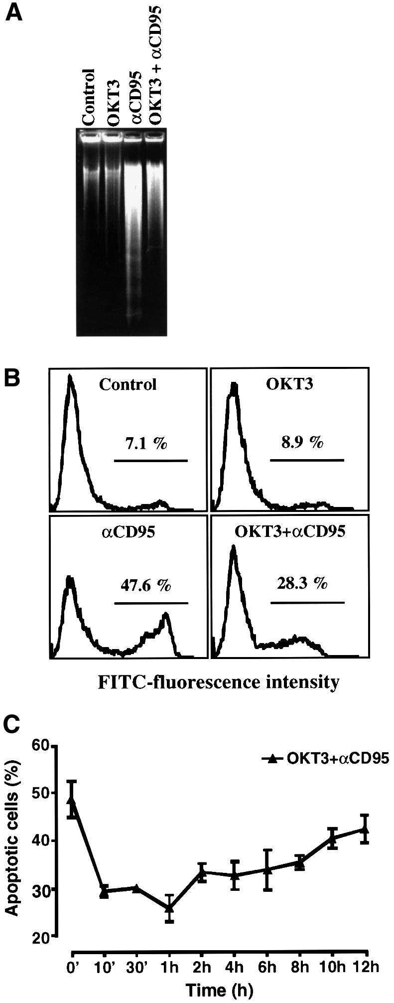
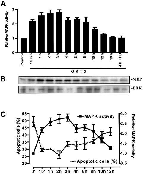
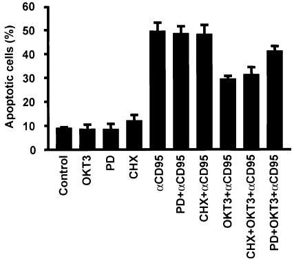
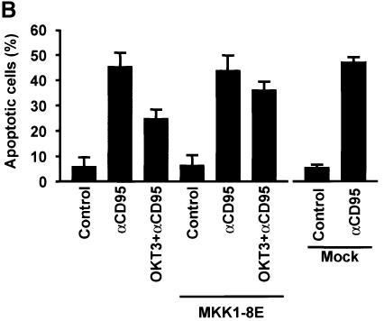
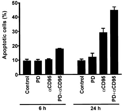
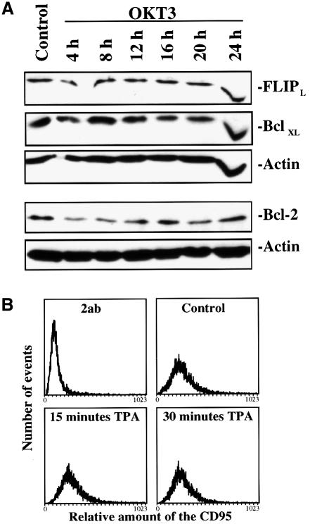
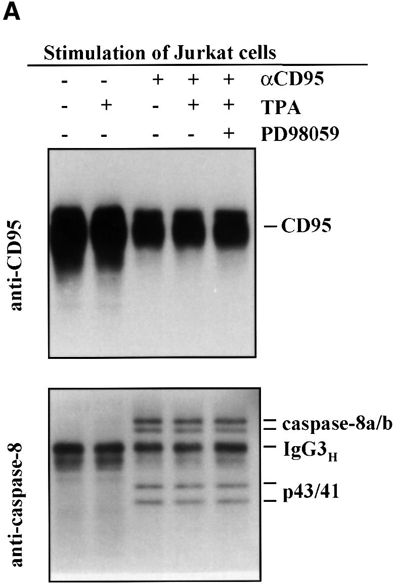
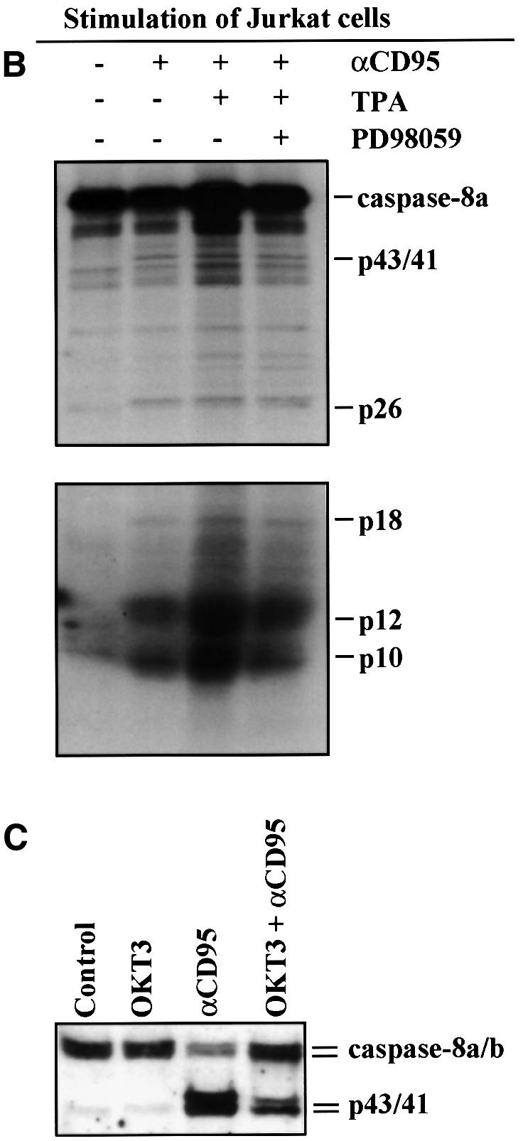
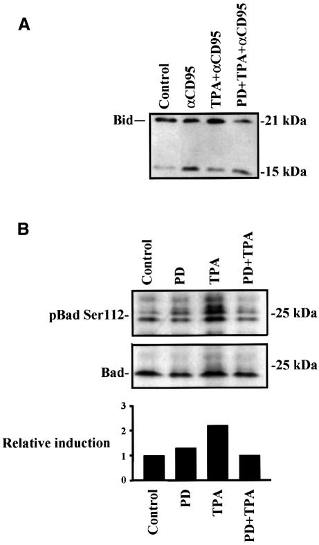
When T cells are activated, the expression of the CD95 ligand is elevated, with the purpose of inducing apoptosis in target cells and to later eliminate the activated T cells. We have shown previously that mitogen-activated protein kinase (MAPK or ERK) signaling suppresses CD95-mediated apoptosis in different cellular systems. In this study we examined whether MAPK signaling controls the persistence and CD95-mediated termination of an immune response in activated T cells. Our results show that activation of Jurkat T cells through the T cell receptor immediately suppresses CD95-mediated apoptosis, and that this suppression is mediated by MAPK activation. During the phase of elevated MAPK activity, the activation of caspase-8 and Bid is inhibited, whereas the assembly of a functional death-inducing signaling complex (DISC) is not affected. These results explain the resistance to CD95 responses observed during the early phase of T cell activation and suggest that MAPK-activation deflects DISC signaling from activating caspase-8 and Bid. The physiological relevance of the results was confirmed in activated primary peripheral T cells, in which inhibition of MAPK signaling markedly sensitized the cells to CD95-mediated apoptosis.


Figures

None
Fig. 1. Activation of Jurkat T cells with an agonistic CD3 antibody, OKT3, suppresses CD95-mediated apoptosis. Jurkat T cells were incubated with medium alone, immobilized anti-CD3 (OKT3) antibody (100 µg/ml), anti-CD95 (anti-Fas, 100 ng/ml) or pre-incubated for 30 min with immobilized OKT3 before addition of anti-CD95. After 2 h aliquots of cells were analyzed by either (A) one-stage DNA gel electrophoresis or (B) annexin V–FITC staining, and the proportion of apoptotic nuclei was determined using a FACScan flow cytometer. Bars indicate the percentage of cells with exposed phoshatidylserine (FITC positive), a feature that is characteristic of apoptotic cells. (C) The OKT3-mediated suppression of CD95-induced apoptosis lasts for 10–12 h after stimulation. Jurkat T cells were pre-incubated with immobilized OKT3 for the indicated time periods prior to incubation with anti-CD95 for 2 h and analyzed as in (A). The data represent mean values (mean ± SEM) from a minimum of three separate experiments.
None
Fig. 2. The kinetics of MAPK activation in Jurkat T cells after OKT3 stimulation corresponds to the observed suppression of CD95-mediated apoptosis. MAPK activation was followed in cell extracts incubated with medium alone, in the presence of immobilized OKT3 or in the presence of the MKK1 inhibitor PD 98059 (30 µM) + OKT3, and the activation was determined by an immunocomplex kinase assay. (A and B) A representative autoradiograph of the immunocomplex kinase assay together with an immunoblot of the immunoprecipitated ERK2, to verify presence of the MAPK, is shown. Multiple samples at the different time points were quantified by phosphoimager analysis. (C) A close correlation between MAPK activity and the amount of apoptotic cells was obtained when the data from the MAPK assays and the percentage of apoptotic cells, treated as described above, were plotted against the incubation time with OKT3. (B and C) The data represent mean values (mean ± SEM) from a minimum of three separate experiments.
None
Fig. 3. OKT3-mediated suppression of CD95-induced apoptosis in Jurkat T cells is independent of protein synthesis and mediated through MAPK activation. Annexin V–FITC staining was used as an indicator for the amount of apoptotic cells. Cells were cultured for 2 h with medium alone, in the presence of anti-CD95 (anti-Fas), immobilized OKT3, PD 98059, CHX (100 µg/ml), OKT3 + anti-CD95, CHX + anti-CD95, CHX + OKT3 + anti-CD95 or PD 98059 + OKT3 + anti-CD95. The cells were pre-incubated with immobilized OKT3 as previously described and/or for 10 min with CHX prior to incubation with anti-CD95. The data represent mean values (mean ± SEM) from a minimum of three separate experiments.
None
Fig. 4. Dominant-negative MKK1 abolishes the anti-CD3-mediated protective effect on CD95-mediated apoptosis. (A) Representative immunofluorescence micrographs of cells transfected with a dominant-negative (MKK1-8E) construct and incubated for 2 h with anti-CD95 in the absence or presence of OKT3. Hoechst staining was used to detect alterations in the nuclei and a monoclonal HA antibody linked to an FITC-conjugated secondary antibody was used to detect the presence of the HA-tagged MKK1 in transfected cells. The arrows indicate the transfected cells. Mock transfected cells were treated as indicated above with or without stimulation of CD95. (B) Percentage of apoptosis in transfected cells after treatment with anti-CD95. The data represent mean values (mean ± SEM) from a minimum of three separate experiments.
None
Fig. 5. Suppression of MAPK activation sensitizes activated human peripheral T cells to CD95-mediated apoptosis. Annexin V–FITC staining was used as an indicator for the amount of apoptotic cells. Activated T cells (day 5–6; 1 × 10⁶ cells/ml) were cultured for 6 or 24 h with medium alone, in the presence of anti-CD95 (anti-Fas, 500 ng/ml), immobilized OKT3, PD 98059, OKT3 + anti-CD95 or PD 98059 + OKT3 + anti-CD95. The cells were pre-incubated with immobilized OKT3 as previously described prior to incubation with anti-CD95. The data represent mean values (mean ± SEM) from a minimum of three separate experiments.
None
Fig. 6. OKT3 stimulation does not affect Bcl-xL, Bcl-2, c-FLIPL and CD95 levels. (A) Jurkat T cells were treated with immobilized OKT3 for the indicated time points and the amount of proteins known to affect apoptosis was assessed by immunoblotting with specific antibodies to Bcl-xL, Bcl-2 and c-FLIPL. Equal loading was confirmed by immunoblotting of the same gel with an actin-specific antibody. A representative immnunoblot from three experiments is shown. (B) To measure the amount of CD95 on the cell surface after MAPK activation, Jurkat T cells were treated as indicated in the figure. The amount of CD95 was then assessed by staining with a specific CD95 antibody (anti-APO-1) followed by an FITC-conjugated secondary antibody and analyzed by flow cytometry. As a negative control we used cells stained with the secondary antibody only.
None
Fig. 7. MAPK activation suppresses overall cleavage of caspase-8 but does not affect formation of a functional DISC. (A) Jurkat T cells were treated as indicated in the figure prior to immunoprecipitation of CD95 from the cell extracts. Subsequently the levels of immunoprecipitated CD95 and associated caspase-8 were detected by western blotting with appropriate antibodies after the respective treatments. A representative immunoblot from three experiments is shown. (B) Jurkat T cells were treated as indicated in the figure prior to immunoprecipitation of CD95 from the cell extracts. Immunoprecipitates were washed four times and incubated with in vitro translated 35S-labeled caspase-8/a. After 24 h the samples were analyzed on 15% SDS–PAGE. The upper part of the gel was exposed for 24 h and the lower part was exposed for 5 days. (C) Jurkat cells were treated as indicated in the figure and the degree of caspase-8 processing in the cells was monitored by western blotting with a caspase-8-specific antibody. Caspase activation can be observed as the appearance of the p43/41 active intermediate fragments of the caspase proforms.
None
Fig. 8. MAPK activation suppresses cleavage of Bid and phosphorylates Bad on Ser112. (A) MAPK-mediated suppression of Bid cleavage. Jurkat T cells were treated as indicated in the figure for 2 h prior to immunoblotting for Bid. The amount of full-length Bid compared with caspase-cleaved p15 was determined by western-blotting. (B) The phosphorylation of Bad on Ser112 was measured by immunoblotting using a phospho-Bad Ser112 antibody after 30 min stimulation with TPA or PD 98059 + TPA. The relative induction (control = 1) was measured by densitometric measurement of the amount of Bad as visualized by an immunoblot using control Bad antibody to the amount of Bad phosphorylated on Ser112.

References

    1. Alderson M.R., Armitage,R.J., Maraskovsky,E., Tough,T.W., Roux,E., Schooley,K., Ramsdell,F. and Lynch,D.H. (1993) Fas transduces activation signals in normal human T lymphocytes. J. Exp. Med., 178, 2231–2235. - PMC - PubMed
    1. Alessi D.R., Cuenda,A., Cohen,P., Dudley,D.T. and Saltiel,A.R. (1995) PD 098059 is a specific inhibitor of the mitogen-activated protein kinase kinase in vitro and in vivo. J. Biol. Chem., 270, 27489–27494. - PubMed
    1. Boldin M.P., Goncharov,T.M., Goltsev,Y.V. and Wallach,D. (1996) Involvement of MACH a novel MORT/Fadd-interacting protease in Fas/APO-1 and TNF receptor induced cell death. Cell, 85, 803–815. - PubMed
    1. Bonni A., Brunet,A., West,A.E., Datta,S.R., Takasu,M.A. and Greenberg,M.E. (1999) Cell survival promoted by the Ras-MAPK signaling pathway by transcription-dependent and -independent mechanisms. Science, 286, 1358–1362. - PubMed
    1. Brunner T. et al. (1995) Cell autonomous Fas (CD95)/Fas-ligand interaction mediates activation-induced apoptosis in T-cell hybridomas. Nature, 373, 441–444. - PubMed

Publication types

MeSH terms

Substances