. 2000 Oct 16;19(20):5429-39.
doi: 10.1093/emboj/19.20.5429.

Activation of B-Raf kinase requires phosphorylation of the conserved residues Thr598 and Ser601

Affiliations

Activation of B-Raf kinase requires phosphorylation of the conserved residues Thr598 and Ser601

B H Zhang et al. EMBO J. .

Abstract

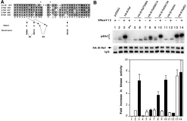
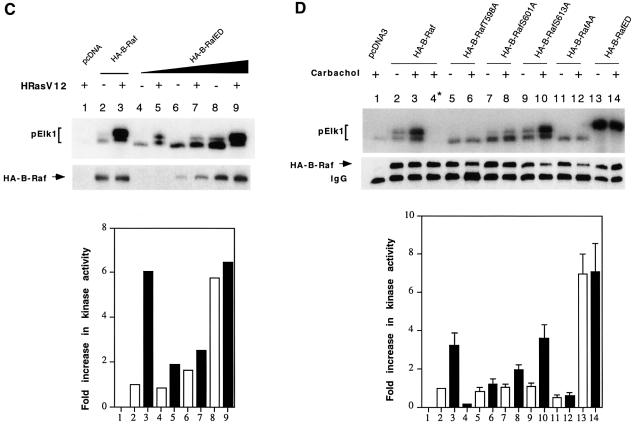
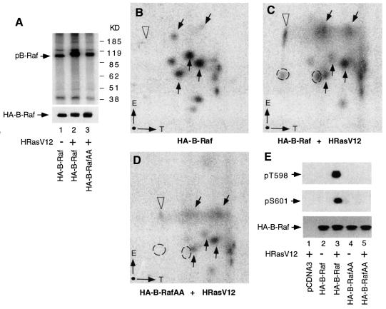
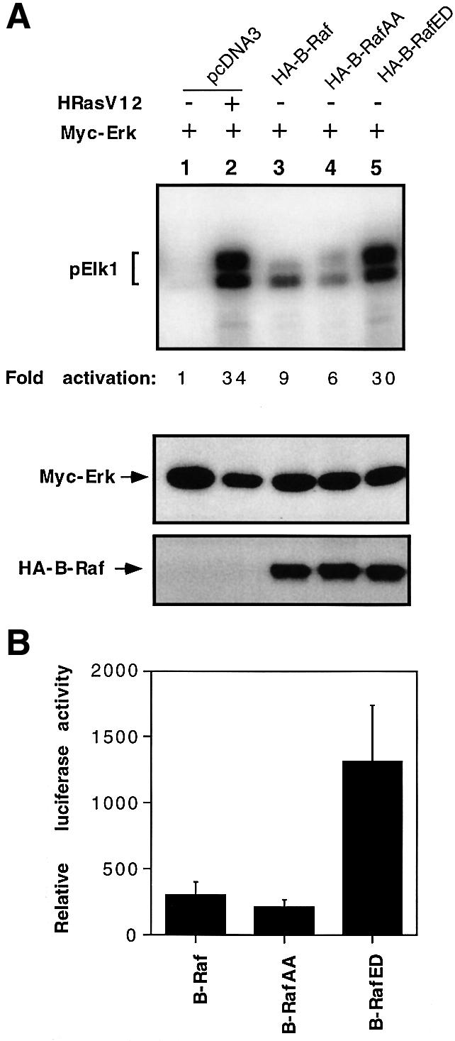
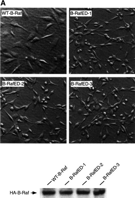
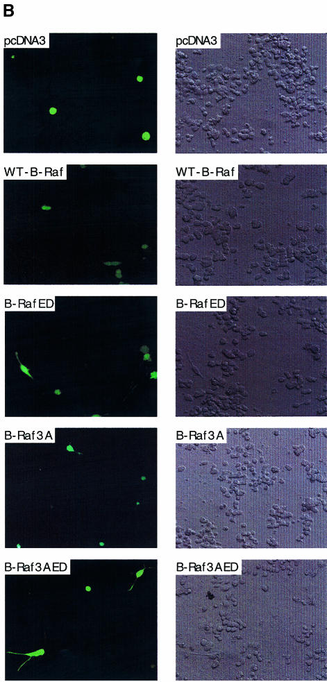
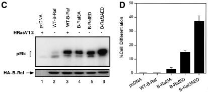
The Raf kinase family serves as a central intermediate to relay signals from Ras to ERK. The precise molecular mechanism for Raf activation is still not fully understood. Here we report that phosphorylation of Thr598 and Ser601, which lie between kinase subdomains VII and VIII, is essential for B-Raf activation by Ras. Substitution of these residues by alanine (B-RafAA) abolished Ras-induced B-Raf activation without altering the association of B-Raf with other signaling proteins. Phosphopeptide mapping and immunoblotting with phospho-specific antibodies confirmed that Thr598 and Ser601 are in vivo phosphorylation sites induced by Ras. Furthermore, replacement of these two sites by acidic residues (B-RafED) renders B-Raf constitutively active. Con sistent with these data, B-RafAA and B-RafED exhibited diminished and enhanced ability, respectively, to stimulate ERK activation and Elk-dependent transcription. Moreover, functional studies revealed that B-RafED was able to promote NIH 3T3 cell transformation and PC12 cell differentiation. Since Thr598 and Ser601 are conserved in all Raf family members from Caenorhabditis elegans to mammals, we propose that phosphorylation of these two residues may be a general mechanism for Raf activation.


Figures

None
Fig. 1. Activation of B-Raf by oncogenic Ras requires both Thr598 and Ser601 phosphorylation. (A) Sequence alignment of Raf kinase activation loop (residues 593–622 for human B-Raf). Residues subjected to site-directed mutagenesis are indicated by arrows. An asterisk denotes the protein kinase C phosphorylation sites. (B) Oncogenic Ras-induced kinase activation of B-Raf and its mutants. Fifty nanograms of HA-tagged pcDNA3 (vector control, lane 1), HA-tagged B-Raf (lanes 2--4), B-RafT598A (lane 5 and 6), B-RafS601A (lanes 7 and 8), B-RafS613A (lanes 9 and 10), B-RafAA (lanes 11 and 12) or B-RafED (lanes 13 and 14) were transiently transfected in COS cells alone (open bars) or with 100 ng of oncogenic Ras (HRasV12, solid bars), as indicated at the top of the panel. The kinase was immunoprecipitated and its activity, reflected by phosphorylation of GST–Elk1 (pElk1), was measured using the coupled assay (see Materials and methods). The upper panel shows autoradiographs of pElk1 that are representative of five independent experiments. The intensity of pElk1 bands was quantified by PhosphorImager (lower panel); results were subtracted from the reading of lane 1 (background) and expressed as fold increase with respect to the cells transfected with wild-type B-Raf without stimulation (lane 2). Lane 4* denotes that GST–MEK was omitted in the assay as a control. Results are mean ± SD from three independent experiments. Western blot of the immunoprecipitated kinase is shown in the middle panel, indicating equivalent protein loadings. (C) Activation of B-RafED by oncogenic Ras. COS cells were transfected with vector (lane 1), 50 ng of wild-type B-Raf (lanes 2 and 3), 5 ng of B-RafED (lanes 4 and 5), 25 ng of B-RafED (lanes 6 and 7) or 50 ng of B-RafED (lanes 8 and 9) in the presence or absence of HRasV12, as indicated at the top of the panel. Results of kinase assay and western blotting of B-Raf using α-HA were representative of two separate experiments. (D) Carbachol-induced kinase activation of B-Raf and its mutants. Cells were co-transfected with the above DNA constructs and hM3. After 24 h, cells were starved in FBS-free medium for 5 h followed by stimulation with carbachol for 5 min. Kinase immunoprecipitation, kinase assay and quantitation of results are as described in (B).
None
Fig. 2. Tryptic phosphopeptide mapping of B-Raf. (A) Metabolic labeling and immunoprecipitation of B-Raf and B-RafAA. COS1 cells grown on a 60 mm plate were transfected with wild-type B-Raf (lane 1), B-Raf + HRasV12 (lane 2) or B-RafAA + HRasV12 (lane 3), labeled with [32P]orthophosphate, and immunoprecipitated with anti-HA antibody. Immunocomplexes were separated by SDS–PAGE and visualized by antoradiography. Positions of size markers are indicated in kilodaltons (KD) on the right. An immunoblot of the above immunoprecipitation using anti-HA antibody is shown at the bottom. (B, C and D) Phosphopeptide mappings for wild-type B-Raf, B-Raf + HRasV12 and B-RafAA + HRasV12, respectively. The 32P-labeled bands were excised from the membrane, digested with trypsin and analyzed by two-dimensional thin-layer electrophoresis. The directions for electrophoresis (E, from cathode to anode) and TLC (T) are indicated in the lower left corner of each panel. Arrows point to positions of phosphopeptides for references between different panels. Dashed circles indicate phosphopeptides present in (C) that are missing in (D). Open arrowheads indicate the Ras-induced phosphopeptide not affected in B-RafAA. (E) Phosphorylation of Thr598 and Ser601 determined by specific anti-phospho Thr598 and Ser601 antibodies. COS cells were transfected with pcDNA (vector, lane 1), wild-type B-Raf (lane 2), co-transfected with wild-type B-Raf and HRasV12 (lane3), B-RafAA (lane 4) or co-transfected B-RafAA and HRasV12 (lane 5). Immunoprecipitates of B-Raf and B-RafAA were isolated and resolved by SDS–PAGE, transferred to membrane, and blotted with anti-phospho Thr598 (top panel) or anti-phospho Ser601 antibody (middle panel). Protein loading was examined by blotting using HA antibody (bottom panel).
None
Fig. 3. B-RafT598A, B-RafS601A or B-RafAA does not alter the association with 14-3-3, HSP90 or MEK. COS cells were transiently transfected with pcDNA3 (vector, lane 1), HA-B-Raf (lane 2), HA-B-RafT598A (lane 3), HA-B-RafS601A (lane 4) or HA-B-RafAA (lane 5). Cells were lysed and the HA-tagged B-Raf was immuno precipitated (IP) with anti-HA and western blotted with anti-HA (B-Raf), anti-14-3-3, anti-HSP90 or anti-MEK antibodies.
None
Fig. 4. Effects of B-Raf, B-RafAA or B-RafED on ERK activity and Elk-dependent transcription. (A) Activation of ERK by B-Raf, B-RafAA or B-RafED. Myc-ERK was co-transfected with pcDNA3, HA-tagged B-Raf, B-RafAA or B-RafED. Co-transfection of Myc-ERK and HRasV12 serves as a positive control (lane 2). Cells were cultured for 24 h followed by starvation for 15 h. Myc-ERK was immuno precipitated with anti-Myc monoclonal antibody 9E10 and assayed for kinase activity using GST–Elk1 as a substrate. ERK kinase activity is shown in the top panel and fold activation was determined by PhosphorImager analysis. Immunoblot detection of Myc-ERK or HA-B-Raf is shown in the middle or bottom panel. (B) Elk1-dependent transcription activation by B-Raf, B-RafAA or B-RafED. COS cells were transfected with Gal4-Elk1 and Gal4-LUC and B-Raf, B-RafAA or B-RafED. A pCMV-LacZ plasmid was co-transfected as an internal control for variations in transfection efficiency. Cells were cultured for 24 h followed by 15 h starvation. Luciferase activity was measured and normalized against the co-transfected β-galactosidase activity and results are mean ± SD from three independent experiments.
None
Fig. 5. B-RafED induces NIH 3T3 cell transformation and PC12 cell differentiation. (A) Morphology of NIH 3T3 cells stably expressing wild-type B-Raf or B-RafED. NIH 3T3 cells were transfected with wild-type B-Raf or B-RafED. Stably transfected cells were selected as described in Materials and methods. Note that morphology for cells expressing B-RafED is from three individual cell colonies. The western blotting for B-Raf expression in stably transfected cells is shown in the lower panel. (B) PC12 cell differentiation induced by B-Raf3A, B-Raf-ED and B-Raf3AED. PC12 cells were co-transfected with pEGFP vector and pcDNA3 (vector control), wild-type B-Raf (WT-B-Raf), B-Raf3A, B-RafED or B-Raf3AED. Two days after transfection, cells were shifted to NGF-minus differentiation medium (DMEM supplemented with 2% horse serum and 1% FBS medium) for 3 days. Cells were examined under the fluorescence microscope for visualization of transfected cells (by GFP) and differentiation (by the presence of neurites). Shown are representative fluorescence images (left) and the corresponding Nomarski images (right) (×200). (C) Kinase activity of B-Raf3A, B-RafED and B-Raf3AED. COS cells were transfected with pcDNA3, WT-B-Raf, B-Raf3A, B-RafED or B-Raf3AED in the presence or absence of HRasV12. Kinase activity (upper panel) and western blotting for B-Raf expression (lower panel) were determined. (D) Quantitation of PC12 cell differentiation induced by B-Raf and its mutants. Results were expressed as a percentage of green cells exhibiting neurite extensions of >2 cell body lengths in total green cells. At least 200 transfected cells were counted.

References

    1. Alessi D.R. and Cohen,P. (1998) Mechanism of activation and function of protein kinase B. Curr. Opin. Genet. Dev., 8, 55–62. - PubMed
    1. Alessi D.R., Saito,Y., Campbell,D.G., Cohen,P., Sithanandam,G., Rapp,U., Ashworth,A., Marshall,C.J. and Cowley,S. (1994) Identification of the sites in MAP kinase kinase-1 phosphorylated by p74raf-1. EMBO J., 13, 1610–1619. - PMC - PubMed
    1. Cai H., Smola,U., Wixler,V., Eisenmann-Tappe,I., Diaz-Meco,M.T., Moscat,J., Rapp,U. and Cooper,G.M. (1997) Role of diacylglycerol-regulated protein kinase C isotypes in growth factor activation of the Raf-1 protein kinase. Mol. Cell. Biol., 17, 732–741. - PMC - PubMed
    1. Carroll M.P. and May,W.S. (1994) Protein kinase C-mediated serine phosphorylation directly activates Raf-1 in murine hematopoietic cells. J. Biol. Chem., 269, 1249–1256. - PubMed
    1. Casamayor A., Morrice,N.A. and Alessi,D.R. (1999) Phosphorylation of Ser-241 is essential for the activity of 3-phosphoinositide-dependent protein kinase-1: identification of five sites of phosphorylation in vivo. Biochem. J., 342, 287–292. - PMC - PubMed

Publication types

MeSH terms