. 2000 Oct 16;19(20):5450-9.
doi: 10.1093/emboj/19.20.5450.

Direct association of ligand-binding and pore domains in homo- and heterotetrameric inositol 1,4,5-trisphosphate receptors

Affiliations

Direct association of ligand-binding and pore domains in homo- and heterotetrameric inositol 1,4,5-trisphosphate receptors

D Boehning et al. EMBO J. .

Abstract

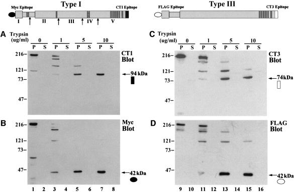
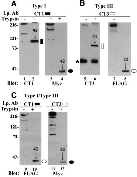
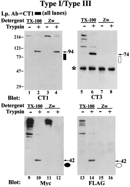
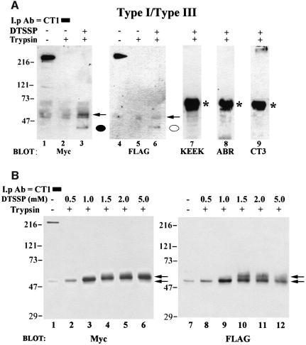
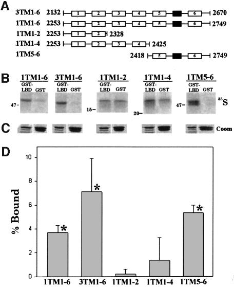
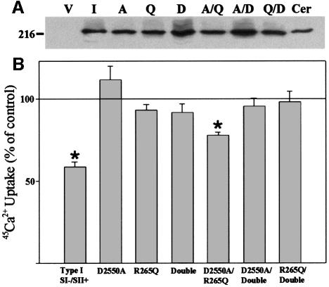
Inositol 1,4,5-trisphosphate receptors (IP(3)Rs) are a family of intracellular Ca(2+) channels that exist as homo- or heterotetramers. In order to determine whether the N-terminal ligand-binding domain is in close physical proximity to the C-terminal pore domain, we prepared microsomal membranes from COS-7 cells expressing recombinant type I and type III IP(3)R isoforms. Trypsin digestion followed by cross-linking and co-immunoprecipitation of peptide fragments suggested an inter-subunit N- and C-terminal interaction in both homo- and heterotetramers. This observation was further supported by the ability of in vitro translated C-terminal peptides to interact specifically with an N-terminal fusion protein. Using a (45)Ca(2+) flux assay, we provide functional evidence that the ligand-binding domain of one subunit can gate the pore domain of an adjacent subunit. We conclude that common structural motifs are shared between the type I and type III IP(3)Rs and propose that the gating mechanism of IP(3)R Ca(2+) channels involves the association of the N-terminus of one subunit with the C-terminus of an adjacent subunit in both homo- and heterotetrameric complexes.


Figures

None
Fig. 1. Peptide fragments generated by limited tryptic digestion of recombinant type I and type III IP3Rs. (Above) Schematic representations of the IP3R expression constructs are illustrated. The indented region represents the ligand-binding pocket, striped bars represent sites of alternative splicing and dark gray bars represent the membrane-spanning regions of the pore domain. N-terminal antibody epitopes are designated by closed (type I) or open (type III) circles, and C-terminal (endogenous) epitopes by closed (type I) or open (type III) squares. Sites that are cleaved by trypsin in the mouse type I receptor are indicated by arrows and the resulting five fragments by Roman numerals I–V (Yoshikawa et al., 1999b). (A–D) Microsomes prepared from cells overexpressing recombinant type I or type III IP3Rs were digested with 0, 1, 5 or 10 µg/ml trypsin. The vesicles were then pelleted and both the pellet (P) and supernatant (S) fractions were subjected to SDS–PAGE (see Materials and methods). (A and B) The type I receptor probed with isoform-specific C-terminal (CT1) and N-terminal (Myc) antibodies, respectively. (C and D) Type III receptor probed with an isoform-specific C-terminal antibody (CT3) and an N-terminal FLAG antibody. The N- and C-terminal protease-resistant fragments are highlighted by an arrow and the epitope is designated as given in the diagram. The molecular weights of the fragments (in kilodaltons) are given.
None
Fig. 2. C-terminal isoform-specific antibodies co-precipitate homo- and heterotypic N-terminal fragments. COS cells were transfected with type I (A), type III (B) and both type I and type III IP3Rs (C). Microsomes prepared from these cells were either mock digested (odd-numbered lanes) or subjected to digestion with 20 µg/ml trypsin (even-numbered lanes) and immunoprecipitated with CT1 (lanes 1–4 and 9–10) or CT3 (lanes 5–8 and 11–12). The antibody used as probe is indicated below each panel. The Myc and FLAG immunoblots illustrate co-precipitating N-terminal peptides. (C) illustrates co-precipitation of heterotypic N-terminal fragments by CT1 or CT3. C- or N-terminal peptide fragments are indicated by molecular weight (in kilodaltons) and an arrow, as well as the schematic epitope designation. An asterisk indicates the IgG band.
None
Fig. 3. Zwittergent disrupts co-precipitation of full-length receptors and peptide fragments. Microsomes prepared from type I/type III co-transfected COS-7 cells were digested with trypsin as described in Figure 2. Microsomes were pelleted and resuspended in solubilization buffer containing 1% Triton X-100 (TX-100) or 1% Zwittergent 3-14 (Zw) and then immunoprecipitated with antibody CT1. Immunoblots were then sequentially probed with CT1, CT3, Myc and FLAG. Molecular weights of C- and N-terminal fragments are indicated by an arrow showing their weight in kilodaltons and the symbol used to designate the epitope in Figure 1. An asterisk indicates the IgG band.
None
Fig. 4. Cross-linking of N- and C-termini by DTSSP. (A) Co-transfected microsomes were subjected to trypsin digestion (lanes 2, 3 and 5–9) or mock digested (lanes 1 and 4) and pelleted. Microsomes in lane 3 and lanes 6–9 were then treated with 0.5 mM DTSSP for 30 min and the reaction was terminated by the addition of 20 mM Tris pH 7.5 as described in Materials and methods. All samples were then solubilized by the addition of 1% Zwittergent, immunoprecipitated with CT1 and subjected to SDS–PAGE. Lanes 1–3 were probed with an anti-Myc antibody and the same immunoblot was stripped and re-probed with a FLAG antibody (lanes 4–6). Cross-linked N-terminal peptides co-precipitating with CT1 are designated by a closed (type I) or open (type III) circle. No DTSSP-specific bands in trypsin treated samples were observed when the same immunoblot was probed with antibodies raised against amino acids 401–414 (KEEK, lane 7) and 1883–1902 (ABR, lane 8) of the type I receptor. Similarly, no immunoreactive bands were observed when the immunoblot was probed with an antibody to the type III C-terminus (CT3, lane 9). (B) Samples were treated as in (A), except that increasing concentrations (0.5–5.0 mM) of cross-linker were used in lanes 2–6 and lanes 8–12. Increasing the DTSSP concentration was accompanied by a shift in mobility of the 42 kDa N-terminal fragment in a dose-dependent manner (indicated by arrows, see text for details). As in (A), when the blot in (B) was stripped and reprobed with KEEK, ABR or CT3 antibodies, no immunoreactive reactive bands were observed in trypsin-treated samples (data not shown). *IgG band.
None
Fig. 5. Recombinant type I IP3R ligand-binding domain interacts specifically with in vitro translated type I and type III C-terminal transmembrane regions. C-terminal type I and type III in vitro translated transmembrane domain constructs representing transmembrane domains 1–6 of the type III IP3R and transmembrane domains 1–6, 1–2, 1–4 and 5–6 of the type I IP3R are schematically diagrammed in (A) (numbered open boxes denote a transmembrane region, and a closed box represents the putative pore loop). Amino acid boundaries (rat sequence) of these peptides are also indicated. The ligand-binding domain encompassing amino acids 1–605 of the type I receptor (SI splice variant) was expressed as a GST fusion protein (GST–LBD1) in E.coli. Either 20 µg of GST or 5 µg of GST–LBD1 were immobilized by the batch method on GST–Sepharose in solubilization buffer and the affinity for the in vitro translated peptides was assayed (see Materials and methods). Immobilized proteins were quenched in SDS–PAGE sample buffer and run on a single 15% SDS–polyacrylamide gel. The gel was stained with Coomassie Blue to confirm equal loading (C) and autoradiographed (B). All lanes in (B) are identical exposures from the same gel (for clarity, input lanes have been omitted). Note that in (B) the molecular weight markers are different for each 35S-labeled peptide (for further details of the transmembrane domain constructs, see Joseph et al., 1997). Radiolabeled bands were quantified by densitometry and specific binding [% Bound; (D)] was calculated as outlined in Materials and methods. The data in (D) are pooled from at least three separate experiments. *Significant specific binding (P <0.001).
None
Fig. 6. Effects on 45Ca2+ flux of co-expressing IP3R constructs defective in ligand binding and/or ion permeation. COS cells were transfected with wild-type or mutant IP3Rs that were defective in ion permeation (D2550A; Boehning and Joseph, 2000), ligand binding (R265Q; Yoshikawa et al., 1996) or both (Double). (A) Immunoblot of 20 µg of COS cell lysate prepared from cells expressing each IP3R construct. Lane V, vector pcDNA3.1; lane I, type I wild type; lane A, D2550A; lane Q, R265Q; lane D, double; lane Cer, 20 µg of cerebellar microsomes as a positive control. All mutations were generated in the type I receptor and the immunoblot was probed with CT1. Expression levels of each construct were not significantly different between multiple experiments (A; data not shown) 45Ca2+ flux in microsomal vesicles expressing recombinant IP3Rs was measured exactly as described previously at a [Ca2+]free of 1.0 µM (Boehning and Joseph, 2000). Data in (B) are plotted as the percentage inhibition of ATP-driven 45Ca2+ uptake by 1.0 µM IP3 (see Materials and methods). No response to IP3 is indicated by a line at 100%. *Significantly different from control (P <0.001).
None
Fig. 7. Model for the inter-subunit association of tetrameric subunits. Each subunit of the tetramer is schematically illustrated by an N-terminal ligand domain (circle) and a C-terminal pore (box). The type I receptor is represented by filled symbols and the type III receptor by open symbols. The results shown in Figures 4 and 5 support the direct association between N- and C-termini, and those in Figure 6 support the gating of one subunit by an adjacent subunit.

References

    1. Berridge M.J. and Irvine,R.F. (1989) Inositol phosphates and cell signaling. Nature, 341, 197–205. - PubMed
    1. Blondel O., Takeda,J., Janssen,H., Seino,S. and Bell,G. (1993) Sequence and functional characterization of a third inositol trisphosphate receptor subtype, IP3R-3, expressed in pancreatic islets, kidney, gastrointestinal tract and other tissues. J. Biol. Chem., 268, 11356–11363. - PubMed
    1. Boehning D. and Joseph,S.K. (2000) Functional properties of recombinant type I and type III inositol 1,4,5-trisphosphate receptor isoforms expressed in COS-7 cells. J. Biol. Chem., 275, 21492–21499. - PubMed
    1. Bronfman M., Loyola,G. and Koenig,C.S. (1998) Isolation of intact organelles by differential centrifugation of digitonin-treated hepatocytes using a table Eppendorf centrifuge. Anal. Biochem., 255, 252–256. - PubMed
    1. Dufour J., Arias,I.M. and Turner,T.J. (1997) Inositol 1,4,5-trisphosphate and calcium regulate the calcium channel function of the hepatic inositol 1,4,5-trisphosphate receptor. J. Biol. Chem., 272, 2675–2681. - PubMed

Publication types

MeSH terms