. 2000 Oct 16;19(20):5460-72.
doi: 10.1093/emboj/19.20.5460.

Mammalian achaete-scute and atonal homologs regulate neuronal versus glial fate determination in the central nervous system

Affiliations

Mammalian achaete-scute and atonal homologs regulate neuronal versus glial fate determination in the central nervous system

K Tomita et al. EMBO J. .

Abstract

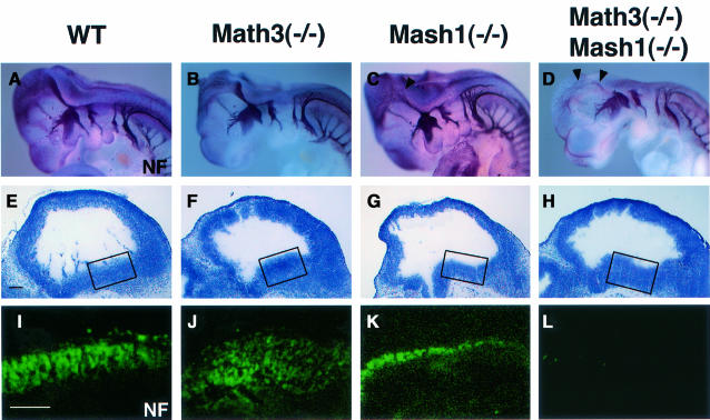
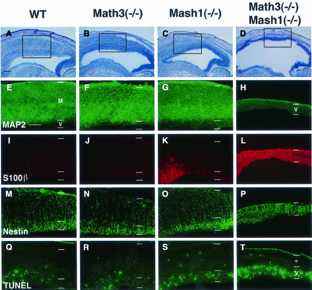
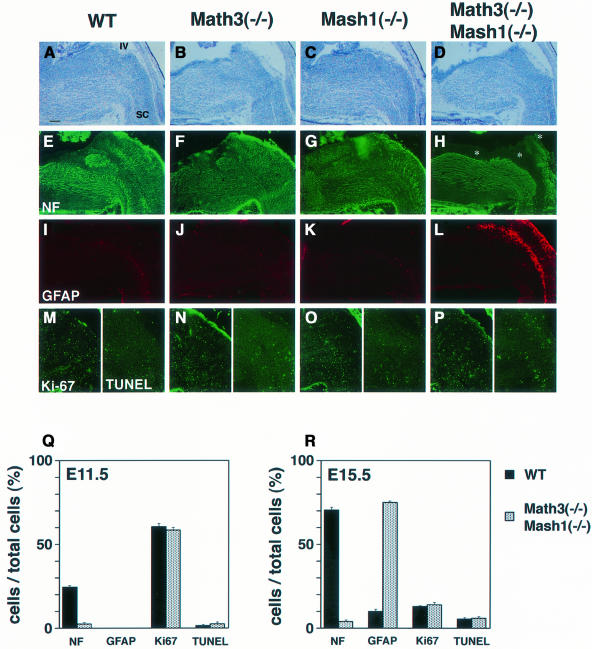
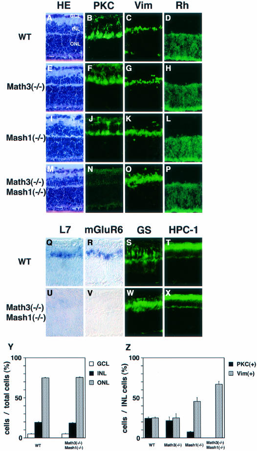
Whereas vertebrate achaete-scute complex (as-c) and atonal (ato) homologs are required for neurogenesis, their neuronal determination activities in the central nervous system (CNS) are not yet supported by loss-of-function studies, probably because of genetic redundancy. Here, to address this problem, we generated mice double mutant for the as-c homolog Mash1 and the ato homolog Math3. Whereas in Mash1 or Math3 single mutants neurogenesis is only weakly affected, in the double mutants tectal neurons, two longitudinal columns of hindbrain neurons and retinal bipolar cells were missing and, instead, those cells that normally differentiate into neurons adopted the glial fate. These results indicated that Mash1 and Math3 direct neuronal versus glial fate determination in the CNS and raised the possibility that downregulation of these bHLH genes is one of the mechanisms to initiate gliogenesis.


Figures

None
Fig. 1. Spatio-temporal expression pattern of Math3 and other bHLH genes. Distribution of bHLH genes on parasagittal sections was examined by in situ hybridization. In all sections, anterior is to the left and dorsal is up. (A–C) Math3 expression in the developing cerebellum at E17.5 (A), P3 (B) and P5 (C). Math3 is expressed in the EGL. (D) At E17.5, Math3 is expressed at the outer region of the EGL, which contains dividing precursors of cerebellar granule cells. (EMath1 is also expressed in the outer region of the EGL. (FNeuroD (brown) is expressed mainly in the inner region of the EGL, which contains postmitotic premigratory cells, whereas Math3 (purple) is expressed mainly in the outer region. (G) At E15.5, Math3 is expressed in the ventricular zone of the dorsal telencephalon but not of the ventral telencephalon. (H) At E12.5, Math3 is expressed at a high level in the ventricular zone of the anterior two thirds of the midbrain. (I) At E15.5, Math3 expression is shifted to the ventricular zone of the posterior midbrain. (J) At E10.5, Math3 is expressed in two longitudinal columns of the hindbrain (asterisks). (K) At E12.5, Math3 expression is observed in two longitudinal columns of the hindbrain (asterisks). (L) At E12.5, Mash1 is expressed in the midbrain. The expression level is higher in the ventricular zone of the anterior two thirds of the midbrain. (M) At E15.5, Mash1 is expressed in the developing midbrain. (N and O) At E10.5 (N) and E12.5 (O), Mash1 is expressed in the ventricular zone of the hindbrain. GE, ganglionic eminence; IV, the fourth ventricle; LV, lateral ventricle; Th, thalamus. Scale bar, 300 μm (A–C and G–O); 30 μm (D–F).
None
Fig. 2. Generation of Math3-deficient mice. (A) Strategy for homologous recombination of Math3 gene by a targeting vector. Schematic representation of the wild-type Math3 gene (top), Math3 targeting vector (middle) and mutant allele (bottom). The neo gene was inserted into the second exon. The closed and open boxes represent the coding and non-coding regions, respectively, of the second exon of the Math3 gene. (B) Southern and northern blot analyses. Genotypes were determined by Southern blot analyses (left and middle). 5′-external probe detected 15 kb wild-type and 6.5 kb mutant bands of XhoI–BamHI-digested genomic DNA. 3′-external probe detected 3.7 kb wild-type and 4.7 kb mutant bands of HincII-digested genomic DNA. Northern blot analysis (right) showed that the functional Math3 mRNA is not present in Math3 homozygous mutant mice. (C) The body weight of wild-type and Math3(–/–) mice. The average with a standard error (n = 13 at P0, 53 at P14 and 26 at >P30) is shown. The growth of Math3(–/–) mice was progressively retarded. (D) Seven-week-old wild-type and Math3(–/–) mice. (E) Hind footprint pattern of wild-type and Math3(–/–) mice. The mutant mice showed ataxic gait.
None
Fig. 3. The cerebellar defects of Math3-deficient mice. (A–F) The wild-type (A, C and E) and Math3(–/–) (B, D and F) cerebellum at E17.5 (A and B), P7 (C and D) and adult (E and F). The Math3(–/–) cerebellar anlage has the EGL and appears normal at E17.5. However, at P7 and adult the Math3(–/–) cerebellum is smaller and the lobule formation is poor. The posterior region is more severely affected in Math3(–/–) cerebellum. (G and HIn situ hybridization of Math1 in E17.5 wild-type (G) and Math3(–/–) (H) cerebellum. Math1 expression appears normal in Math3(–/–) cerebellum. (I and JIn situ hybridization of NeuroD in E17.5 wild-type (I) and Math3(–/–) (J) cerebellum. NeuroD expression appears normal in Math3(–/–) cerebellum. (K and L) Histology of wild-type (K) and Math3(–/–) (L) cerebellum at P14. The molecular layer (ML) and IGL of Math3(–/–) cerebellum are smaller, suggesting that granule cell number is reduced. The Purkinje cell layer (PCL) appears normal in Math3(–/–) cerebellum. (M and N) Staining with anti-Ki-67 antibody. Cells in the outer region of the EGL are mitotic in both wild-type (M) and Math3(–/–) (N) cerebellum at P14. (O and P) TUNEL assay. Only some cells are TUNEL+ in the wild-type EGL (O) whereas many cells are TUNEL+ in the Math3(–/–) EGL (P), indicating that many Math3(–/–) precursors are dying in the EGL. (Q and R) Histology of wild-type (Q) and Math3(–/–) (R) cerebellum at P30. The ML and IGL of Math3(–/–) cerebellum are still smaller, suggesting that granule cell number is reduced. (S and T) Staining with anti-calbindin antibody of wild-type (S) and Math3(–/–) (T) cerebellum at P30. The Purkinje cell layer appears normal in Math3(–/–) cerebellum. In all sections, anterior is to the left and dorsal is up. Scale bar, 200 μm (A and B); 500 μm (C and D); 800 μm (E and F); 100 μm (G–J); 50 μm (K–T).
None
Fig. 4. The midbrain defects of Math3-Mash1 mutant mice. (AD) Whole-mount immunostaining with anti-neurofilament (NF) antibody of E10.5 embryos of wild type (A), Math3(–/–) (B), Mash1(–/–) (C) and Math3(–/–)-Mash1(–/–) (D). Neurite extension is slightly reduced in Mash1(–/–) (C, arrowhead) and more severely reduced in Math3(–/–)-Mash1(–/–) (D, arrowheads). (EH) Histology of E11.5 midbrain of wild type (E), Math3(–/–) (F), Mash1(–/–) (G) and Math3(–/–)-Mash1(–/–) (H). (IL) Immunohistochemical staining with anti-NF antibody of the boxed regions in (E–H). Neurons are normally generated in Math3(–/–) (J). However, there are fewer neurons in Mash1(–/–) (K) and virtually no neurons in Math3(–/–)-Mash1(–/–) (L). Thus, neuronal differentiation is blocked in Math3(–/–)-Mash1(–/–) midbrain. In all sections, anterior is to the left and dorsal is up. Scale bar, 150 μm (E–L).
None
Fig. 5. The defects of the tectum of Math3-Mash1 mutant mice. (AD) Histology of E15.5 tectum. The tectum is slightly thinner in Math3(–/–) (B) and Mash1(–/–) (C) and much thinner in Math3(–/–)-Mash1(–/–) (D) than in the wild type (A). (EH) Immunohistochemistry with anti-MAP2 antibody of the boxed regions in (A–D). The wild-type (E), Math3(–/–) (F) and Mash1(–/–) tectum (G) consist of the ventricular zone (V) and the mantle layer (M), which contains massive MAP2+ neurons. In contrast, Math3(–/–)-Mash1(–/–) tectum consists of only the ventricular zone and lacks neurons (H). (IL) Immunohistochemistry with anti-S100β antibody. There are no S100β+ cells in the wild-type (I) and Math3(–/–) tectum (J) whereas some cells expressed S100β in the Mash1(–/–) tectum (K). Strikingly, in Math3(–/–)-Mash1(–/–) tectum (L) the majority of the cells expressed S100β, indicating that gliogenesis is significantly enhanced in the double mutants. (MP) Immunohistochemistry with anti-Nestin antibody. The double-mutant tectum (P) consists only of the ventricular zone (Nestin+). (QT) TUNEL assay. Only some cells are TUNEL+ in the wild-type (Q) and Math3(–/–) (R) tectum. In contrast, there are slightly more TUNEL+ cells in Mash1(–/–) (S) and many more TUNEL+ cells in Math3(–/–)-Mash1(–/–) (T). Thus, double-mutant neural precursor cells are blocked from neuronal differentiation and result in extensive apoptosis. The asterisk in (T) indicates non-neural tissues including the skull. In all sections, anterior is to the left and dorsal is up. Scale bar, 300 μm.
None
Fig. 6. The defects of the hindbrain of Math3-Mash1 mutant mice. (AD) Histology of wild-type (A), Math3(–/–) (B), Mash1(–/–) (C) and Math3(–/–)-Mash1(–/–) hindbrain (D) at E15.5. (EH) Immunohistochemistry with anti-NF antibody. Neurons are generated in the whole region of wild-type (E), Math3(–/–) (F) and Mash1(–/–) hindbrain (G). In contrast, neurons are missing in the two longitudinal columns of Math3(–/–)-Mash1(–/–) hindbrain (H, asterisks). (IL) Immunohistochemistry with anti-GFAP antibody of adjacent sections of (E–H). Whereas only a few glial cells are present in wild-type (I), Math3(–/–) (J) and Mash1(–/–) (K) hindbrain, many glial cells are generated in the regions that lack neurons in Math3(–/–)-Mash1(–/–) hindbrain (L). (MP) Ki-67 (left) and TUNEL staining (right). Each panel shows the right half of (A–D). No significant difference in Ki-67 and TUNEL staining is observed in the wild-type and mutant hindbrain. In all sections, anterior is to the left and dorsal is up. IV, the fourth ventricle; SC, spinal cord. Scale bar, 250 μm. (Q and R) Quantification of the cells in the longitudinal columns of the hindbrain. The cell number of the two longitudinal columns was counted in a section (16 μm thick) of at least three independent samples of the wild-type and Math3(–/–)-Mash1(–/–) embryos at E11.5 (Q) and E15.5 (R). The ratios of NF+, GFAP+, Ki-67+ and TUNEL+ cells per the total cells in the columns are shown with bars to represent the standard error.
None
Fig. 7. The retinal explant cultures from mutant embryos. Retinal explants prepared from E15.5 embryos of wild-type (AD and QT), Math3(–/–) (EH), Mash1(–/–) (IL) and Math3(–/–)-Mash1(–/–) (MP and UX) were cultured for 2 weeks and subjected to immunohistochemistry and in situ hybridization. HE staining (A, E, I and M) indicates that the cell number of each layer is not affected in all the mutants. However, whereas there are the normal number of bipolar cells (PKC+) in Math3(–/–) (F), there are fewer in Mash1(–/–) (J) and virtually no bipolar cells in Math3(–/–)-Mash1(–/–) (N). In contrast, the number of Müller glial cells (Vim+) is slightly increased in Mash1(–/–) (K) and significantly increased in Math3(–/–)-Mash1(–/–) (O). The number of rods (Rh+) (D, H, L and P) appears normal in all the mutants. (Q–X) In situ hybridization of L7 (Q and U) and mGluR6 (R and V) (bipolar cell markers) and immunohistochemistry with anti-glutamine synthetase (S and W) (a Müller glial marker) and anti-HPC-1 antibodies (T and X) (an amacrine cell marker). Both L7 and mGluR6 expression is lost whereas GS+ cells are increased in the double mutants. Amacrine cells (HPC-1+) are not affected (X). Scale bar, 25 μm. (Y and Z) Quantification of retinal cells. The cell number was counted in a section (16 μm thick, 500 μm wide) of the central region of at least three independent samples from each genotype, and the relative ratios of retinal cells were determined. The percentage of GCL, INL and ONL cells per the total retinal cells (Y) and that of PKC+ and Vim+ cells per the total INL cells (Z) are shown with bars to represent the standard error.

References

    1. Akazawa C., Sasai,Y., Nakanishi,S. and Kageyama,R. (1992) Molecular characterization of a rat negative regulator with a basic helix-loop-helix structure predominantly expressed in the developing nervous system. J. Biol. Chem., 267, 21879–21885. - PubMed
    1. Akazawa C., Ishibashi,M., Shimizu,C., Nakanishi,S. and Kageyama,R. (1995) A mammalian helix-loop-helix factor structurally related to the product of Drosophila proneural gene atonal is a positive transcriptional regulator expressed in the developing nervous system. J. Biol. Chem., 270, 8730–8738. - PubMed
    1. Anderson D.J. (1999) Lineages and transcription factors in the specification of vertebrate primary sensory neurons. Curr. Opin. Neurobiol., 9, 517–524. - PubMed
    1. Anderson D.J. and Jan,Y.N. (1997) The determination of the neuronal phenotype. In Cowan,W.M. (ed.), Molecular and Cellular Approaches to Neural Development. Oxford University Press, New York, NY, pp. 26–63.
    1. Ben-Arie N., Bellen,H.J, Armstrong,D.L., McCall,A.E., Gordadze,P.R., Guo,Q., Matzuk,M.M. and Zoghbi,H.Y. (1997) Math1 is essential for genesis of cerebellar granule neurons. Nature, 390, 169–172. - PubMed

Publication types

MeSH terms