. 2000 Oct 16;19(20):5502-13.
doi: 10.1093/emboj/19.20.5502.

Importin-11, a nuclear import receptor for the ubiquitin-conjugating enzyme, UbcM2

Affiliations

Importin-11, a nuclear import receptor for the ubiquitin-conjugating enzyme, UbcM2

S M Plafker et al. EMBO J. .

Abstract

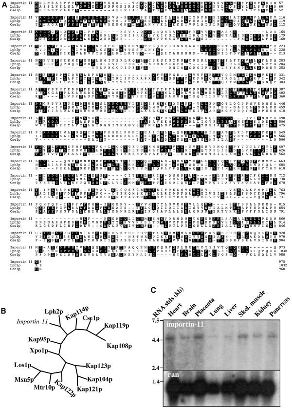
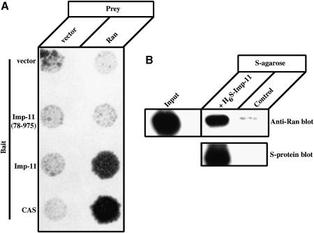
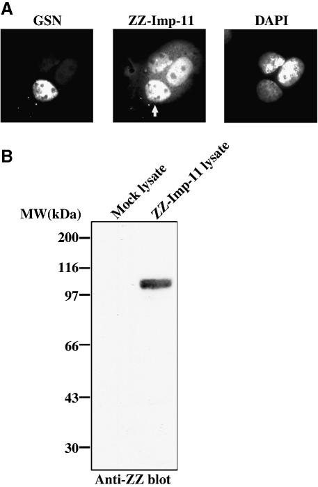
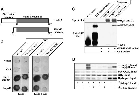
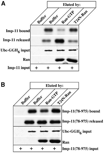
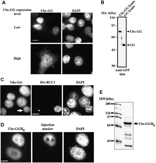
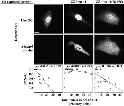
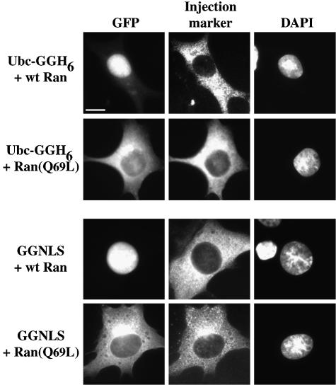
Importins are members of a family of transport receptors (karyopherins) that mediate the nucleocytoplasmic transport of protein and RNA cargoes. We identified importin-11 as a potential new human member of this family, on the basis of limited similarity to the Saccharomyces cerevisiae protein, Lph2p, and cloned the complete open reading frame. Importin-11 interacts with the Ran GTPase, and constitutively shuttles between the nuclear and cytoplasmic compartments. A yeast dihybrid screen identified UbcM2, an E2-type ubiquitin-conjugating enzyme, as a binding partner and potential transport cargo for importin-11. Importin-11 and UbcM2 interact directly, and the complex is disassembled by Ran:GTP but not by Ran:GDP. UbcM2 is constitutively nuclear and shuttles between the nuclear and cytoplasmic compartments. Nuclear import of UbcM2 requires Ran and importin-11, and is inhibited by wheatgerm agglutinin, energy depletion or dominant interfering mutants of Ran and importin-beta. These data establish importin-11 as a new member of the karyopherin family of transport receptors, and identify UbcM2 as a nuclear member of the E2 ubiquitin-conjugating enzyme family.


Figures

None
Fig. 1. Importin-11 is closely related to Lph2p, and is expressed in many human tissues. (A) Amino acid sequences of importin-11 and S.cerevisiae karyopherins Lph2p and Cse1p were aligned using the Clustal program. Identical residues common to two out of three sequences are shaded. (B) Unrooted phylogenetic tree of importin-11 and the 14 S.cerevisiae karyopherins, generated using PAUP and TreeView. (C) Northern blotting of mRNAs from human tissues was performed with a probe generated using the ESTAA082435 plasmid as a template. The same blot was hybridized with a Ran probe as a loading control.
None
Fig. 2. Importin-11 binds Ran directly and this interaction requires the N-terminal 77 residues of importin-11. (A) HF7c (MATa) yeast that express the indicated bait proteins as GAL4 DBD fusions were mated with the W303 (MATα) strain expressing the VP16 TA domain either alone (vector) or as a fusion with Ran. Diploid yeast was selected on Leu/Trp plates and replica-plated onto Leu/Trp/His plates. (B) Ran(Q69L)-His6 was mixed with S-protein–agarose beads alone (control) or with beads plus H6-S-Imp-11. Bound proteins were separated by SDS–PAGE and detected by immunoblotting with a monoclonal antibody to Ran and peroxidase-conjugated, anti-mouse secondary, or with peroxidase-conjugated S-protein. A sample representing 2.5% of the Ran(Q69L)-His6 added to each sample is also shown (Input).
None
Fig. 3. Importin-11 shuttles in and out of the nucleus. (A) Transfected donor BHK cells, expressing zz-tagged importin-11 (ZZ-Imp-11), were fused to GSN2 acceptor cells. The fused cells were grown in medium containing cycloheximide, then fixed, permeabilized and immunostained with a Texas Red-conjugated antibody to detect ZZ-Imp-11. DNA was stained with DAPI. Left panel shows the green channel (GSN); middle panel shows the red channel (ZZ-Imp-11); right panel shows the DNA staining. The white arrow in the middle panel denotes the acceptor nucleus within the fused cell. (B) Immunoblot of lysates prepared from mock-transfected and ZZ-Imp-11-transfected BHK cells. The blot was probed with an HRP-conjugated, rabbit anti-goat antibody.
None
Fig. 4. Importin-11 interacts with an E2-type ubiquitin-conjugating enzyme, UbcM2. (A) Schematic representation of UbcM2, and of a truncation of the enzyme lacking the first 52 amino acid residues [UbcM2(53–207)]. N-terminal extension domain is demarcated by the white box and the catalytic domain by the shaded box. (B) Conjugation assays were carried out as in Figure 2. Diploid yeast were replica-plated onto selective medium (LWH) and similar plates containing 3-AT (LWH+3AT). (C) GST–Ubc or GST was mixed with protein-S–agarose beads plus H6-S-Imp-11 and GST–Ubc was mixed with beads alone (control). Bound proteins were immunoblotted with an anti-GST antibody, or with peroxidase-conjugated S-protein. An aliquot representing 50% of the GST proteins added to each sample is also shown. (D35S-labeled importin-11 and importin-β, expressed by in vitro transcription–translation, were incubated with recombinant Ubc-GGH6, GFP-His6 (GH6) or His6-importin-α (H6-Imp-α), immobilized on Ni2+–agarose beads, or with beads alone. Proteins remaining associated with the beads were resolved by SDS–PAGE and detected by staining (Coomassie Brilliant Blue) or fluorography.
None
Fig. 5. The importin-11–UbcM2 complex is dissociated specifically by Ran:GTP. (A35S-labeled importin-11, expressed by in vitro transcription–translation, was combined with either 1 µg of recombinant Ubc-GGH6 immobilized on Ni2+–agarose beads or with beads alone. Bead-associated proteins were eluted by buffer, or by Ran loaded with GTP (Ran:GTP) or a mutant of Ran that cannot bind nucleotide (T24N-Ran). Proteins remaining associated with the beads (Imp-11 bound and Ubc-GGH6 input) and aliquots of those eluted (Imp-11 released and Ran) were resolved by SDS–PAGE and detected by staining with Coomassie Brilliant Blue or fluorography. (B) As for (A), but using the N-terminal mutant, Imp-11(78–975).
None
Fig. 6. UbcM2 is a constitutively shuttling, nuclear protein. (A) BHK cells were transfected with plasmid encoding Ubc-GG then fixed, permeabilized and analyzed by fluorescence microscopy as described in Materials and methods. Cells expressing relatively low amounts of protein were imaged at 500 ms exposures (Low) and those expressing larger amounts at 150 ms (High). (B) Cells were transfected with plasmids encoding Ubc-GG or a GFP-GFP fusion (GG). Protein extracts were immunoblotted for GFP. Full-length forms of each fusion protein are denoted by arrows. (C) Fusion assays were carried out on donor BHK cells expressing Ubc-GG, and acceptor tsBN2/RCC1 cells that express HA-tagged RCC1. After fusion, cells were processed for immunofluorescence using 12CA5 antibody and a Texas Red-conjugated secondary. Left panel shows the GFP fluorescence (Ubc-GG); middle panel shows Texas Red fluorescence (HA-RCC1); right panel shows DNA staining (DAPI). The white arrow in the left panel denotes the acceptor nucleus within the fused cell (n = 20). (D) Binucleated BHK cells were micro-injected into one nucleus with a mixture of Ubc-GGH6 (3 µM) and TRITC-labeled dextran (Injection marker) and incubated for 30 min before fixation (n = 40). Bars, 10 µm. (E) Coomassie Blue-stained gel of the Ubc-GGH6 protein used for all micro-injection experiments. Full-length Ubc-GGH6 is denoted with an arrow.
None
Fig. 7. Nuclear accumulation of UbcM2 is dependent on functional importin-11. BHK cells ectopically expressing Ubc-GG alone (–) or together with zz-tagged importin-11 (ZZ-Imp-11), the mutant ZZ-Imp-11(78–975) or importin-β were processed for immunofluorescence as described for Figure 3. GFP was imaged under conditions such that no pixels were saturated, and quantitated to obtain total fluorescence (N+C) and nuclear fluorescence (N) for each cell. Graphs show fractional nuclear GFP fluorescence for a range of expression levels of the Ubc-GG, in the presence or absence of the co-expressed importins. The data in the graphs were compiled from 10–20 cells in each condition for two separate experiments. Bar, 10 µm. (See Supplementary data for method of image analysis and importin-β graph.)
None
Fig. 8. Nuclear import of UbcM2 is Ran dependent. BHK cells were micro-injected into the cytoplasm with mixtures of Ubc-GGH6 (2.5 µM), TRITC-labeled dextran (1 mg/ml) plus either wild-type Ran (60 µM) or mutant Ran(Q69L) (50 µM). Cells were incubated for 30 min at 37°C before fixation. Control injections were done with GGNLS (19 µM). Left panel shows GFP fluorescence (GFP); middle panel shows the TRITC fluorescence (Injection marker); right panel shows the DNA staining (DAPI). Bar, 10 µm; 30–40 cells were injected, with similar results.
None
Fig. 9. Importin-11 can mediate the import of UbcM2 in vitro. (A) Aliquots of the cytosolic extracts used in (B), (D) and (E) were analyzed by immunoblotting with 12CA5 anti-HA antibody. Importin-11 migrates as a doublet. (B) Representative images of permeabilized HeLa cells overlayed with mixtures of Ubc-GGH6 (1.2 µM) and assay buffer + 4.5 µM Ran (Buffer), or cytosolic extracts containing no importin-11 (Mock cytosol), HA3-tagged importin-β (Imp-β cytosol) or HA3-tagged importin-11 (Imp-11 cytosol). (C) Relative levels of Ubc-GGH6 nuclear accumulation. Data were collected and pooled from two experiments and represent between 100 and 200 cells/condition. (D) Photomicrographs of cells incubated with Ubc-GGH6, Imp-11 cytosol, and either an energy regenerating system (+energy) or the inhibitor apyrase (+apyrase). (E) Digitonin-permeabilized HeLa cells were overlayed with mixtures of recombinant Ubc-GGH6, Imp-11 cytosol, energy and either no inhibitor (–) (a), mutant Ran:GTP [Ran(Q69L)] (6 µM) (b), wheatgerm agglutinin (0.28 mg/ml) (c) or importin-β (45–462) (2 µM) (d). The corresponding DAPI-stained nuclei for each sample are also shown (e–h). Bars, 10 µm.

References

    1. Black B.E., Levesque,L., Holaska,J.M., Wood,T.C. and Paschal,B.M. (1999) Identification of an NTF2-related factor that binds Ran-GTP and regulates nuclear protein export. Mol. Cell. Biol., 19, 8616–8624. - PMC - PubMed
    1. Borer R.A., Lehner,C.F., Eppenberger,H.M. and Nigg,E.A. (1989) Major nucleolar proteins shuttle between nucleus and cytoplasm. Cell, 56, 379–390. - PubMed
    1. Chen P., Johnson,P., Sommer,T., Jentsch,S. and Hochstrasser,M. (1993) Multiple ubiquitin-conjugating enzymes participate in the in vivo degradation of the yeast MATα2 repressor. Cell, 74, 357–369. - PubMed
    1. Chi N.C. and Adam,S.A. (1997) Functional domains in nuclear import factor p97 for binding the nuclear localization sequence receptor and the nuclear pore. Mol. Biol. Cell, 8, 945–956. - PMC - PubMed
    1. Chook Y.M. and Blobel,G. (1999) Structure of the nuclear transport complex karyopherin-2-Ran.GppNHp. Nature, 399, 230–237. - PubMed

Publication types

MeSH terms