. 2000 Oct;106(8):951-61.
doi: 10.1172/JCI10905.

Edg-1, the G protein-coupled receptor for sphingosine-1-phosphate, is essential for vascular maturation

Affiliations

Edg-1, the G protein-coupled receptor for sphingosine-1-phosphate, is essential for vascular maturation

Y Liu et al. J Clin Invest. 2000 Oct.

Abstract

Sphingolipid signaling pathways have been implicated in many critical cellular events. Sphingosine-1-phosphate (SPP), a sphingolipid metabolite found in high concentrations in platelets and blood, stimulates members of the endothelial differentiation gene (Edg) family of G protein-coupled receptors and triggers diverse effects, including cell growth, survival, migration, and morphogenesis. To determine the in vivo functions of the SPP/Edg signaling pathway, we disrupted the Edg1 gene in mice. Edg1(-/-) mice exhibited embryonic hemorrhage leading to intrauterine death between E12.5 and E14.5. Vasculogenesis and angiogenesis appeared normal in the mutant embryos. However, vascular maturation was incomplete due to a deficiency of vascular smooth muscle cells/pericytes. We also show that Edg-1 mediates an SPP-induced migration response that is defective in mutant cells due to an inability to activate the small GTPase, Rac. Our data reveal Edg-1 to be the first G protein-coupled receptor required for blood vessel formation and show that sphingolipid signaling is essential during mammalian development.


Figures

Figure 1
Figure 1
Targeted disruption and embryonic expression of the Edg1 gene. (a) Schematic representation of the Edg1 targeting strategy. The structure of the mouse Edg1 locus is shown at the top, the structure of the Edg1 targeting vector in the middle, and the predicted structure of the homologous recombined locus on the bottom. RT-5′ and RT-3′, primers for RT-PCR. B, BamHI; Bgl, BglII; PBSK, pBluescript vector. (b) Genotyping of mouse offspring from the Edg1 heterozygous mating. Wild-type Edg1 locus yielded a 9.5-kb BamHI band. Disrupted Edg1 locus yielded a 2.5-kb BamHI band. No Edg1–/– mice were found born alive. (c) RT-PCR analysis of total RNA from E12.5 mouse embryos by using RT-5′ and RT-3′. Edg1+/+ and Edg1+/– RNA yielded the predicted 630-bp amplification product. No amplification product was detected from Edg1–/– RNA. (d, e) Whole-mount of Edg1+/– E9.5 and E10.5 embryos stained with X-Gal. H, heart; DA, dorsal aorta; ISA, intersomatic arteries; CP, capillaries; TC, telencephalon; ACV, anterior cardinal vein. (f) Longitudinal section of dorsal aorta (DA) from E10.5 Edg1+/– embryo. LacZ staining is seen in arterial ECs (AEC). (g) Longitudinal section of posterior cardinal vein (PCV) from E10.5 Edg1+/– embryo. LacZ staining is seen in arterial endothelial cells (AEC) but not in venous endothelial cells (VEC). (h) Transverse section of dorsal aorta from E12.5 Edg1+/– embryo. Vascular ECs and VSMCs are stained. EC, endothelial cell; VSMC, vascular smooth muscle cell. Scale bars = 50 μm.
Figure 2
Figure 2
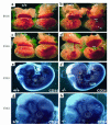
Phenotype of Edg1–/– embryos and normal vascular network in the Edg1–/– embryos. Photomicrographs of E12.5 and E13.5 embryos with the amnion, yolk sac, and placenta intact (a and c), or with extraembryonic membranes removed (b and d). Edg1–/– embryos show normal yolk sac vasculature but with less blood (arrows). Yolk sacs of Edg1–/– embryos display progressive edema. E12.5 Edg1–/– embryo shows intraembryonic hemorrhages in the body and limbs. FL, front limb; HL, hind limb. E13.5 Edg1–/– embryo demonstrates severe intraembryonic hemorrhages and edema. Both E12.5 and E13.5 Edg1–/– embryos display pericardial cavity (PCC) edema. (eh) E12.5 wild-type and Edg1–/– embryos were stained with an anti-CD34 mAb and visualized by low-power (e and f) or higher-power (g and h) magnification. Note the normal vascular patterning, capillary plexus, and capillary sprouting (black arrowheads) in the Edg1–/– embryos. Small blood vessels in the forebrain of Edg1–/– embryos are slightly dilated (white arrowheads).
Figure 3
Figure 3
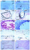
Vascular smooth muscle defects in the Edg1–/– embryos. (a and b) Aortae of E12.5 embryos, sectioned longitudinally and stained with anti-SMαA antibody. Note the lack of SMαA-positive smooth muscle cells (bracket) on the dorsal side of aorta in the Edg1–/– embryo. (c and d) Transverse sections of aortae from E12.5 embryos stained with anti-SMαA. Smooth muscle cells have accumulated at the ventral site of the aorta in the Edg1–/– embryo. Note the discontinuous endothelial cell (EC) layer (bracket) in the Edg1–/– embryo. Many blood cells have leaked out to the surrounding tissues in the Edg1–/– embryo (arrowheads). BC, blood cells. (e and f) H&E staining of aorta from E11.5 and 12.5 Edg1–/– embryos. Arrows point to ECs. Note their normal, flattened morphology in e and abnormal, cuboidal morphology in f. (g and h) Cranial arteries from E12.5 embryos stained with anti-SMαA antibody. Note the clustering of smooth muscle cells and nearly naked endothelial tube from the Edg1–/– embryo. (i and j) Sections of intestine from E12.5 embryos stained with anti-SMαA. Note that that coverage of intestine by smooth muscle is similar in control and mutant embryos. Scale bars = 1 mm (a and b); 50 μm (c and d); 50 μm (e and f); 50 μm (g and h); 500 μm (i and j).
Figure 4
Figure 4
Reduced VSMCs and pericytes in the Edg1–/– vessels. (a–d) EM microscopic analyses of representative small blood vessels from the limb (a and b) and brain capillaries (c and d) from E12.5 wild-type and Edg1–/– embryos. Reduced number of VSMCs (bracket in b) and the lack of capillary pericytes (PC) were found in the Edg1–/– embryos. Notice the abnormally rounded EC nucleus in the Edg1–/– capillary (d). (e and f) EC junctions (arrows) in wild-type and mutant embryos. Note the normal EC junction (EJ) in the Edg1–/– embryo (f). BC, blood cell. ×2,000 (a–d); ×50,000 (e and f).
Figure 5
Figure 5
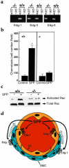
Migration and Rac activation defects in Edg1–/– embryonic fibroblasts. (a) RT-PCR analysis of Edg1, -3, and -5 expression was carried out with total RNA isolated from Edg1+/+, Edg1+/–, and Edg1–/– embryonic fibroblasts. (b) SPP chemotactic responses of embryonic fibroblasts. Serum-starved Edg1+/+ and Edg1–/– fibroblasts were allowed to migrate toward a gradient produced by SPP (100 nM). Control (Cont.) indicates medium without serum was used as the chemoattractant. Chemotaxis was measured as described in Methods. Data are means ± SD of triplicate determinations. AStatistically significant difference compared with the control, determined by Student’s t test (P < 0.01). (c) Rac activation in fibroblasts. Edg1+/+ and Edg1–/– fibroblasts were serum-starved and then treated with SPP for 5 minutes. The cell lysates were used both for affinity precipitation with the PAK-1–conjugated agarose to pull down activated, GTP-bound Rac (top panel) and without fractionation to determine total Rac levels (bottom panel) by SDS-PAGE and immunoblotting. (d) Model of Edg-1 functions in blood vessel development. The Edg1 knockout demonstrates that Edg-1 is essential for vascular maturation by impairing the recruitment of smooth muscle cells to vessel walls. SPP, found in blood, may directly stimulate Edg-1 on VSMCs, facilitating their migration to vessels walls. In a second mechanism, which does not exclude the first, SPP could stimulate Edg-1 expressed on endothelial cells, which in turn recruit may VSMCs. EC, endothelial cell; SMC, smooth muscle cell.

Comment in

References

    1. Shayman JA. Perspectives in basic science: sphingolipids. Kidney Int. 2000;58:11–26. - PubMed
    1. Huwiler A, Kolter T, Pfeilschifter J, Sandhoff K. Physiology and pathophysiology of sphingolipid metabolism and signaling. Biochim Biophys Acta. 2000;1485:63–99. - PubMed
    1. Spiegel S, Merrill AH., Jr Sphingolipid metabolism and cell growth regulation. FASEB J. 1996;10:1388–1397. - PubMed
    1. Lee MJ, et al. Sphingosine-1-phosphate as a ligand for the G protein-coupled receptor EDG-1. Science. 1998;279:1552–1555. - PubMed
    1. Hla T, Maciag T. An abundant transcript induced in differentiating human endothelial cells encodes a polypeptide with structural similarities to G-protein–coupled receptors. J Biol Chem. 1990;265:9308–9313. - PubMed

MeSH terms