. 2000 Oct;106(8):963-71.
doi: 10.1172/JCI10828.

Pax3 is required for enteric ganglia formation and functions with Sox10 to modulate expression of c-ret

Affiliations

Pax3 is required for enteric ganglia formation and functions with Sox10 to modulate expression of c-ret

D Lang et al. J Clin Invest. 2000 Oct.

Abstract

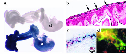
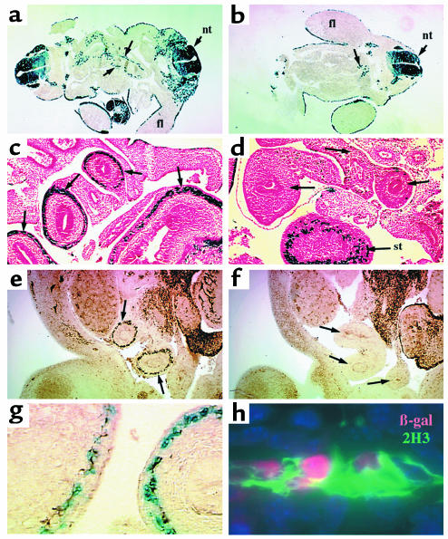
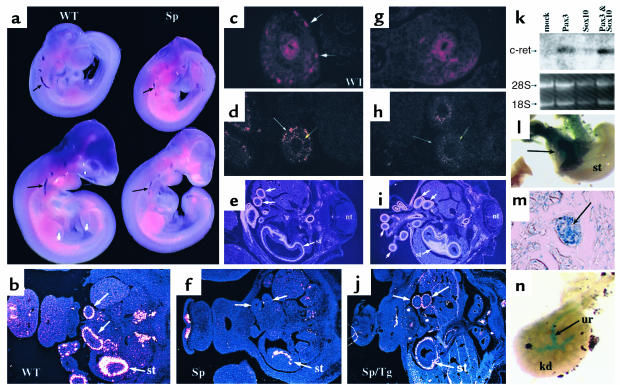
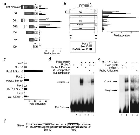
Hirschsprung disease and Waardenburg syndrome are human genetic diseases characterized by distinct neural crest defects. Patients with Hirschsprung disease suffer from gastrointestinal motility disorders, whereas Waardenburg syndrome consists of defective melanocyte function, deafness, and craniofacial abnormalities. Mutations responsible for Hirschsprung disease and Waardenburg syndrome have been identified, and some patients have been described with characteristics of both disorders. Here, we demonstrate that PAX3, which is often mutated in Waardenburg syndrome, is required for normal enteric ganglia formation. Pax3 can bind to and activate expression of the c-RET gene, which is often mutated in Hirschsprung disease. Pax3 functions with Sox10 to activate transcription of c-RET, and SOX10 mutations result in Waardenburg-Hirschsprung syndrome. Thus, Pax3, Sox10, and c-Ret are components of a neural crest development pathway, and interruption of this pathway at various stages results in neural crest-related human genetic syndromes.


Figures

Figure 1
Figure 1
A subset of Pax3-expressing cells are fated to become enteric ganglia. Neural crest–specific regulatory elements of the Pax3 promoter were used to make transgenic mice expressing Cre recombinase. (a) After mating with Cre reporter mice, Pax3-expressing neural crest cells express β-galactosidase (stained blue) and populate the stomach (st) and intestines. The gastrointestinal tract from an E14.5 transgenic embryo is blue (bottom), whereas tissue from a Cre reporter embryo without the P3pro-Cre transgene processed in parallel is not stained (top). (b) Transverse section through a labeled loop of bowel reveals β-galactosidase–expressing cells in the region populated by enteric ganglia (arrows). The section is counterstained with eosin. (c) Coimmunohistochemistry of an adjacent section to that shown in b reveals neurofilament expression (2H3 antibody, red) colocalizing with β-galactosidase (blue). (d) Higher-power image of a similar section to that shown in c colabeled for neurofilament (2H3, green) and β-galactosidase (red) expression. Colocalization results in a yellow signal, identifying labeled cells as enteric ganglia.
Figure 2
Figure 2
Enteric ganglia are deficient in Splotch intestine. (a and b) Transverse sections of E10.5 wild-type (a) and Sp–/– (b) Cx43-LacZ transgenic embryos are shown after staining for β-galactosidase activity (blue). β-Galactosidase–positive cells are seen lining the primitive gut in wild-type (arrows, a) but not Sp–/– (b) embryos, although a small number of blue cells are seen near the proximal foregut (arrow, b). (c and d) β-Galactosidase detection in E12.5 wild-type (c) and Sp–/– (d) transverse sections at the abdominal level reveals expression in the wall of the stomach (st) and all loops of bowel (arrows, c) in wild-type embryos. No expression is seen in the gut distal to the stomach in Sp–/– embryos (arrows, d). (e and f) Neurofilament immunohistochemistry of E12.5 wild-type (e) and Sp–/– (f) sections shows loss of neurofilament expression in Sp–/– embryos in a pattern similar to that seen for β-galactosidase (compare arrows, e and f). (g) Costaining for β-galactosidase (blue) and neurofilament (brown) shows overlapping expression patterns in the bowel wall of wild-type embryos. (h) Higher power of nuclear β-galactosidase (red) and neurofilament (2H3, green) stained wild-type intestine confirms that Cx43-lacZ transgene labels enteric neurons. nt, neural tube. fl, forelimb.
Figure 3
Figure 3
Pax3 is required for normal c-ret expression. (a) Whole-mount in situ hybridization reveals c-ret expression in wild-type (WT) E9.5 (top) and E10 (bottom) embryos. c-ret expression is reduced in Splotch (Sp) embryos (arrows). (b) Radioactive in situ hybridization reveals c-ret expression in the stomach (st) and loops of bowel in wild-type (WT) embryos at E12.5. (f) Expression is missing from intestine distal to the stomach (st) in Splotch (Sp) embryos (arrows). (j) Splotch embryos carrying a transgene that replaces Pax3 expression in neural crest cells (Sp/Tg) have normal c-ret expression in the intestine (arrows). (c and g) c-Ret protein expression is evident in the bowel of wild-type E12.5 embryos (arrows, c) but not in Splotch intestine (g). (d and h) Ednrb mRNA expression is detected by radioactive in situ hybridization in the enteric ganglia of E12.5 wild-type embryos (white arrow, d) and in the surrounding mesenchyme (yellow arrow). Ednrb expression is lost the region of normal enteric ganglia formation of Splotch embryos (white arrow, h) but retained in surrounding mesenchyme (yellow arrow). (e and i) SM22α expression, used as a marker of smooth muscle, is normal in wild-type (e) and Splotch (i) embryos. nt, neural tube. (k) Northern analysis of RNA derived from transfected P19 embryocarcinoma cells reveals increased c-ret expression after transfection of Pax3 (lane 2) or Pax3 with Sox10 (lane 4) compared with mock transfection (lane 1). Ethidium bromide–stained gel is shown below to confirm similar loading of RNA in each lane. (l) Transgenic RET-lacZ embryo reveals β-galactosidase activity in portions of stomach (st, arrow) and intestine. (m) Section through intestine of sample shown in l reveals β-galactosidase activity in enteric ganglia. (n) RET-lacZ transgenic kidney (kd) reveals β-galactosidase activity in the ureter (ur).
Figure 4
Figure 4
A Pax3- and Sox10- responsive enhancer in the c-RET gene. (a) Cotransfection of 293T cells with reporter constructs composed of portions of the c-RET upstream genomic sequence (see Methods) cloned upstream of a luciferase gene with pCMV-Pax3, pCMV-Sox10, or both reveals a 750-bp enhancer element (open box) and a more proximal repressor element (compare full-length Ret promoter to construct D15). All transfections were normalized for transfection efficiency (see Methods) and are expressed as fold activation compared with transfection without Pax3 or Sox10 (mean ± SD; n =12 for each condition). (b) Further analysis of the 750-bp enhancer was performed by modifying construct D15. Deletion of 45 bp at the 3′ end significantly reduced activity (compare constructs 615 and 570). Mutation in a putative Pax3 binding site (indicated by “X” in construct 615mut) had a similar effect, although activation by Sox10 alone was not affected by this mutation. Residual Pax3 responsiveness present in construct 570 is eliminated by deletion of 20 bp at the 5′ end of the enhancer (compare constructs 570 and 550). The 3′ and 5′ response elements are indicated by dotted lines and filled bars labeled A and B, respectively. Cotransfection results are expressed as in a, n=12 for each condition. (c) The response of construct 615 (depicted in b) is specific for Pax3 and is activated to a much lesser degree by Pax2, Pax6, and Pax9. Although low-level activation is apparent after cotransfection with Pax2 or Pax6, no synergistic activation with cotransfected Sox10 is evident. Cotransfection results are expressed as in a; n=8 for each condition. Pax3 and Sox10 bind to the c-RET enhancer. (d) EMSA using a 45-bp probe derived from the 3′ response element in the 750-bp c-RET enhancer (site A in part b) reveals specific binding by Pax3 (lane 3). Pax3 does not bind to a similar probe in which the putative Pax3 binding site has been replaced by an AscI restriction site (lane 4; and see Methods). Unlabeled competitor wild-type probe is able to compete for binding (lane 5), whereas mutated probe competes far less efficiently (lane 6), indicating binding specificity. (e) Sox10 also binds to site A, and mutation in the putative Sox10 binding site significantly reduces binding. In vitro translated Sox10 forms two complexes in EMSA experiments with probe A (lanes 2 and 3), whereas the reticulocyte lysate without Sox10 does not bind (lane 1). Mutation in the putative Sox10 binding site (see Methods) reduces Sox10 binding (lane 4). Lanes 1 and 2 are from a different gel than lanes 3 and 4. (f) The sequence of probe A is shown with Sox10 and Pax3 binding sites boxed.

References

    1. LeDouarin, N.M. 1982. The neural crest. Cambridge University Press. London, United Kingdom. 259 pp.
    1. McKusick, V.A. 1998. Online mendelian inheritance in man, OMIM™. http://www.ncbi.nlm.nih.gov/Omim. Johns Hopkins University and National Center for Biotechnology Information, National Library of Medicine. Baltimore, Maryland, USA, and Bethesda, Maryland, USA.
    1. Gershon MD. Disorders of enteric neuronal development: insights from transgenic mice. Am J Physiol. 1999;277:G262–G267. - PubMed
    1. Pachnis V, Mankoo B, Costantini F. Expression of the c-ret proto-oncogene during mouse embryogenesis. Development. 1993;119:1005–1017. - PubMed
    1. Romeo G, et al. Point mutations affecting the tyrosine kinase domain of the RET proto-oncogene in Hirschsprung’s disease. Nature. 1994;367:377–378. - PubMed

Publication types

MeSH terms