. 2000 Oct 16;192(8):1081-92.
doi: 10.1084/jem.192.8.1081.

Apoptosis control in syncytia induced by the HIV type 1-envelope glycoprotein complex: role of mitochondria and caspases

Affiliations

Apoptosis control in syncytia induced by the HIV type 1-envelope glycoprotein complex: role of mitochondria and caspases

K F Ferri et al. J Exp Med. .

Abstract

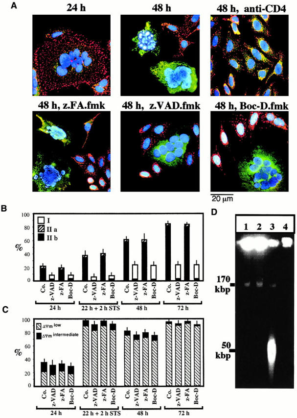
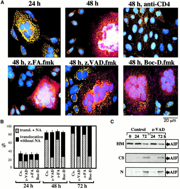
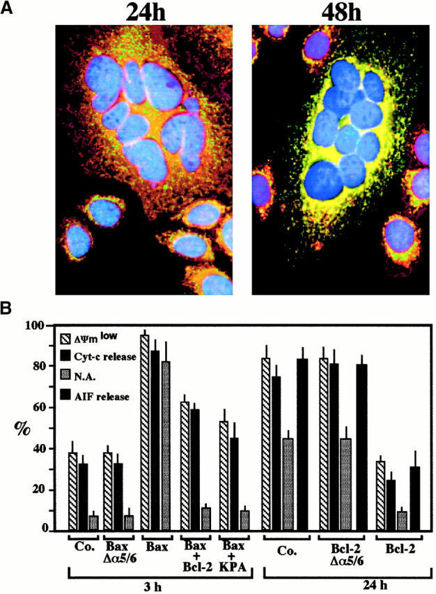
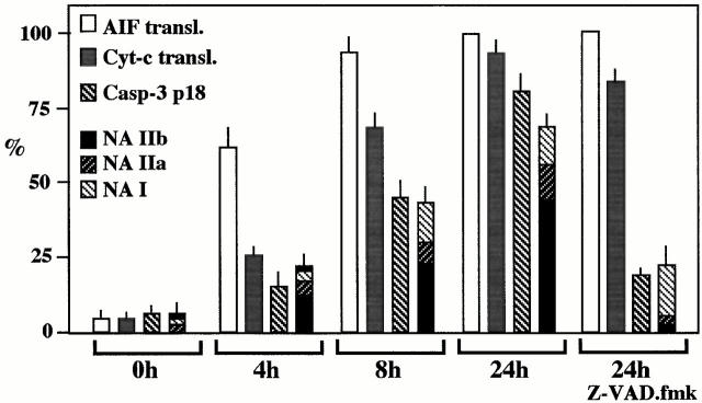
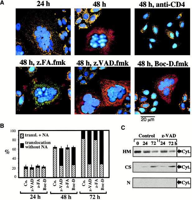
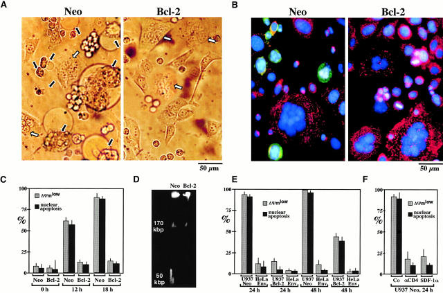
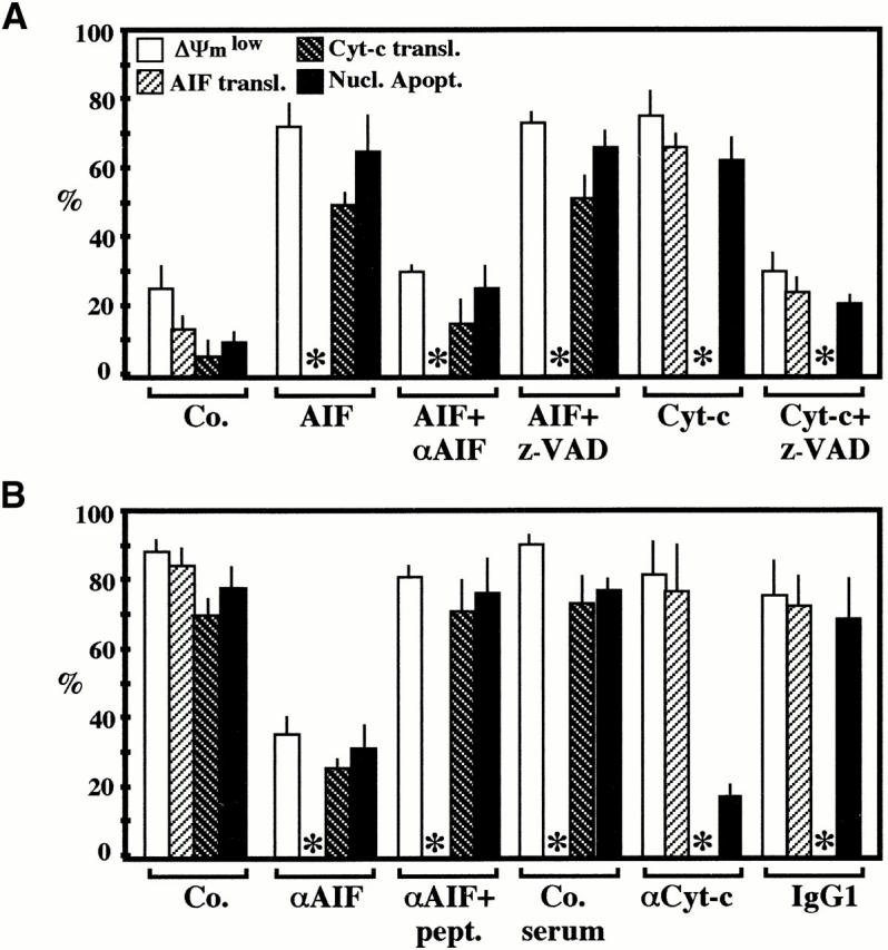
Syncytia arising from the fusion of cells expressing a lymphotropic HIV type 1-encoded envelope glycoprotein complex (Env) with cells expressing the CD4/CXC chemokine receptor 4 complex spontaneously undergo cell death. Here we show that this process is accompanied by caspase activation and signs of mitochondrial membrane permeabilization (MMP), including the release of intermembrane proteins such as cytochrome c (Cyt-c) and apoptosis-inducing factor (AIF) from mitochondria. In Env-induced syncytia, caspase inhibition did not suppress AIF- and Cyt-c translocation, yet it prevented all signs of nuclear apoptosis. Translocation of Bax to mitochondria led to MMP, which was inhibited by microinjected Bcl-2 protein or bcl-2 transfection. Bcl-2 also prevented the subsequent nuclear chromatin condensation and DNA fragmentation. The release of AIF occurred before that of Cyt-c and before caspase activation. Microinjection of AIF into syncytia sufficed to trigger rapid, caspase-independent Cyt-c release. Neutralization of endogenous AIF by injection of an antibody prevented all signs of spontaneous apoptosis occurring in syncytia, including the Cyt-c release and nuclear apoptosis. In contrast, Cyt-c neutralization only prevented nuclear apoptosis, and did not affect AIF release. Our results establish that the following molecular sequence governs apoptosis of Env-induced syncytia: Bax-mediated/Bcl-2-inhibited MMP --> AIF release --> Cyt-c release --> caspase activation --> nuclear apoptosis.


Figures

Figure 1
Figure 1
The effect of caspase inhibitors on nuclear apoptosis and ΔΨm collapse induced by the interaction between Env and CD4. (A) Fluorescence micrographs of HeLa Env cells and HeLa CD4 cells cocultured at a 1:1 ratio for the indicated period (24 or 48 h), in the absence or presence of Boc-D.fmk, Z-VAD.fmk, Z-FA.fmk, or anti-CD4 Ab. Cells were stained with Hoechst 33342 (blue fluorescence) and the ΔΨm-sensitive dye JC-1 (red fluorescence of mitochondria with a high ΔΨm, green fluorescence of mitochondria with a low ΔΨm). Syncytia representing the dominant phenotype (>50% of cells) are shown. (B) Frequency of syncytia with chromatin condensation after various periods of coculture (24, 48, or 72 h). As a positive control of apoptosis induction, the apoptosis inducer staurosporin (STS) was added to 22-h-old syncytia for 2 h. The stage of chromatin condensation (I, IIa, or IIb) was determined as described in Materials and Methods and reference 45. Co., control. (C) Kinetic analysis of ΔΨm loss. The frequency of cells exhibiting a homogeneously ΔΨm high, ΔΨm low, or an intermediate phenotype (ΔΨm intermediate, red JC-1 fluorescence in peripheral mitochondria, green fluorescence in perinuclear mitochondria) was determined. Results are the means of 7 independent experiments (mean ± SEM) in which at least 200 cells were counted for each data point. (D) DNA fragmentation induced by the coculture of HeLa CD4 and HeLa Env cells. Cells were either cultured individually (lane 1, HeLa CD4; lane 2, HeLa Env) or cocultured to allow for the generation of syncytia in the absence (lane 3) or presence of Z-VAD.fmk (lane 4). Syncytia were recovered after 72 h of culture, followed by pulse field gel electrophoresis of nuclear DNA.
Figure 2
Figure 2
Caspase-independent AIF translocation in Env/CD4-induced syncytia. (A) Syncytia at different stages of coculture (performed as described in the legend to Fig. 1) were fixed and permeabilized, followed by immunostaining with Abs specific for AIF (revealed by PE, red fluorescence) and the mitochondrial matrix protein hsp60 (green fluorescence), as well as counterstaining with Hoechst 33342 (blue fluorescence). Note that AIF and hsp60 are colocalized (yellow punctate fluorescence, blend of green plus red) in 24-h syncytia, as well as in individual cells in which cell fusion was blocked by the anti-CD4 Leu3A mAb. Diffuse red cytoplasmic fluorescence and purple nuclear fluorescence (blend of red plus blue) are indicative of the AIF translocation. As an internal control of caspase inhibition efficacy, nuclear chromatin condensation is inhibited by Boc-D.fmk and Z-VAD.fmk but not by Z-FA. fmk. (B) Kinetic analysis of AIF translocation (transl.) and nuclear apoptosis (NA), as determined by immunofluorescence. Results are the means of 6 independent experiments (mean ± SEM) in which a minimum of 200 syncytia was examined for each data point. Co., control. (C) AIF translocation determined by subcellular fractionation. A mixture of HeLa Env cells and HeLa CD4 cells was cocultured during the indicated intervals (0 = no coculture) in the presence or absence of Z-VAD.fmk, lysed, and separated into nuclei (N), mitochondria-enriched heavy membranes (HM), and organelle-free cytosols (CS), as described in Materials and Methods. Each fraction was subjected to SDS-PAGE, Western blot, and immunodetection of AIF.
Figure 5
Figure 5
Bax/Bcl-2–mediated control of MMP in Env/CD4-induced syncytia. (A) Subcellular redistribution of Bax during spontaneous apoptosis in syncytia. Syncytia were obtained by the coculture of HeLa Env and HeLa CD4 cells for 24 or 48 h, followed by staining with anti-COX (green fluorescence), an anti-Bax mAb (revealed by PE, red fluorescence), and Hoechst 33342 (blue fluorescence). At 24 h, 82 ± 3% of syncytia manifest a preponderantly nonmitochondrial distribution of Bax, whereas at 48 h, 61 ± 5% cells demonstrate a mainly mitochondrial localization of Bax (88 ± 4% at 72 h, n = 3). (B) Microinjection of recombinant proteins from the Bax/Bcl-2 family. 24-h-old syncytia were microinjected with recombinant Bax, Bcl-2, inactive mutant proteins (BaxΔα5/6, Bcl-2Δα5/6), and/or Koenig's polyanion (KPA), followed by determination of the frequency of cells with a low ΔΨm (green JC-1 fluorescence), Cyt-c translocation, AIF translocation (only determined after 24 h), and nuclear chromatin condensation. Results are the means of three experiments (mean ± SEM). N.A., nuclear apoptosis; Co., control.
Figure 4
Figure 4
Kinetics of AIF translocation, Cyt-c translocation, and caspase-3 activation in HIV-1–infected syncytia. CD4-expressing HeLa cells were cocultured with chronically HIV-1–infected H9/IIIB cells at a 2:1 ratio. At different time points, syncytia were fixed and permeabilized to assess the translocation of AIF (as described in the legend to Fig. 2) or that of Cyt-c (as described in the legend to Fig. 3), as well as the activation of caspase-3, using an antiserum specific for the active caspase-3 p18 subunit. Moreover, the frequency of syncytia exhibiting different stages of nuclear apoptosis was assessed (as described in the legend to Fig. 1). Results are the means of three experiments (mean ± SEM). NA, nuclear apoptosis; transl., translocation.
Figure 3
Figure 3
Caspase-independent Cyt-c translocation in syncytia. (A) Syncytia arising from the coculture of HeLa Env and HeLa CD4 cells, treated as described in the legends to Fig. 1 and Fig. 2, were stained with mAbs specific for Cyt-c (revealed by PE) and COX (revealed by FITC) as well as with Hoechst 33342. Representative photomicrographs are shown. Note the diffuse red fluorescence indicative of Cyt-c release from mitochondria occurring in the majority of 48-h-old syncytia. (B) Kinetic analysis of AIF, Cyt-c release, and nuclear apoptosis. Results (obtained as described in A) are the means of six independent experiments (mean ± SEM). NA, nuclear apoptosis; transl., translocation; Co., control. (C) Cyt-c translocation determined by immunoblotting. A mixture of HeLa Env cells and HeLa CD4 cells was cocultured during the indicated intervals in the presence or absence of Z-VAD.fmk, lysed, subjected to subcellular fractionation (HM, heavy membranes; CS, cytosols; N, nuclei), and immunodetection of Cyt-c (same samples as described in the legend to Fig. 2).
Figure 6
Figure 6
Bcl-2–mediated inhibition of apoptosis induced by the Env–CD4 interaction in a heterologous system. (A) CD4-expressing Jurkat cells transfected with the Bcl-2 gene or an empty vector conferring Neo resistance were fused with HeLa Env cells at a 1:1 ratio. After 18 h of coculture, phase–contrast images of syncytia were obtained. Note that Bcl-2 overexpression does not impede the formation of syncytia, yet prevents cytoplasmic blebbing. White and black arrows indicate normal and apoptotic syncytia, respectively. (B) JC-1/Hoechst 33342 staining patterns of Jurkat/HeLa Env syncytia (same experiment as described in A). (C) Kinetic analysis of apoptosis induction by fusion of Bcl-2 or Neo Jurkat cells to HeLa Env cells. The frequency of cells demonstrating a JC-1–detectable ΔΨm reduction and nuclear apoptosis was determined after the indicated period of coculture (mean ± SEM, n = 3). (D) DNA fragmentation pattern induced by coculture of HeLa Env cells with Neo Jurkat or Bcl-2 cells. Cells were recovered after 18 h and nuclear DNA was analyzed by pulse field gel electrophoresis. (E) Bcl-2–mediated inhibition of HeLa Env–induced apoptosis of U937 cells. U937 cells transfected with Bcl-2 or Neo were cocultured with HeLa Env cells. This manipulation does not lead to the formation of syncytia, although cell-to-cell contacts are established. The frequency of cells acquiring a low ΔΨm and nuclear apoptosis (NA) was assessed at various time points (n = 3, mean ± SEM) for the two cell types involved. (F) Apoptosis of U937 cells interacting with HeLa Env cells involves CD4 and chemokine receptors. U937 Neo cells were cocultured with HeLa Env cells for 24 h, either in the absence (Co) or presence of Leu3a (5 μg/ml) or SDF-1α (10 μg/ml), followed by determination of the frequency of cells with a low ΔΨm and nuclear apoptosis (n = 3, mean ± SEM).
Figure 7
Figure 7
Molecular hierarchy between the mitochondrial release of AIF and Cyt-c. 24-h-old syncytia, generated by coculture of HeLa Env and HeLa CD4 cells, were microinjected with the indicated combination of AIF, Cyt-c and/or anti-AIF, and anti–Cyt-c Abs, followed by an additional culture period of 3 (A) or 24 h (B), optionally in the presence of Z-VAD.fmk, as indicated. Unfixed cells were then stained with Hoechst 33324 and JC-1 or, alternatively, cells were fixed and stained with anti-AIF plus anti-hsp60 or anti–Cyt-c plus anti-COX to determine the nuclear and mitochondrial parameters of apoptosis. After microinjection of anti-AIF or anti–Cyt-c Abs, the subcellular localization of AIF or Cyt-c, respectively, could not be determined (asterisks). Each point represents the mean of at least 200 microinjected syncytia (n = 3, mean ± SEM). Nucl. Apopt., nuclear apoptosis; transl., translocation; Co., control; pept., peptide.

References

    1. Fauci A.S. Host factors and the pathogenesis of HIV-induced disease. Nature. 1996;384:529–534. - PubMed
    1. Gougeon M.L., Montagnier L. Programmed cell death as a mechanism of CD4 and CD8 T cell depletion in AIDS. Molecular control and effect of highly active anti-retroviral therapy. Ann. NY Acad. Sci. 1999;887:199–212. - PubMed
    1. Li C.J., Friedman D.J., Wang C., Metelev V., Pardee A.B. Induction of apoptosis in uninfected lymphocytes by HIV-1 Tat protein. Science. 1995;268:429–431. - PubMed
    1. Westendorp M.O., Shatrov V.A., Schulze-Osthoff K., Frank R., Kraft M., Los M., Krammer P.H., Dröge W., Lehrmann V. HIV-1 Tat potentiates TNF-induced NF-κB activation and cytotoxicity by altering the cellular redox state. EMBO (Eur. Mol. Biol. Organ.) J. 1995;14:546–554. - PMC - PubMed
    1. Stewart S.A., Poon B., Jowett J.B.M., Chen I.S.Y. Human immunodeficiency virus type 1 Vpr induces apoptosis following cell cycle arrest. J. Virol. 1997;71:5579–5592. - PMC - PubMed

Publication types

MeSH terms