. 2000 Oct 16;192(8):1175-82.
doi: 10.1084/jem.192.8.1175.

Nuclear factor kappa B is required for the development of marginal zone B lymphocytes

Affiliations

Nuclear factor kappa B is required for the development of marginal zone B lymphocytes

A Cariappa et al. J Exp Med. .

Abstract

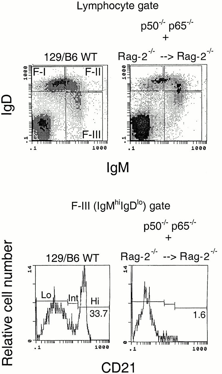
Although immunoglobulin (Ig)M(hi)IgD(lo/-)CD21(hi) marginal zone B cells represent a significant proportion of naive peripheral splenic B lymphocytes, few of the genes that regulate their development have been identified. This subset of peripheral B cells fails to emerge in mice that lack nuclear factor (NF)-kappa Bp50. Less drastic reductions in marginal zone B cell numbers are also seen in the spleens of recombination activating gene (Rag)-2(-/-) mice reconstituted with NF-kappa Bp65(-/-) fetal liver cells and in c-Rel(-/-) mice. In contrast, steady-state levels of IgD(hi) splenic follicular B cells are not significantly reduced in the absence of NF-kappa Bp50, NF-kappa Bp65, or c-Rel. Reconstitution of B cells in Rag-2(-/-) mice with a mixture of p50(-/-)/p65(-/-) fetal liver cells and Rag-2(-/-) bone marrow cells revealed that the generation of marginal zone B cells requires the expression of NF-kappa B in developing B cells, as opposed to supporting cells.


Figures

Figure 1
Figure 1
NF-κBp50 is required for MZ B cell generation. (a) Immunofluorescence performed on splenic cryosections with anti-IgM–FITC and MOMA-1–PE revealed a marked reduction in MZ B cells in the p50−/− mouse (right). Multiple sections revealed similar results. WT, wild-type. (b) Splenic IgMhiIgDhi and IgDhiIgMlo follicular B cell populations appear grossly normal in the p50−/− mouse. (c) IgMhiIgDloCD21hi MZ B cells are markedly reduced in p50−/− mice. Three wild-type and three p50−/− mice were analyzed in the above studies. The results shown are representative. (d) IgMhiIgDloCD1dhi and IgMhiIgDloCD38hi MZ B cells are markedly reduced in p50−/− mice. Three wild-type and three p50−/− mice were analyzed in the above studies. The results shown are representative.
Figure 1
Figure 1
NF-κBp50 is required for MZ B cell generation. (a) Immunofluorescence performed on splenic cryosections with anti-IgM–FITC and MOMA-1–PE revealed a marked reduction in MZ B cells in the p50−/− mouse (right). Multiple sections revealed similar results. WT, wild-type. (b) Splenic IgMhiIgDhi and IgDhiIgMlo follicular B cell populations appear grossly normal in the p50−/− mouse. (c) IgMhiIgDloCD21hi MZ B cells are markedly reduced in p50−/− mice. Three wild-type and three p50−/− mice were analyzed in the above studies. The results shown are representative. (d) IgMhiIgDloCD1dhi and IgMhiIgDloCD38hi MZ B cells are markedly reduced in p50−/− mice. Three wild-type and three p50−/− mice were analyzed in the above studies. The results shown are representative.
Figure 2
Figure 2
A T cell–independent type II antigen is captured in vivo by wild-type (WT) MZ B cells but not by any of the B cell populations in p50−/− spleens. Splenocytes were isolated from wild-type and p50−/− mice that were or were not immunized with TNP-Ficoll. In immunized mice, cells that could be stained with anti-TNP antibodies were readily visualized within the CD45R+CD21hiCD23lo MZ B cell gate. Significant numbers of TNP-staining cells were not seen in any of the splenic B cell pools in p50−/− mice.
Figure 3
Figure 3
NF-κBp65 and c-Rel contribute to MZ B cell development. (a) IgMhiIgDloCD21hi MZ B cells are markedly reduced in Rag-2−/− mice reconstituted with p65−/− fetal liver and Rag-2−/− bone marrow cells (bottom panels). Splenic IgDhiIgMlo follicular B cell numbers appear grossly normal (top panels). Two p65−/− chimeras were analyzed in these studies. The results shown are representative. WT, wild-type. (b) IgMhiIgDloCD21hi MZ B cells are markedly reduced in c-Rel−/− mice (bottom panels), but splenic IgDhiIgMlo follicular B cell numbers appear grossly normal (top panels). Two control and two c-Rel−/− mice were analyzed. The results shown are representative.
Figure 3
Figure 3
NF-κBp65 and c-Rel contribute to MZ B cell development. (a) IgMhiIgDloCD21hi MZ B cells are markedly reduced in Rag-2−/− mice reconstituted with p65−/− fetal liver and Rag-2−/− bone marrow cells (bottom panels). Splenic IgDhiIgMlo follicular B cell numbers appear grossly normal (top panels). Two p65−/− chimeras were analyzed in these studies. The results shown are representative. WT, wild-type. (b) IgMhiIgDloCD21hi MZ B cells are markedly reduced in c-Rel−/− mice (bottom panels), but splenic IgDhiIgMlo follicular B cell numbers appear grossly normal (top panels). Two control and two c-Rel−/− mice were analyzed. The results shown are representative.
Figure 4
Figure 4
NF-κBp50 must be expressed in developing lymphocytes for MZ B cell generation. Rag-2−/− mice were reconstituted with fetal liver from p50−/−/p65−/− mice, and bone marrow from Rag-2−/− mice. Reconstitution results in clearly identifiable IgMhiIgDlo, IgMhiIgDhi, and IgDhi IgMlo subpopulations (top panels). In the IgMhiIgDlo fraction, CD21hi cells were markedly reduced in the p50−/−/p65−/− chimeras (bottom panels). Six separate p50−/−/p65−/− chimeric mice were analyzed in these studies. The results shown are representative. WT, wild-type.
Figure 5
Figure 5
Absolute numbers of splenic B cells in wild-type (WT) and mutant mice. (a) Absolute numbers of newly formed, follicular, and MZ B cells in wild-type, p50−/−, c-Rel−/−, and Rag-2−/− mice reconstituted with mixtures including wild-type, p65−/−, and p50−/−/p65−/− fetal liver cells, respectively, in combination with Rag-2−/− bone marrow cells. (b) Absolute numbers of MZ B cells in the above mice depicted using an expanded scale.
Figure 5
Figure 5
Absolute numbers of splenic B cells in wild-type (WT) and mutant mice. (a) Absolute numbers of newly formed, follicular, and MZ B cells in wild-type, p50−/−, c-Rel−/−, and Rag-2−/− mice reconstituted with mixtures including wild-type, p65−/−, and p50−/−/p65−/− fetal liver cells, respectively, in combination with Rag-2−/− bone marrow cells. (b) Absolute numbers of MZ B cells in the above mice depicted using an expanded scale.

References

    1. MacLennan I.C.M., Gray D. Antigen driven selection of virgin and memory B cells. Immunol. Rev. 1986;91:61–85. - PubMed
    1. Kraal G. Cells in the marginal zone of the spleen. Int. Rev. Cytol. 1992;132:31–74. - PubMed
    1. Oliver A.M., Martin F., Gartland G.L., Carter R.H., Kearney J.F. Marginal zone B cells exhibit unique activation, proliferative and immunoglobulin secretory responses. Eur. J. Immunol. 1997;27:2366–2374. - PubMed
    1. Oliver A.M., Martin F., Kearney J.F. IgMhighCD21high lymphocytes enriched in the splenic marginal zone generate effector cells more rapidly than the bulk of follicular B cells. J. Immunol. 1999;162:7198–7207. - PubMed
    1. Makowska A., Faizunnessa N.N., Anderson P., Midtvedt T., Cardell S. CD1high B cellsa population of mixed origin. Eur. J. Immunol. 1999;29:3285–3295. - PubMed

Publication types

MeSH terms