

Secretion of RTX leukotoxin by Actinobacillus actinomycetemcomitans
- PMID: 11035711
- PMCID: PMC97685
- DOI: 10.1128/IAI.68.11.6094-6100.2000
Secretion of RTX leukotoxin by Actinobacillus actinomycetemcomitans
Abstract
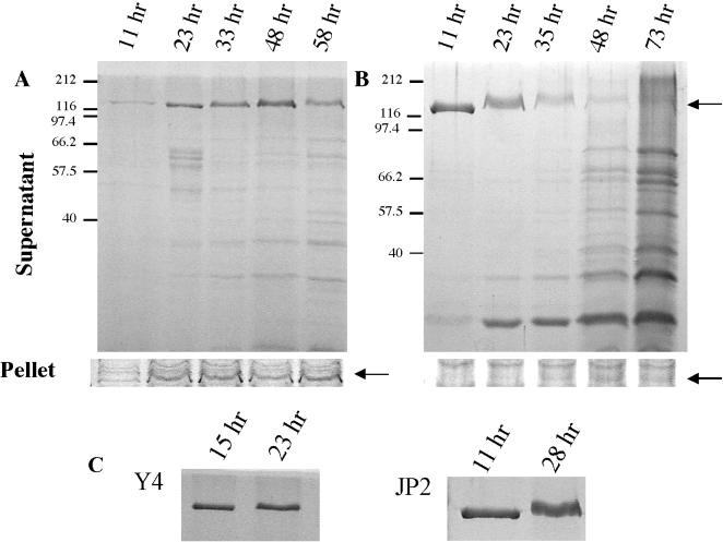
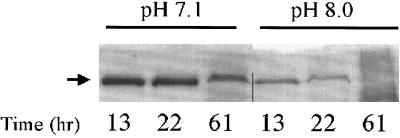
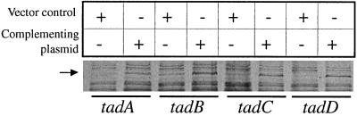
Actinobacillus actinomycetemcomitans, the etiologic agent for localized juvenile periodontitis and certain other human infections, such as endocarditis, expresses a leukotoxin that acts on polymorphonuclear leukocytes and macrophages. Leukotoxin is a member of the highly conserved repeat toxin (RTX) family of bacterial toxins expressed by a variety of pathogenic bacteria. While the RTX toxins of other bacterial species are secreted, the leukotoxin of A. actinomycetemcomitans is thought to remain associated with the bacterial cell. We have examined leukotoxin production and localization in rough (adherent) and smooth (nonadherent) strains of A. actinomycetemcomitans. We found that leukotoxin expressed by the rough, adherent, clinical isolate CU1000N is indeed cell associated, as expected. However, we were surprised to find that smooth, nonadherent strains of A. actinomycetemcomitans, including Y4, JP2 (a strain expressing a high level of toxin), and CU1060N (an isogenic smooth variant of CU1000N), secrete an abundance of leukotoxin into the culture supernatants during early stages of growth. After longer times of incubation, leukotoxin disappears from the supernatants, and its loss is accompanied by the appearance of a number of low-molecular-weight polypeptides. The secreted leukotoxin is active, as evidenced by its ability to kill HL-60 cells in vitro. We found that the growth phase and initial pH of the growth medium significantly affect the abundance of secreted leukotoxin, and we have developed a rapid (<2 h) method to partially purify large amounts of leukotoxin. Remarkably, mutations in the tad genes, which are required for tight nonspecific adherence of A. actinomycetemcomitans to surfaces, cause leukotoxin to be released from the bacterial cell. These studies show that A. actinomycetemcomitans has the potential to secrete abundant leukotoxin. It is therefore appropriate to consider a possible role for leukotoxin secretion in the pathogenesis of A. actinomycetemcomitans.
Figures
Similar articles
-
cis Elements and trans factors are both important in strain-specific regulation of the leukotoxin gene in Actinobacillus actinomycetemcomitans.Infect Immun. 1996 Sep;64(9):3451-60. doi: 10.1128/iai.64.9.3451-3460.1996. Infect Immun. 1996. PMID: 8751884 Free PMC article.
-
Association of Actinobacillus actinomycetemcomitans leukotoxin with nucleic acids on the bacterial cell surface.Infect Immun. 1993 Nov;61(11):4878-84. doi: 10.1128/iai.61.11.4878-4884.1993. Infect Immun. 1993. PMID: 8406888 Free PMC article.
-
Purification of secreted leukotoxin (LtxA) from Actinobacillus actinomycetemcomitans.Protein Expr Purif. 2002 Aug;25(3):465-71. doi: 10.1016/s1046-5928(02)00037-2. Protein Expr Purif. 2002. PMID: 12182827
-
Aggregatibacter actinomycetemcomitans leukotoxin: a powerful tool with capacity to cause imbalance in the host inflammatory response.Toxins (Basel). 2011 Mar;3(3):242-59. doi: 10.3390/toxins3030242. Epub 2011 Mar 18. Toxins (Basel). 2011. PMID: 22069708 Free PMC article. Review.
-
Molecular pathogenicity of the oral opportunistic pathogen Actinobacillus actinomycetemcomitans.Annu Rev Microbiol. 2003;57:29-55. doi: 10.1146/annurev.micro.57.030502.090908. Annu Rev Microbiol. 2003. PMID: 14527274 Review.
Cited by
-
Leukotoxin confers beta-hemolytic activity to Actinobacillus actinomycetemcomitans.Infect Immun. 2006 Apr;74(4):2015-21. doi: 10.1128/IAI.74.4.2015-2021.2006. Infect Immun. 2006. PMID: 16552030 Free PMC article.
-
Screen for leukotoxin mutants in Aggregatibacter actinomycetemcomitans: genes of the phosphotransferase system are required for leukotoxin biosynthesis.Infect Immun. 2008 Aug;76(8):3561-8. doi: 10.1128/IAI.01687-07. Epub 2008 Jun 9. Infect Immun. 2008. PMID: 18541661 Free PMC article.
-
Epigallocatechin gallate inhibits leukotoxin release by Aggregatibacter actinomycetemcomitans by promoting association with the bacterial membrane.Mol Oral Microbiol. 2020 Jan;35(1):29-39. doi: 10.1111/omi.12275. Epub 2019 Dec 26. Mol Oral Microbiol. 2020. PMID: 31816197 Free PMC article.
-
Toxin-triggered liposomes for the controlled release of antibiotics to treat infections associated with the gram-negative bacterium, Aggregatibacter actinomycetemcomitans.Colloids Surf B Biointerfaces. 2024 Jun;238:113870. doi: 10.1016/j.colsurfb.2024.113870. Epub 2024 Mar 21. Colloids Surf B Biointerfaces. 2024. PMID: 38555763 Free PMC article.
-
LL-37 opsonizes and inhibits biofilm formation of Aggregatibacter actinomycetemcomitans at subbactericidal concentrations.Infect Immun. 2013 Oct;81(10):3577-85. doi: 10.1128/IAI.01288-12. Epub 2013 Jul 8. Infect Immun. 2013. PMID: 23836819 Free PMC article.
References
-
- Ausubel F M, Brent R, Kingston R E, Moore D D, Seidman J G, Smith J A, Struhl K, editors. Current protocols in molecular biology. New York, N.Y: John Wiley & Sons; 1989.
-
- Berthold P, Forti D, Kieba I R, Rosenbloom J, Taichman N S, Lally E T. Electron immunocytochemical localization of Actinobacillus actinomycetemcomitans leukotoxin. Oral Microbiol Immunol. 1992;7:24–27. - PubMed
-
- Bhakdi S, Tranum-Jensen J. Damage to mammalian cells by proteins that form transmembrane pores. Rev Physiol Biochem Pharmacol. 1987;107:147–223. - PubMed
Publication types
MeSH terms
Substances
LinkOut - more resources
Full Text Sources
Other Literature Sources
