. 2000 Nov;68(11):6133-8.
doi: 10.1128/IAI.68.11.6133-6138.2000.

Antigenic variation of Anaplasma marginale by expression of MSP2 mosaics

Affiliations

Antigenic variation of Anaplasma marginale by expression of MSP2 mosaics

A F Barbet et al. Infect Immun. 2000 Nov.

Abstract

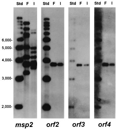
Anaplasma marginale is a tick-borne pathogen, one of several closely related ehrlichial organisms that cause disease in animals and humans. These Ehrlichia species have complex life cycles that require, in addition to replication and development within the tick vector, evasion of the immune system in order to persist in the mammalian reservoir host. This complexity requires efficient use of the small ehrlichial genome. A. marginale and related ehrlichiae express immunoprotective, variable outer membrane proteins that have similar structures and are encoded by polymorphic multigene families. We show here that the major outer membrane protein of A. marginale, MSP2, is encoded on a polycistronic mRNA. The genomic expression site for this mRNA is polymorphic and encodes numerous amino acid sequence variants in bloodstream populations of A. marginale. A potential mechanism for persistence is segmental gene conversion of the expression site to link hypervariable msp2 sequences to the promoter and polycistron.


Figures

FIG. 1
FIG. 1
Structure and variability of the msp2 polycistronic expression site. (A) Diagram of the four ORFs comprising the polycistronic expression site for msp2, showing the molecular weights (M.W.) of encoded proteins, primary (1°) and secondary (2°) RT-PCR products generated from msp2 mRNA, location of probe used in the RNase protection assay (RPA probe, see Fig. 2), and FspI cleavage sites (see Fig. 4 and 5). P, predicted promoter region, 72 to 108 bp 5′ to orf4; T, predicted prokaryotic terminator sequence, GTAGACCAGC....TAGTCGTCAC, 149 to 200 bp 3′ to msp2. (B) PLOTSIMILARITY profile of nucleotide sequence variability between msp2 expression sites of Florida (F) and South Idaho (I-1) strains of A. marginale. A similarity score of 1.0 indicates identical sequence in a sliding window of 10 nucleotides, and a decreasing score from 1.0 to 0.0 indicates increasing variation. (C) PLOTSIMILARITY profile of expression site variability in the South Idaho strain of A. marginale examined at two time points 10 days apart in acute rickettsemia (I-1 and I-2).
FIG. 2
FIG. 2
Analysis of msp2 transcript structure by RT-PCR, primer extension analysis, and RNase protection assay (RPA). For RT-PCR, total DNase-treated RNA of Florida strain A. marginale was reverse transcribed into DNA using oligonucleotide primer AB198, which anneals to the 3′ end of msp2. The cDNA was amplified in a primary PCR with primers AB765 and AB766, which generated a product of 3.2 kbp. The primary PCR product was amplified in a secondary or nested PCR with primer combinations AB192 and AB764, AB689 and AB764, and AB192 and AB688 to generate products of 2.0, 1.5, and 0.7 kbp, respectively. See Fig. 1 for the locations of RT-PCR products. S, molecular size standards; +, with reverse transcriptase; −, negative control reactions without reverse transcriptase. For primer extension, oligonucleotide primer AB784, which anneals 153 nucleotides 3′ to the ATG initiation codon of orf4, was radiolabeled with 32P and extended in sequencing reactions using reverse transcriptase and either total RNA of A. marginale or denatured, PCR-amplified genomic DNA containing orf2 to orf4, msp2, and flanking regions as templates. The order of sequencing reactions T, G, C, and A, is shown above the DNA lanes and was the same in the RNA lanes. A strong stop was detected in RNA at the A (underlined) in sequence TGCAACCCACACACCCATAAGG, with evidence also for a minority of transcripts continuing to the next base, C (italic). This corresponds to the coding-strand sequence CCTTATGGGTGTGTGGGTTGCA (see the text). For the RNase protection assay, a 32P-labeled antisense RNA probe of 722 nucleotides (317 nucleotides of the msp2 gene, 307 nucleotides of orf2 and intercistronic spacer, 98 nucleotides of plasmid vector) was allowed to hybridize to various amounts (10, 3, and 0.1 μg) of total RNA of A. marginale and carrier yeast RNA or to yeast RNA alone (lane 0) and then unprotected single-stranded probe was digested with RNase and analyzed by denaturing polyacrylamide gel electrophoresis. C, probe plus yeast RNA, not digested with RNase. The positions of molecular size standards are shown on the left. A band of 624 bp containing the A. marginale sequences within the probe (msp2 and orf2) was the predominant fragment protected.
FIG. 3
FIG. 3
Multiple different msp2 variants are present in the polycistronic expression site in each population of A. marginale. The expression site was amplified by PCR using primers which annealed 288 bp 3′ to the termination codon of msp2 (AB752) and to the intercistronic sequence between orf3 and orf4 (AB750) to generate a product of 2.9 kbp from A. marginale genomic DNA that contained msp2, orf2, and orf3. The PCR product was cloned in pCR-XL-TOPO vector (Invitrogen), and independent colonies containing a 2.9-kbp insert were selected for sequencing of cloned plasmid DNA. The hypervariable region of the msp2 gene was sequenced on both strands in 106 independent clones derived by PCR amplification from genomic DNA of the F, I-1, and I-2 A. marginale populations. DNA sequences were translated to amino acids, and the different variant sequences were aligned with PILEUP. The predominant sequence variants are shown. The percentage of each sequence variant in that population is indicated in brackets, e.g., the major sequence variant detected in the South Idaho A. marginale population I-1 was variant A, which was found in 48% of the independent clones of the expression site. Identical amino acids shared between all variants are indicated by dashes and shown on the bottom row of the alignment.
FIG. 4
FIG. 4
Structure of msp2 and orf2 to orf4 in genomic DNA of Florida and South Idaho strains of A. marginale. Southern blots of Florida (F) or South Idaho (I) genomic DNA digested with the restriction enzyme FspI and hybridized with probes specific for msp2, orf2, orf3, or orf4 are shown. FspI cleaves 41 nucleotides 5′ to orf4 and 268 nucleotides 3′ to msp2 to release a fragment of 3.76 kbp containing the complete polycistronic msp2 expression site sequence (see Fig. 1) from both Florida and South Idaho genomic DNAs. Molecular size standards are shown in the left lane of each blot. Multiple msp2-related sequences are detected in genomic DNA of both strains; only msp2 sequences located in the expression site are contiguous with orf2 to orf4.
FIG. 5
FIG. 5
Comparison of genes encoding the msp2 hypervariable region in Florida (F) and Florida relapse (F-rel) strains of A. marginale. Hypervariable-region sequences were obtained from independent clones of the msp2 expression site (ES) in F (10 clones) and F-rel (11 clones) genomic DNA. From this DNA sequence information, different oligonucleotide probes to the hypervariable region were synthesized to use in Southern blotting. These probes contained sequence either unique to one set of expression site clones (the AB871 and AB872 sequences were both observed in 2 of 11 clones of the F-rel expression site and 0 of 10 clones of the F expression site) or were present in different proportions (the AB847 sequence was present in 5 of 10 expression site clones in the F strain and 1 of 11 expression site clones in the F-rel strain). A. marginale genomic DNA of the F or F-rel strains was digested with FspI to release the 3.76-kbp expression site (arrow), separated by gel electrophoresis, and probed with the indicated fluorescein-labeled synthetic oligonucleotides. With probe AB872, an extra (expression site) band of 3.76 kbp is detected in F-rel compared to F genomic DNA probed with the same sequence. A 4.4-kbp band is detected in both F and F-rel DNA, which may contain a template copy for conversion of the expression site. The expression site band is less intense than the 4.4-kbp band because the AB872 sequence is present in the expression site of only some organisms in the F-rel population of A. marginale (approximately 18% [2 of 11] of the total would be predicted from the fraction of F-rel expression site clones containing the AB872 sequence). This figure is approximate because of the small number of clones analyzed and the possibility of representation bias following amplification and cloning of the expression site. A similar result was obtained with a different F-rel-specific probe, AB871, except that there are two potential template copies of the AB871 sequence on 4.4- and 3.5-kbp fragments. The AB847 blot is an example of the converse situation: the AB847 sequence is well represented in the expression site of the F population of A. marginale but has nearly disappeared from the expression site in F-rel A. marginale. Potential template copies for the AB847 expression site sequence are on 11- and 7.2-kbp fragments.

References

    1. Alleman A R, Kamper S M, Viseshakul N, Barbet A F. Analysis of the Anaplasma marginale genome by pulsed-field electrophoresis. J Gen Microbiol. 1993;139:2439–2444. - PubMed
    1. Alleman A R, Palmer G H, McGuire T C, McElwain T F, Perryman L E, Barbet A F. Anaplasma marginale major surface protein 3 is encoded by a polymorphic, multigene family. Infect Immun. 1997;65:156–163. - PMC - PubMed
    1. Andersson S G, Zomorodipour A, Andersson J O, Sicheritz-Ponten T, Alsmark U C, Podowski R M, Naslund A K, Eriksson A S, Winkler H H, Kurland C G. The genome sequence of Rickettsia prowazekii and the origin of mitochondria. Nature. 1998;396:133–140. - PubMed
    1. Barbet A F, Kamper S M. The importance of mosaic genes to trypanosome survival. Parasitol Today. 1993;9:63–66. - PubMed
    1. Barbet A F, Palmer G H, Myler P J, McGuire T C. Characterization of an immunoprotective protein complex of Anaplasma marginale by cloning and expression of the gene coding for polypeptide Am105L. Infect Immun. 1987;55:2428–2435. - PMC - PubMed

Publication types

Substances

Associated data

LinkOut - more resources