. 2000 Nov;68(11):6378-83.
doi: 10.1128/IAI.68.11.6378-6383.2000.

Characterization of the catalytic domain of Clostridium novyi alpha-toxin

Affiliations

Characterization of the catalytic domain of Clostridium novyi alpha-toxin

C Busch et al. Infect Immun. 2000 Nov.

Abstract

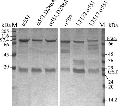
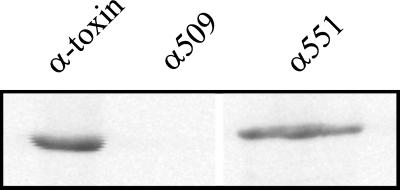
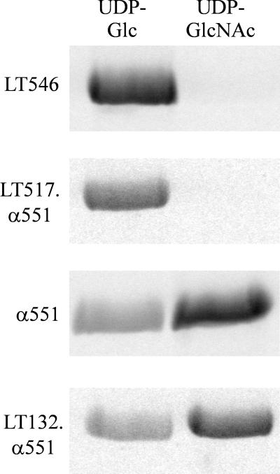
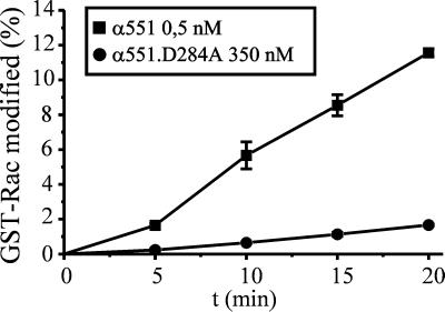
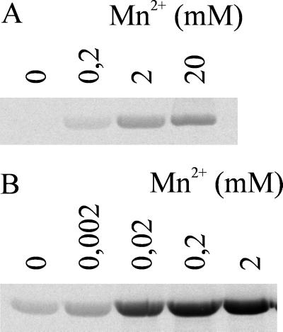
Clostridium novyi alpha-toxin belongs to the family of large clostridial cytotoxins which act on cells through the modification of small GTP-binding proteins. We present here an analysis of the catalytic domain of alpha-toxin. A NH(2)-terminal 551-amino-acid fragment, alpha 551, was found to contain the full enzyme activity of the holotoxin, whereas a slightly shortened fragment encompassing 509 amino acids showed no detectable enzyme activity. Further characterization of the enzymatically active fragment alpha 551 revealed a substrate specificity for both UDP-N-acetylglucosamine and UDP-glucose. A Michaelis-Menten constant of 17 microM was determined for the substrate UDP-N-acetylglucosamine, while that for UDP-glucose was about 20 times higher, indicating a weaker affinity of the toxin for the latter substrate. Mutation of the aspartic acids of a conserved motif DXD within alpha 551 reduced enzyme activity >700-fold and inhibited cytotoxicity after microinjection in cells. Inhibition of enzyme activity of the DXD mutant could be partially overcome by increased concentrations of manganese ions, suggesting the involvement of these aspartic acids in Mn(2+) binding. By construction of chimeras of enzymatically active fragments of C. sordellii lethal toxin and C. novyi alpha-toxin, we located the region involved in nucleotide-sugar specificity to amino acids 133 through 517.


Figures

FIG. 1
FIG. 1
Purified recombinant toxin fragments and chimeras. N-terminal toxin fragments, mutated fragments, and chimeric toxin fragments were constructed as GST fusion proteins, expressed in E. coli, and purified by affinity chromatography. Toxin fragments and mutants (Frag.) are shown as GST fusion proteins, α551 (lane 1), α551.D286A (lane 2), α551.D288A (lane 3), α509 (lane 4), LT132.α551 (lane 5), and LT517.α551 (lane 6); approximately 3 μg was loaded in lane 1, 2 μg was loaded in lanes 2 to 5, and 1 μg was loaded in lane 6.
FIG. 2
FIG. 2
Glucosylation of Rac by alpha-toxin and fragments. Recombinant GST-Rac (1 μg) was glucosylated by alpha-toxin, α509, or α551 (100 nM each) in the presence of UDP–[14C]N-acetylglucosamine for 30 min. Labeled proteins were then analyzed by SDS-PAGE and phosphorimaging (shown).
FIG. 3
FIG. 3
Nucleotide-sugar specificity of alpha-toxin fragments and chimeric toxin fragments. Recombinant GST-Rac (2 μg) was glucosylated by α551, LT546, LT132.α551, or LT517.α551 in the presence of UDP-[14C]glucose or UDP–[14C]N-acetylglucosamine as indicated. Labeled proteins were then analyzed by SDS-PAGE and phosphorimaging (shown).
FIG. 4
FIG. 4
Substrate specificity of α551 and lethal toxin–alpha-toxin chimeras. Recombinant Rho, Rac, Cdc42, and Ras (2 μg each) were glucosylated by α551 or LT132.α551 in the presence of UDP–[14C]N-acetylglucosamine or by LT546 or LT517.α551 in the presence of UDP-[14C]glucose for 30 min. Labeled proteins were analyzed by SDS-PAGE and phosphorimaging (shown).
FIG. 5
FIG. 5
Time course of the glucosylation of GST-Rac by α551 and α551.D284A. GST-Rac (1 μg) was incubated with purified NH2-terminal toxin fragment α551 (0.5 nM, ■) or mutant α551.D284A (350 nM, ●) in the presence of UDP–[14C]N-acetylglucosamine (10 μM) for the indicated times. Labeled proteins were analyzed by SDS-PAGE and phosphorimaging.
FIG. 6
FIG. 6
Manganese ion dependency of enzyme activity of α551 and α551.D286A. GST-Rac (1 μg) was incubated with α551 (1 nM; B) or α551.D286A (500 nM; A) in the presence of UDP–[14C]N-acetylglucosamine (10 μM) with the indicated concentrations of MnCl2 for 30 min. Labeled proteins were then analyzed by SDS-PAGE and phosphorimaging (shown).
FIG. 7
FIG. 7
Microinjection of α551 and α551.D286A in HeLa cells. HeLa cells were injected with control buffer (A) or with buffer containing 1 μM α551 (B) or α551.D284A (C). Photographs were taken 30 min (B) or 4 h (A and C) after microinjection.

References

    1. Bette P, Frevert J, Mauler F, Suttorp N, Habermann E. Pharmacological and biochemical studies of cytotoxicity of Clostridium novyi type A alpha-toxin. Infect Immun. 1989;57:2507–2513. - PMC - PubMed
    1. Bette P, Oksche A, Mauler F, von Eichel-Streiber C, Popoff M R, Habermann E. A comparative biochemical, pharmacological and immunological study of Clostridium novyi α-toxin, C. difficile toxin B, and C. sordellii lethal toxin. Toxicon. 1991;29:877–887. - PubMed
    1. Busch C, Hofmann F, Gerhard R, Aktories K. Involvement of a conserved tryptophan residue in the UDP-glucose binding of large clostridial cytotoxin glycosyltransferases. J Biol Chem. 2000;275:13228–13234. - PubMed
    1. Busch C, Hofmann F, Selzer J, Munro J, Jeckel D, Aktories K. A common motif of eukaryotic glycosyltransferases is essential for the enzyme activity of large clostridial cytotoxins. J Biol Chem. 1998;273:19566–19572. - PubMed
    1. Charnock S J, Davies G J. Structure of the nucleotide-diphospho-sugar transferase, SpsA from Bacillus subtilis, in native and nucleotide-complexed forms. Biochemistry. 1999;38:6380–6385. - PubMed

Publication types

MeSH terms

LinkOut - more resources