. 2000 Nov;68(11):6431-40.
doi: 10.1128/IAI.68.11.6431-6440.2000.

Temporal pore formation-mediated egress from macrophages and alveolar epithelial cells by Legionella pneumophila

Affiliations

Temporal pore formation-mediated egress from macrophages and alveolar epithelial cells by Legionella pneumophila

O A Alli et al. Infect Immun. 2000 Nov.

Abstract

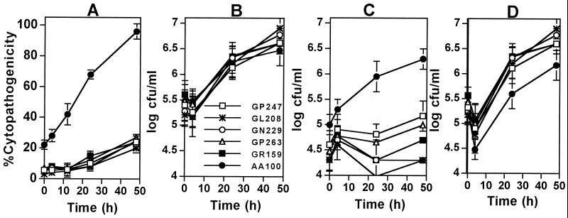
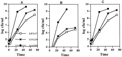
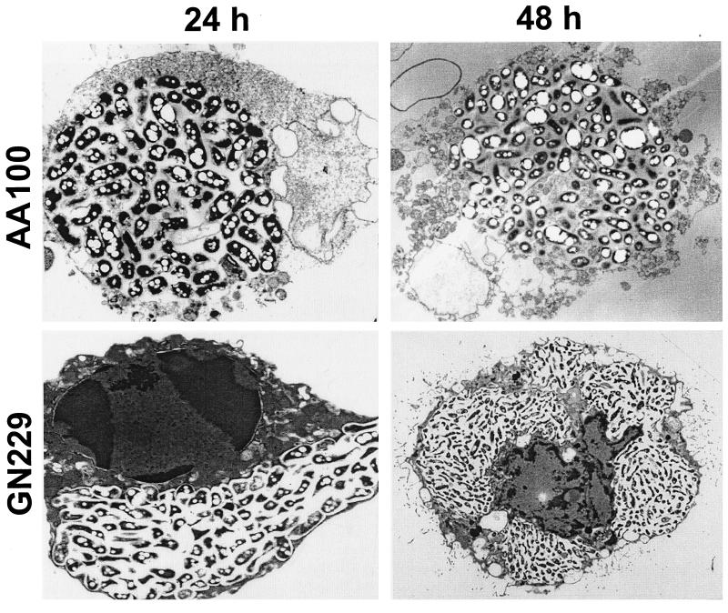
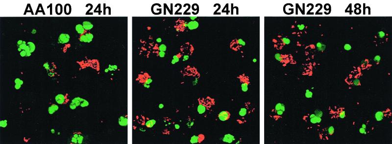
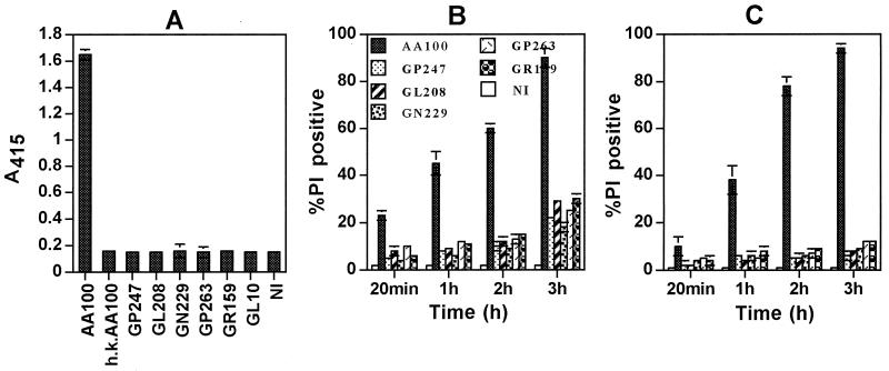
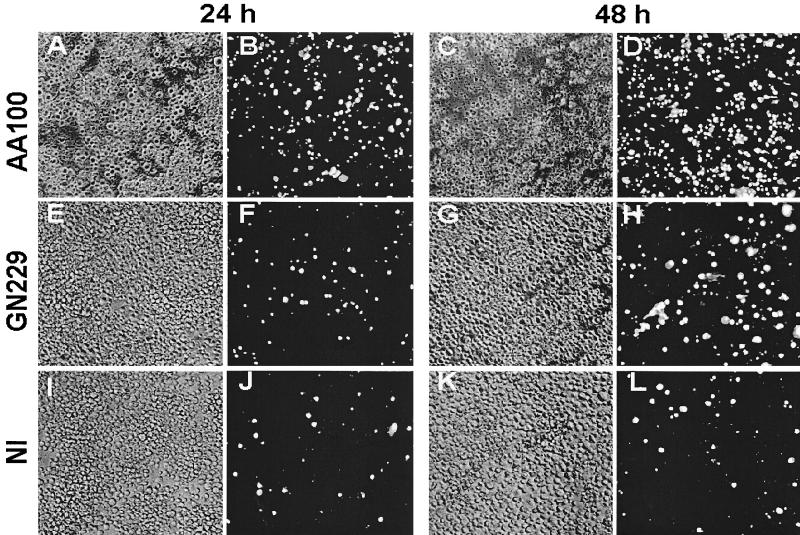
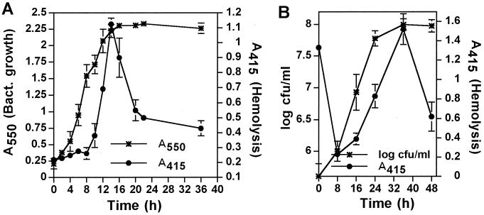
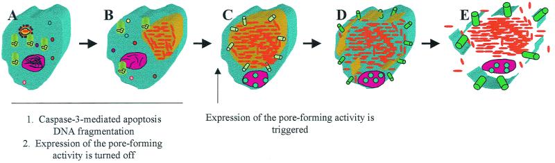
Legionella pneumophila does not induce apoptosis in the protozoan host, but induces pore formation-mediated cytolysis after termination of intracellular replication (L.-Y. Gao and Y. Abu Kwaik, Environ. Microbiol. 2:79-90, 2000). In contrast to this single mode of killing of protozoa, we have recently proposed a biphasic model by which L. pneumophila kills macrophages, in which the first phase is manifested through the induction of apoptosis during early stages of the infection, followed by an independent and temporal induction of necrosis during late stages of intracellular replication. Here we show that, similar to the protozoan host, the induction of necrosis and cytolysis of macrophages by L. pneumophila is mediated by the pore-forming toxin or activity. This activity is temporally and maximally expressed only upon termination of bacterial replication and correlates with cytolysis of macrophages and alveolar epithelial cells in vitro. We have identified five L. pneumophila mutants defective in the pore-forming activity. The phagosomes harboring the mutants do not colocalize with the late endosomal or lysosomal marker Lamp-1, and the mutants replicate intracellularly similar to the parental strain. Interestingly, despite their prolific intracellular replication, the mutants are defective in cytotoxicity and are "trapped" within and fail to lyse and egress from macrophages and alveolar epithelial cells upon termination of intracellular replication. However, the mutants are subsequently released from the host cell, most likely due to apoptotic death of the host cell. Data derived from cytotoxicity assays, confocal laser scanning microscopy, and electron microscopy confirm the defect in the mutants to induce necrosis of macrophages and the failure to egress from the host cell. Importantly, the mutants are completely defective in acute lethality (24 to 48 h) to intratracheally inoculated A/J mice. We conclude that the pore-forming activity of L. pneumophila is not required for phagosomal trafficking or for intracellular replication. This activity is expressed upon termination of bacterial replication and is essential to induce cytolysis of infected macrophages to allow egress of intracellular bacteria. In addition, this activity plays a major role in pulmonary immunopathology in vivo.


Figures

FIG. 1
FIG. 1
The L. pneumophila mutants are defective in killing and exiting U937 macrophages, but not in intracellular replication. (A) Cytopathogenicity to infected cells (MOI of 5) was determined by Alamar blue assays and compared to that of the noninfected cells. (B) Growth kinetics within U937 macrophages. The indicated numbers of bacteria represent the combined numbers of intracellular bacteria and bacteria released into the supernatant. (C) Bacteria released into the tissue culture medium. (D) Intracellular bacteria. Values are the means of triplicate samples, and error bars represent standard deviations.
FIG. 2
FIG. 2
Intracellular growth kinetics and egress of U937 macrophages infected at an MOI of 0.5. (A) Growth kinetics within U937 macrophages. The indicated numbers of bacteria represent the combined numbers of intracellular bacteria and bacteria released into the supernatant. (B) Bacteria released into the tissue culture medium. (C) Intracellular bacteria. Values are the means of triplicate samples, and standard deviations are not shown due to their small values.
FIG. 3
FIG. 3
The rib mutants' defect in cytolysis of the host cell is due to a defect in necrosis-mediated killing. Representative transmission electron micrographs of infected U937 macrophages at 24 h and 48 h postinfection by the wild-type strain AA100 and the GN229 mutant. The original magnifications were ×7,000 and ×5,000 for the 24- and 48-h infections, respectively.
FIG. 4
FIG. 4
The defect in the rib mutants to induce cytolysis of macrophages is not due to a defect in induction of apoptosis. Representative TUNEL assays were performed for infections by strain AA100 or the GN229 mutant at 24 and 48 h postinfection. The apoptotic nuclei are shown in green, while the bacteria were detected with an antibody and are shown in red. The 48-h time point is not shown for AA100, due to complete lysis and loss of the monolayers. Stacked images of multiple 0.5-μm confocal z-sections are shown.
FIG. 5
FIG. 5
The rib mutants of L. pneumophila are defective in expression of the pore-forming toxin or activity. (A) Contact-dependent hemolysis of sRBCs. GL10 is a dotA icmWXYZ mutant derivative of AA100 (24). h.k., heat-killed AA100; NI, noninfected; A415, measurement of optical density of the released hemoglobin at a wavelength of 415 nm. (B and C) Rapid contact-dependent pore formation in U937 macrophages and WI-26 alveolar epithelial cells (C) at an MOI of 500, measured by permeability to PI and expressed as percent PI positive. At least 100 cells were examined for each of the multiple samples. Values are the means of triplicate samples, and error bars represent standard deviations.
FIG. 6
FIG. 6
The mutants are defective in cytotoxicity. Phase-contrast images and PI staining of U937 macrophages examined at 24 h (left two columns) and 48 h (right two columns) postinfection are shown. Phase-contrast images are shown in panels A, C, E, G, I, and K, and the corresponding PI staining is shown in panels B, D, F, H, J, and L, respectively. NI, noninfected.
FIG. 7
FIG. 7
Growth-phase-dependent expression of the pore-forming activity by L. pneumophila in vitro and intracellularly. (A and B) Contact-dependent hemolysis of sRBCs by in vitro-grown L. pneumophila (A) or intracellular bacteria isolated from U937 macrophages (B). Infection of the cells in panel B was performed with in vitro-grown bacteria that had reached their maximal growth and hemolysis (14 h in panel A). At the indicated time points, the bacterial growth was determined by the A550 in panel A or by the CFU in panel B (left y axis), and hemolytic activity was determined (right y axis) with an equivalent number of bacteria at all time points. Values are the means of triplicate samples, and error bars represent standard deviations.
FIG. 8
FIG. 8
A model of growth-phase-dependent cytolysis of mammalian cells by L. pneumophila upon termination of intracellular bacterial replication to egress from the spent host cell. During early stages of formation of the mitochondria and rough endoplasmic reticulum-surrounded phagosome (A) and during exponential intracellular replication (B), expression of the pore-forming activity is turned off, but caspase-3-mediated apoptosis is triggered. Upon transition to the postexponential phase of growth, expression of the pore-forming activity is triggered, which results in insertions of pores in the phagosomal membrane first (C), leading to its disruption (D). This is followed by insertions of the pores in the plasma membrane (E), leading to osmotic lysis of the cell and release of the intracellular bacteria.

References

    1. Abu Kwaik Y. Fatal attraction of mammalian cells to Legionella pneumophila. Mol Microbiol. 1998;30:689–696. - PubMed
    1. Abu Kwaik Y. Induced expression of the Legionella pneumophila gene encoding a 20-kilodalton protein during intracellular infection. Infect Immun. 1998;66:203–212. - PMC - PubMed
    1. Abu Kwaik Y. The phagosome containing Legionella pneumophila within the protozoan Hartmanella vermiformis is surrounded by the rough endoplasmic reticulum. Appl Environ Microbiol. 1996;62:2022–2028. - PMC - PubMed
    1. Abu Kwaik Y, Eisenstein B I, Engleberg N C. Phenotypic modulation by Legionella pneumophila upon infection of macrophages. Infect Immun. 1993;61:1320–1329. - PMC - PubMed
    1. Abu Kwaik Y, Engleberg N C. Cloning and molecular characterization of a Legionella pneumophila gene induced by intracellular infection and by various in vitro stress stimuli. Mol Microbiol. 1994;13:243–251. - PubMed

Publication types

LinkOut - more resources