. 2000 Nov;68(11):6449-56.
doi: 10.1128/IAI.68.11.6449-6456.2000.

Key role for DsbA in cell-to-cell spread of Shigella flexneri, permitting secretion of Ipa proteins into interepithelial protrusions

Affiliations

Key role for DsbA in cell-to-cell spread of Shigella flexneri, permitting secretion of Ipa proteins into interepithelial protrusions

J Yu et al. Infect Immun. 2000 Nov.

Abstract

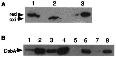
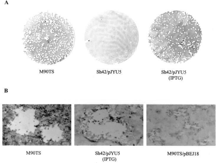
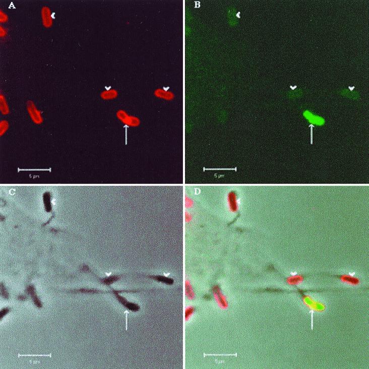
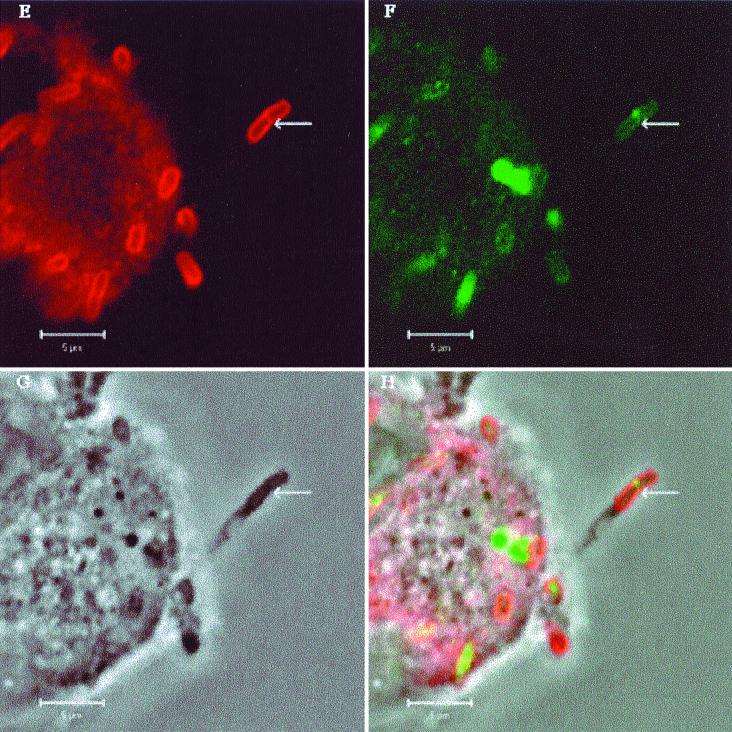
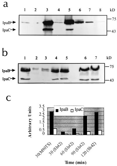
DsbA, a disulfide bond catalyst, is necessary for realization of the pathogenic potential of Shigella flexneri. Sh42, a mutant strain differing from wild-type M90TS solely because it expresses nonfunctional DsbA33G (substitution for 33C at the active site), secreted less IpaB and IpaC than M90TS in response to various stimuli in vitro. A kinetic study demonstrated that Sh42 responded more slowly to Congo red than M90TS. By modulating relative concentrations of functional and nonfunctional DsbA within bacteria, functional enzyme has been shown to be necessary for intercellular spread. By confocal microscopy, M90TS dividing in protrusions was shown to secrete Ipa proteins from the septation furrow, anticipating lysis of protrusions, while Sh42 showed minimal Ipa secretion in this location. In the light of a previous demonstration that DsbA is not necessary for entry of epithelial cells, we conclude that a role in virulence of this disulfide bond catalyst lies in facilitating secretion of Ipa proteins specifically within epithelial protrusions, in turn allowing cell-to-cell spread of S. flexneri.


Figures

FIG. 1
FIG. 1
Expression of DsbA from S. flexneri. (A) Analysis of the redox status of wild-type and mutant DsbA33G during laboratory growth. Lanes 1 and 2, DsbA from M90TS, treated with reducing (lane 1) and nonreducing (lane 2) buffer; lane 3, DsbA33G from Sh42 treated with nonreducing buffer. Anti-DsbA polyclonal antiserum was used for the immunoblot. (B) Expression of recombinant DsbA from plasmids during bacterial growth. Lanes 1 and 5, M90TS/pBEJ18 (noninduced); lanes 2 and 6, M90TS/pBEJ18 (induced); lanes 3 and 7, Sh42/pJYU5 (noninduced); lanes 4 and 8, Sh42/pJYU5 (induced). DsbA in lanes 1 to 4 was detected by anti-DsbA antiserum and in lanes 5 to 8 was detected by anti-His6 antibody (Sigma). red, reduced form; oxi, oxidized form.
FIG. 2
FIG. 2
Plaque assays. CFU (106) from each strain were used to infect CaCo-2 cell monolayers in a 35-mm-diameter culture dish. (A) Whole plates; (B) individual plaques.
FIG. 3
FIG. 3
Localization of IpaB in S. flexneri-infected HeLa cells by double fluorescence labeling and confocal microscopy. (A to D) HeLa cells infected with M90TS. Arrows indicate protrusions containing a dividing bacterium; arrowheads indicate nondividing bacteria in protrusions. (E to H) HeLa cells infected with Sh42. Arrows indicate protrusions containing Sh42. Other bacteria lie within the cell or on the surface. (A and E) Bacteria binding rabbit anti-Shigella LPS antiserum or goat anti-rabbit IgG conjugated with Texas red. (B and F) Localization of IpaB with mouse monoclonal antibody H16 or FITC-conjugated goat anti-mouse IgG. Panels C and G are phase-contrast images of the fields shown in panels A and B and E and F, respectively. (D and F) Overlays of A, B, and C and E, F, and G, respectively.
FIG. 3
FIG. 3
Localization of IpaB in S. flexneri-infected HeLa cells by double fluorescence labeling and confocal microscopy. (A to D) HeLa cells infected with M90TS. Arrows indicate protrusions containing a dividing bacterium; arrowheads indicate nondividing bacteria in protrusions. (E to H) HeLa cells infected with Sh42. Arrows indicate protrusions containing Sh42. Other bacteria lie within the cell or on the surface. (A and E) Bacteria binding rabbit anti-Shigella LPS antiserum or goat anti-rabbit IgG conjugated with Texas red. (B and F) Localization of IpaB with mouse monoclonal antibody H16 or FITC-conjugated goat anti-mouse IgG. Panels C and G are phase-contrast images of the fields shown in panels A and B and E and F, respectively. (D and F) Overlays of A, B, and C and E, F, and G, respectively.
FIG. 4
FIG. 4
Secreted Ipa proteins from S. flexneri in vitro. (a) Secreted Ipa proteins in response to different stimuli. Lanes 1, 3, 5, and 7, M90TS; lanes 2, 4, 6, and 8, Sh42; lanes 1 and 2, unactivated controls; lanes 3 and 4, Congo red-activated samples; lanes 5 and 6, fibronectin-activated samples; lanes 7 and 8, laminin-activated samples. IpaB and IpaC are indicated by arrows, and molecular mass markers are indicated. (b) Kinetics of the secretion of Ipa proteins in response to Congo red. Lane 1, secreted Ipa proteins from M90TS at 30 min; lanes 2, 3, 4, and 5, secreted Ipa proteins from Sh42 at 30, 60, 90, and 120 min, respectively; lanes 6 and 7, cell-associated Ipa proteins from M90TS and Sh42, respectively. (c) Semiquantitation of the secreted Ipa proteins shown in panel b (lanes 1 to 5) by image densitometry.

References

    1. Adam T, Giry M, Boquet P, Sansonetti P J. Rho-dependent membrane folding causes Shigella entry into epithelial cells. EMBO J. 1996;15:3315–3321. - PMC - PubMed
    1. Allaoui A, Mounier J, Prevost M C, Sansonetti P J, Parsot C. icsB: a Shigella flexneri virulence gene necessary for the lysis of protrusions during intercellular spread. Mol Microbiol. 1992;6:1605–1616. - PubMed
    1. Bahrani F K, Sansonetti P J, Parsot C. Secretion of Ipa proteins by Shigella flexneri: inducer molecules and kinetics of activation. Infect Immun. 1997;65:4005–4010. - PMC - PubMed
    1. Bardwell J C A, McGovern K, Beckwith J. Identification of a protein required for disulphide bond formation in vivo. Cell. 1991;67:581–589. - PubMed
    1. Barzu S, Benjelloun-Touimi Z, Phalipon A, Sansonetti P J, Parsot C. Functional analysis of the Shigella flexneri IpaC invasin by insertional mutagenesis. Infect Immun. 1997;65:1599–1605. - PMC - PubMed

Publication types

Substances

LinkOut - more resources