. 2000 Oct 24;97(22):12204-9.
doi: 10.1073/pnas.200348397.

Pinpointing when T cell costimulatory receptor CTLA-4 must be engaged to dampen diabetogenic T cells

Affiliations

Pinpointing when T cell costimulatory receptor CTLA-4 must be engaged to dampen diabetogenic T cells

F Luhder et al. Proc Natl Acad Sci U S A. .

Abstract

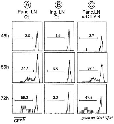
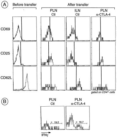
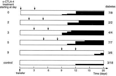
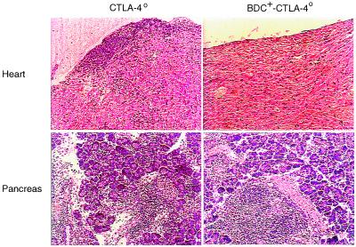
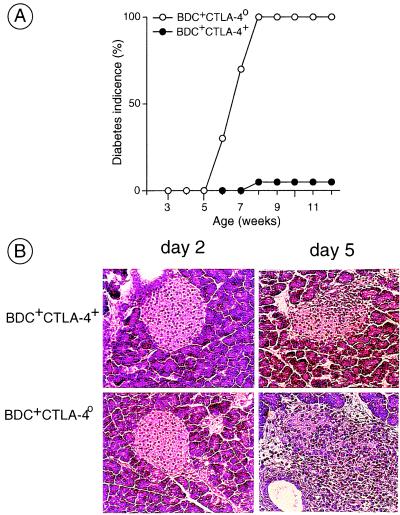
Engagement of the T cell costimulatory receptor CTLA-4 can potently down-regulate an immune response. For example, in a T cell receptor transgenic mouse model of autoimmune diabetes, CTLA-4 interactions keep pancreatic islet-reactive T cells in check, evidenced by the finding that mAb blockade of CTLA-4 rapidly provokes diabetes in animals that would not normally succumb until many months later. Interestingly, this effect is only observed early in the course of disease, before insulitis is stably entrenched. Here, we have exploited a highly synchronous and easily manipulable transfer system to determine precisely when CTLA-4 must be engaged to check the diabetogenicity of islet-reactive T cells. Our results indicate that CTLA-4 interactions during initial priming of the T cells in the pancreatic lymph nodes are not determinant. Rather, the critical interactions occur when the T cells secondarily reencounter their antigen in the target organ, the pancreatic islets. In addition, we made use of CTLA-4-deficient mice to bolster our interpretation that CTLA-4 engagement has a dampening rather than an enhancing influence on diabetes progression.


Figures

Figure 1
Figure 1
No detectable effect of anti-CTLA-4 treatment on division of transferred BDC2.5 T cells in the PLNs. CFSE-labeled splenocytes from juvenile BDC2.5/NOD mice were transferred into adult Cαo/o/NOD animals that had been treated with anti-CTLA-4 mAb or PBS 2 h before transfer. PLNs and inguinal lymph node were removed from the recipients at the indicated times after transfer, and cells were stained with mAbs against CD4 and Vβ4 (the trangene-encoded TCR β-chain). Shown are histograms of CFSE staining for gated CD4+Vβ4+ cells. The reduction in CFSE staining intensity signifies that cell proliferation has taken place, and the proportion of divided cells is indicated.
Figure 2
Figure 2
Activation of BDC2.5 T cells after transfer into anti-CTLA-4-treated and control recipients. (A) Splenocytes from juvenile BDC2.5/NOD mice were pooled and an aliquot was stained for CD4, CD8, and early (CD25 and CD69) and late (CD62L) activation markers. Cells were transferred into adult Cαo/o/NOD mice that were treated with anti-CTLA-4 as described in the legend to Fig. 1. At day 3, single-cell suspensions from PLNs and inguinal lymph nodes were stained for CD4, CD8, and various activation markers; histograms gated on CD4+ cells are shown. (B) Same as in A except intracellular staining for IFN-γ was performed in place of staining for activation markers.
Figure 3
Figure 3
Diabetes development after treatment with anti-CTLA-4 mAb initiated at various times. Cαo/o/NOD recipients of BDC2.5 splenocytes were treated with anti-CTLA-4 mAb (2 × 200 μg) or control mAb or PBS starting on different days after transfer, as listed on the left. Arrows indicate the times of anti-CTLA-4 injection; solid black shading shows the incidence of diabetes; and the numbers on the right represent the final diabetes frequency. Inexplicably, in a few experiments, all mice became diabetic, whether they were treated with anti-CTLA-4 or not; data from these experiments were excluded from the results shown above.
Figure 4
Figure 4
Organ infiltration in CTLA-4-deficient mice in the presence or absence of the BDC2.5 TCR transgenes. Organs were removed from 3-week-old CTLA-4o/o/NOD mice and 6-week-old BDC2.5-positive CTLA-4o/o/NOD mice, and paraffin sections were stained with hematoxylin/eosin. Only heart and pancreas sections are shown. The pictures are representative of three mice of each type.
Figure 5
Figure 5
Diabetes promotion by CTLA-4-deficient BDC2.5 T cells. (A) CTLA-4o/+ mice were crossed three times to BDC2.5/NOD mice and intercrossed to obtain BDC2.5-positive CTLA-4o/o/NOD animals (selected for H-2g7 homozygosity). These offspring were followed for diabetes. (B) Splenocytes from BDC2.5-positive CTLA-4o/o/NOD mice and CTLA-4+ control littermates were transferred into Cαo/o/NOD recipients. Pancreata from individual recipients were removed at days 2 or 5 after transfer, and paraffin sections stained with hematoxylin/eosin were examined by histology.

References

    1. Oosterwegel M A, Greenwald R J, Mandelbrot D A, Lorsbach R B, Sharpe A H. Curr Opin Immunol. 1999;11:294–300. - PubMed
    1. Chambers C A, Allison J P. Curr Opin Cell Biol. 1999;11:203–210. - PubMed
    1. McAdam A J, Schweitzer A N, Sharpe A H. Immunol Rev. 1998;165:231–247. - PubMed
    1. Linsley P S, Greene J L, Tan P, Bradshaw J, Ledbetter J A, Anasetti C, Damle N K. J Exp Med. 1992;176:1595–1604. - PMC - PubMed
    1. Wu Y, Guo Y, Huang A, Zheng P, Liu Y. J Exp Med. 1997;185:1327–1335. - PMC - PubMed

Publication types