. 2000 Oct 24;97(22):12250-5.
doi: 10.1073/pnas.200366597.

Tonsillar memory B cells, latently infected with Epstein-Barr virus, express the restricted pattern of latent genes previously found only in Epstein-Barr virus-associated tumors

Affiliations

Tonsillar memory B cells, latently infected with Epstein-Barr virus, express the restricted pattern of latent genes previously found only in Epstein-Barr virus-associated tumors

G J Babcock et al. Proc Natl Acad Sci U S A. .

Abstract

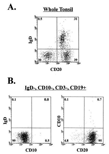
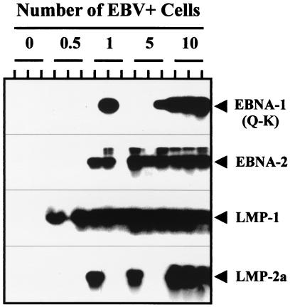
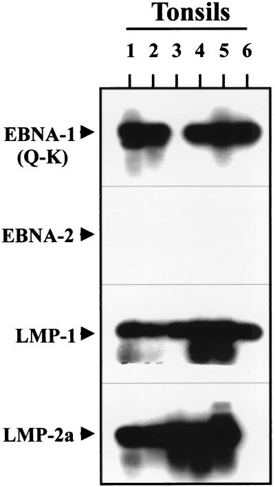
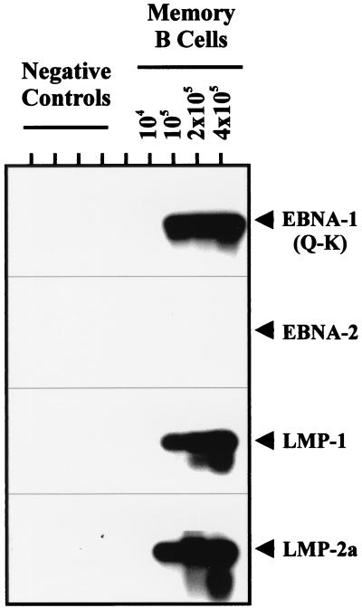
Epstein-Barr virus (EBV) establishes a life-long persistent infection in most of the human population. In the peripheral blood, EBV is restricted to memory B cells that are resting and express limited genetic information. We have proposed that these memory cells are the site of long-term persistent infection. We now show that memory cells in the tonsil express the genes for EBV nuclear antigen 1 (EBNA1) (from the Qp promoter), latent membrane protein 1 (LMP1), and LMP2a but do not express EBNA2 or the EBNA3s. This pattern of latent gene expression has only been seen previously in EBV-associated tumors such as nasopharyngeal carcinoma, Hodgkin's disease (HD), and T/NK lymphomas. Normal circulating memory B cells frequently reenter secondary lymphoid tissue, where they receive signals essential for their survival. Specifically they require signals from antigen-specific T helper cells and from antigen itself. LMP1 and LMP2 are known to be able to generate these signals in a ligand-independent fashion. We suggest, therefore, that the transcription pattern we have found in latently infected, tonsillar, memory B cells is used because it allows for the expression of LMP1, LMP2a, and EBNA1 in the absence of the immunogenic and growth-promoting EBNA2 and EBNA3 molecules. LMP1 and LMP2a are produced to provide the surrogate rescue and survival signals needed to allow latently infected memory cells to persist, and EBNA1 is produced to allow replication of the viral episome.


Figures

Figure 1
Figure 1
FACS analysis of the purified memory population. Whole tonsillar lymphocytes were depleted of IgD+ naive B cells and CD3+ T cells by MACS. The depletions were 99% efficient. The resulting population was stained for CD10 and IgD expression, and the memory B cells (IgD−, CD10−) were separated from the germinal center B cells (IgD−, CD10+) by FACS. (A) Unfractionated tonsil cells stained with FITC anti-CD20 (a pan-B cell marker) and PE-coupled anti-IgD. (B) FACS reanalysis of the purified populations with FITC anti-CD10 and PE-coupled anti-IgD (Left) or FITC anti-CD20 and PE-coupled anti-CD10 (Right). Note that the purified population is 95% pure B cells, based on restaining for CD20, and contains only 0.1% IgD+ (naive) and 0.5–0.8% CD10+ (germinal center) B cells.
Figure 2
Figure 2
Sensitivity controls for the RT-PCR analysis. EBV-positive cells from a cell line were spiked into 5 × 10⁶ EBV-negative tonsillar lymphocytes at the numbers indicated before RNA extraction and cDNA synthesis. Each number of cells was tested independently in triplicate. The cell lines used were IB4 for EBNA2, LMP1, and LMP2a and RAEL for EBNA1(Q-K).
Figure 3
Figure 3
RT-PCR analysis for EBV latent gene expression in tonsillar memory B cells. RT-PCR analysis for the latent genes EBNA-1(Q-K), EBNA-2, LMP-1, and LMP-2a was performed on 106 purified memory B cells from six independent tonsils. For details see Materials and Methods. The migration points for the PCR products are indicated by arrows.
Figure 4
Figure 4
RT-PCR analysis of titrated memory cells. Memory cells were isolated, and RT-PCR analysis for EBNA-1(Q-K), EBNA-2, LMP-1, and LMP-2a was performed. The number of cells tested for each dilution is shown. For each dilution tested the number of cells was brought up to 5 × 10⁶ by the addition of EBV-negative tonsillar lymphocytes before RNA extraction and cDNA synthesis. EBV-negative tonsillar lymphocytes (5 × 10⁶) were also used as the negative control. The migration points for the PCR products are indicated by arrows.
Figure 5
Figure 5
RT-PCR analysis of tonsillar lymphocytes fractionated on the basis of sIg expression. Purified memory cells were labeled with FITC-coupled anti-IgG, IgM, and IgA, and cells from the positive and negative fractions were isolated by FACS. RT-PCR was performed on 106 cells of each population, as described in Fig. 3 and Materials and Methods. The positive controls were five cells from an EBV-positive cell line brought up to 5 × 10⁶ by the addition of EBV-negative tonsillar lymphocytes before RNA extraction and cDNA synthesis. EBV-negative tonsillar lymphocytes (5 × 10⁶) also were used as the negative control.

References

    1. Rickinson A B, Kieff E. In: Virology. Fields B N, Knipe D M, Howley P M, editors. Vol. 2. New York: Raven; 1996. pp. 2397–2446.
    1. Babcock G J, Decker L L, Freeman R B, Thorley-Lawson D A. J Exp Med. 1999;190:567–576. - PMC - PubMed
    1. Chen F, Zou J Z, di, R. L, Winberg G, Hu L F, Klein E, Klein G, Ernberg I. J Virol. 1995;69:3752–3758. - PMC - PubMed
    1. Qu L, Rowe D T. J Virol. 1992;66:3715–3724. - PMC - PubMed
    1. Tierney R J, Steven N, Young L S, Rickinson A B. J Virol. 1994;68:7374–7385. - PMC - PubMed

Publication types