. 2000 Oct 24;97(22):12210-5.
doi: 10.1073/pnas.210391497.

Antimicrobial activity of MHC class I-restricted CD8+ T cells in human tuberculosis

Affiliations

Antimicrobial activity of MHC class I-restricted CD8+ T cells in human tuberculosis

S Cho et al. Proc Natl Acad Sci U S A. .

Abstract

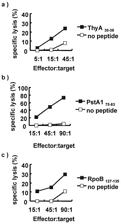
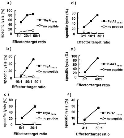
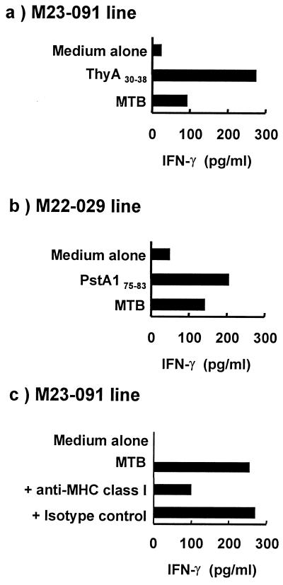
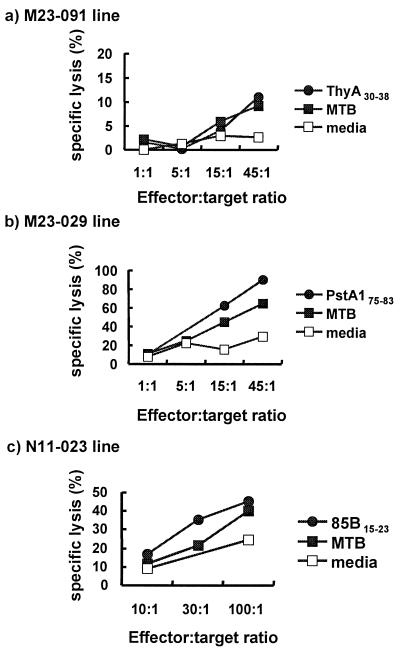
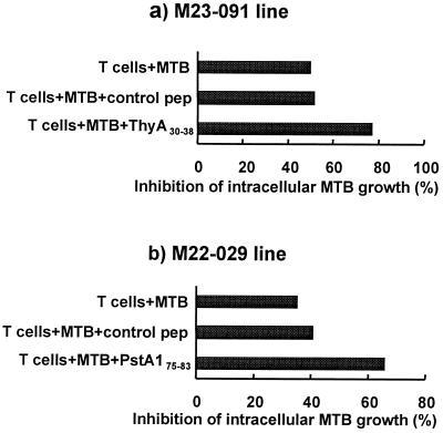
Studies of mouse models of tuberculosis (TB) infection have indicated a central role for MHC class I-restricted CD8+ T cells in protective immunity. To define antigens and epitopes of Mycobacterium tuberculosis (MTB) proteins that are presented by infected cells to CD8+ T cells, we screened 40 MTB proteins for HLA class I A*0201-binding motifs. Peptides that bound with high affinity to purified HLA molecules were subsequently analyzed for recognition by CD8+ cytotoxic T lymphocytes. We identified three epitopes recognized by CD8+ T cells from patients recovering from TB infection. Those three epitopes were derived from three different antigens: thymidylate synthase (ThyA(30-38)), RNA polymerase beta-subunit (RpoB(127-135)), and a putative phosphate transport system permease protein A-1 (PstA1(75-83)). In addition, CD8+ T cell lines specific for three peptides (ThyA(30-38), PstA1(75-83), and 85B(15-23)) were generated from peripheral blood mononuclear cells of normal HLA-A*0201 donors. These CD8+ T cell lines specifically recognized MTB-infected macrophages, as demonstrated by production of IFN-gamma and lysis of the infected target cells. Finally, CD8+ cytotoxic T lymphocytes reduced the viability of the intracellular MTB, providing evidence that CD8+ T cell recognition of MHC class I-restricted epitopes of these MTB antigens can contribute to effective immunity against the pathogen.


Figures

Figure 1
Figure 1
CTL responses from TB patients. PBMC from TB patients (HLA-A*0201) were stimulated with individual MTB peptides for 1 week and restimulated with peptide-pulsed autologous monocytes for an additional week. The cytolytic activity of these cultured PBMC was tested on day 14 by 51Cr release assay. (a) CTL activity specific for 9-mer peptide derived from thymidylate synthase (amino acids 30–38). (b) CTL activity specific for 9-mer peptide derived from RNA polymerase β-subunit (amino acids 127–135). (c) CTL activity specific for 9-mer peptide derived from PstA1 (amino acids 75–83).
Figure 2
Figure 2
Cytolytic activity of CTL lines generated from PBMC of uninfected subjects by in vitro immunization. PBMC from MTB-uninfected HLA-A*0201 donors were in vitro immunized to generate CTL lines specific for peptides, ThyA30–38 (a–c) and PstA175–83. (d–f). Cell lines (a) N1–091, (c) M7–091, and (f) M7–029 were generated by the method of Tsai et al. (16). Cell lines (b) M23–091, (d) M22–029, and (e) M23–029 were generated by the method of Plebanski et al. (17).
Figure 3
Figure 3
IFN-γ release by MTB peptide-specific CTL after stimulation by MTB-infected and MTB peptide-pulsed cells. CD8+ T cell lines specific for (a and c) ThyA30–38 peptide (M23–091). (b) PstA175–83 peptide were coincubated with MTB-infected THP-1 cells at a ratio of 20:1. In some cultures, the peptide, either ThyA30–38 or PstA175–83, was added to the cultures. The supernatant was collected and assayed for IFN-γ. (c) The MHC restriction was evaluated by using the anti-HLA class I antibody (W6/32). The isotype-matched antibody (IgG2a) was used as a control.
Figure 4
Figure 4
Cytotoxicity of CD8+ T cell lines against MTB-infected macrophages. Macrophages from a heterologous HLA-A*0201 donor were infected with MTB at a multiplicity of infection of 5–10 bacilli per cell (a and b), or THP-1 were infected at a multiplicity of infection of 5–10 bacilli per cell (c). MTB-infected and corresponding peptide-pulsed THP-1 cells were tested for lysis by 51Cr release assay by incubation with (a) ThyA30–38 peptide-specific line (M23–091), (b) PstA175–83 peptide-specific line, and (c) 85B15–23 peptide-specific line (N11–023).
Figure 5
Figure 5
Inhibition of the growth of intracellular MTB by peptide-specific CD8+ T cells. MTB peptide-specific CD8+ T cell lines were coincubated with MTB-infected THP-1 cells in the presence or absence of MTB-derived peptides or a control peptide at an effector-to-target ratio of 20:1. Cells were lysed and CFU determined. The CFU of infected THP-1 without T cells at 24 h was 167,000 for the experiment in which M23–091 was tested and 66,400 for the experiment in which M22.029 was tested.

References

    1. Bloom B R, Murray C J L. Science. 1992;257:1055–1064. - PubMed
    1. Flynn J L, Goldstein M M, Triebold K J, Koller B, Bloom B R. Proc Natl Acad Sci USA. 1992;89:12013–12017. - PMC - PubMed
    1. Muller I, Cobbold S P, Waldmann H, Kaufmann S H E. Infect Immun. 1987;55:2037–2041. - PMC - PubMed
    1. Serbina N V, Liu C C, Scanga C A, Flynn J L. J Immunol. 2000;165:353–363. - PubMed
    1. Behar S M, Dascher C C, Grusby M J, Wang C R, Brenner M B. J Exp Med. 1999;189:1973–1980. - PMC - PubMed

Publication types

Substances