. 2000 Nov 7;97(23):12625-30.
doi: 10.1073/pnas.220247197.

PERK mediates cell-cycle exit during the mammalian unfolded protein response

Affiliations

PERK mediates cell-cycle exit during the mammalian unfolded protein response

J W Brewer et al. Proc Natl Acad Sci U S A. .

Abstract

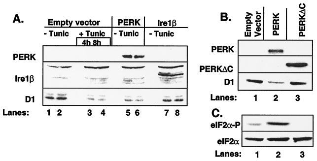
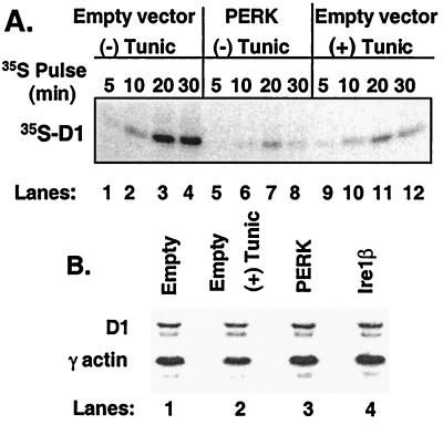
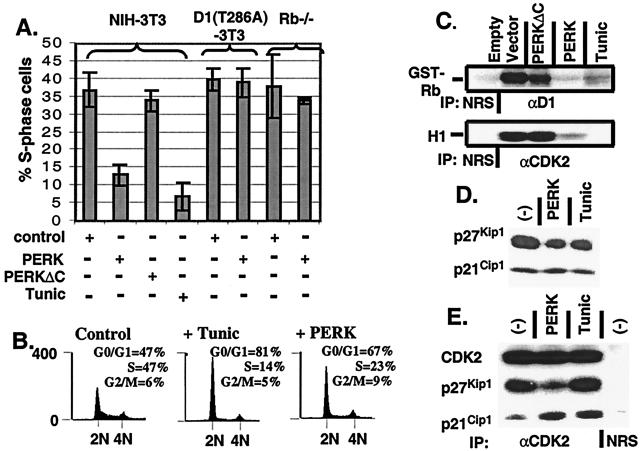
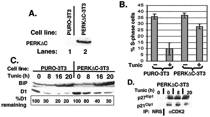
The accumulation of unfolded proteins in the endoplasmic reticulum (ER) triggers the unfolded protein response (UPR)-signaling pathway. The UPR coordinates the induction of ER chaperones with decreased protein synthesis and growth arrest in the G(1) phase of the cell cycle. Three ER transmembrane protein kinases (Ire1alpha, Ire1beta, and PERK) have been implicated as proximal effectors of the mammalian UPR. We now demonstrate that activation of PERK signals the loss of cyclin D1 during the UPR, culminating in cell-cycle arrest. Overexpression of wild-type PERK inhibited cyclin D1 synthesis in the absence of ER stress, thereby inducing a G(1) phase arrest. PERK expression was associated with increased phosphorylation of the translation elongation initiation factor 2alpha (eIF2alpha), an event previously shown to block cyclin D1 translation. Conversely, a truncated form of PERK lacking its kinase domain acted as a dominant negative when overexpressed in cells, attenuating both cyclin D1 loss and cell-cycle arrest during the UPR without compromising induction of ER chaperones. These data demonstrate that PERK serves as a critical effector of UPR-induced growth arrest, linking stress in the ER to control of cell-cycle progression.


Figures

Figure 1
Figure 1
PERK regulates accumulation of cyclin D1. (A) NIH 3T3 cells infected with empty virus (lanes 1–4), virus encoding PERK (lanes 5–6), or virus encoding Ire1β (lanes 7–8) were left untreated or were treated with 0.5 μg/ml tunicamycin (lanes 3–4). Equivalent amounts of cellular protein were resolved on denaturing polyacrylamide gels and subjected to immunoblot analysis with the 9E10 antibody, which recognizes a c-myc epitope tag expressed at the C terminus of both PERK and Ire1β or a cyclin D1-specific monoclonal antibody. Sites of antibody binding were visualized by enhanced chemiluminescence. (B and C) After infection of NIH 3T3 cells with virus encoding PERK (lane 2), PERKΔC (lane 3) or empty virus (lane 1), equivalent amounts of total protein were resolved on a denaturing polyacrylamide gel and transferred to nitrocellulose membrane. Membranes were subsequently probed with the 9E10 antibody (PERK and PERKΔC) or antibodies specific for cyclin D1, serine 51-phosphorylated eIF2α, or total eIF2α. Sites of antibody binding were visualized by enhanced chemiluminescence.
Figure 2
Figure 2
Inhibition of cyclin D1 translation by PERK. (A) After infection of NIH 3T3 cells with virus encoding PERK (lanes 5–8) or empty virus (lanes 1–4 and 9–12), cells were left untreated or were treated with 0.5 μg/ml tunicamycin for 5 h and were subsequently pulse labeled with 35S-methionine for the indicated intervals. Cyclin D1 was immunoprecipitated from lysates, resolved on a denaturing gel, and visualized by autoradiography. (B) Total RNA was isolated from NIH 3T3 cells infected with PERK (lane 3), Ire1β (lane 4), empty virus (lane 1), or empty virus with tunicamycin treatment for 6 h and subjected to Northern blot analysis with probes specific for cyclin D1 and γ-actin.
Figure 3
Figure 3
Enforced overexpression of cyclin D1 prevents PERK-dependent cell-cycle arrest. (A) NIH 3T3 cells, cyclin D1-T286A derivatives, and Rb−/− MEFs were infected with the viruses indicated at the bottom of the graph. Empty virus-infected cells were left untreated or treated with tunicamycin for 20 h. During the last 2 h of tunicamycin treatment, all cell populations were pulsed with BrdUrd and processed for immunofluorescence with a BrdUrd-specific monoclonal antibody except for Rb−/− MEFs in which the S-phase fraction was determined by flow cytometry, as in B. The number of BrdUrd-positive cells from a minimum of three independent experiments were quantitated and are expressed relative to the total population of cells. (B) NIH 3T3 cells were infected with virus encoding PERK or empty virus (control) and left uninfected or treated with 0.5 μg/ml tunicamycin for 20 h. Forty-eight hours after infection (or 20 h after addition of tunicamycin), cells were stained with propidium iodide and assayed for DNA content by flow cytometry (14). (C) NIH 3T3 cells were infected the indicated retroviruses, lysed, and immune complexes were recovered with either a cyclin D1 antibody or a CDK2 antibody and analyzed for protein kinase activity by using retinoblastoma protein or histone H1 as the substrate respectively. (D) Whole-cell lysates prepared from NIH 3T3 cells left untreated, treated with 0.5 μg/ml tunicamycin for 20 h, or infected with virus encoding PERK were subjected to a direct immunoblot analysis by using either p21Cip1- or p27Kip1-specific antibodies. (E) Whole-cell lysates prepared from cells treated as in D were precipitated with antibodies to CDK2. Denatured immune complexes separated on gels were blotted with antibodies to CDK2, p21Cip1, or p27Kip1.
Figure 4
Figure 4
PERK is required for UPR-dependent cell-cycle arrest. (A) NIH 3T3 cells were infected with virus encoding PERKΔC and selected with 7.5 μg/ml puromycin for 4 days. Expression of PERKΔC was confirmed by immunoblot analysis. (B) Parental NIH 3T3 or cells expressing PERKΔC were treated with tunicamycin for 20 h and pulsed with BrdUrd during the last 2.5 h of tunicamycin treatment. The percentage of S-phase cells is expressed as the number of BrdUrd-positive cells vs. the total number of cells. (C) Equivalent amounts of total protein prepared from cells treated as above were resolved on a denaturing polyacrylamide gel and blotted with antibodies specific for BiP and cyclin D1. Sites of antibody binding were visualized by enhanced chemiluminescence. (D) Whole-cell lysates prepared from PERKΔC-3T3 cells treated with 0.5 μg/ml tunicamycin for the indicated intervals were precipitated with antibodies to CDK2. Denatured immune complexes were separated on gels and blotted with antibodies to p21Cip1 or p27Kip1.

Comment in

  • Pausing to decide.
    Niwa M, Walter P. Niwa M, et al. Proc Natl Acad Sci U S A. 2000 Nov 7;97(23):12396-7. doi: 10.1073/pnas.250476097. Proc Natl Acad Sci U S A. 2000. PMID: 11058174 Free PMC article. Review. No abstract available.

References

    1. Sidrauski C, Chapman R, Walter P. Trends Cell Biol. 1998;8:245–249. - PubMed
    1. Lee A S. Curr Opin Cell Biol. 1992;4:267–273. - PubMed
    1. Wang X Z, Lawson B, Brewer J W, Zinszner H, Sanjay A, Mi L J, Boorstein R, Kreibich G, Hendershot L M, et al. Mol Cell Biol. 1996;16:4273–4280. - PMC - PubMed
    1. Brostrom C O, Brostrom M A. Prog Nucleic Acid Res Mol Biol. 1998;58:79–125. - PubMed
    1. Melero J A, Fincham V. J Cell Physiol. 1978;88:355–365. - PubMed

Publication types

LinkOut - more resources