. 2000 Oct 24;97(22):12227-32.
doi: 10.1073/pnas.220399597.

Suppression of angiogenesis by lentiviral delivery of PEX, a noncatalytic fragment of matrix metalloproteinase 2

Affiliations

Suppression of angiogenesis by lentiviral delivery of PEX, a noncatalytic fragment of matrix metalloproteinase 2

A Pfeifer et al. Proc Natl Acad Sci U S A. .

Abstract

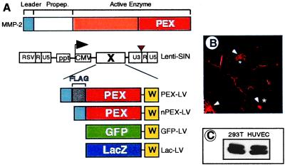
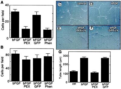
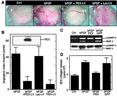
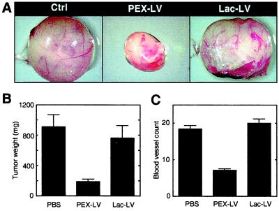
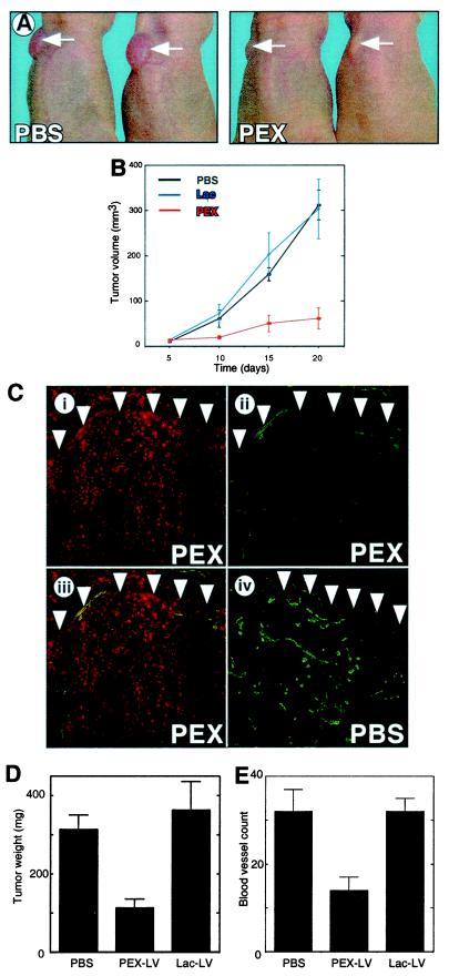
Modulation of the balance between pro- and antiangiogenic factors holds great promise for the treatment of a broad spectrum of human disease ranging from ischemic heart disease to cancer. This requires both the identification of angiogenic regulators and their efficient delivery to target organs. Here, we demonstrate the use of a noncatalytic fragment of matrix metalloproteinase 2 (termed PEX) delivered by lentiviral vectors in different angiogenesis models. Transduction of human endothelial cells with PEX virus suppressed endothelial invasion and formation of capillary-like structures without affecting chemotaxis in vitro. Lentiviral delivery of PEX blocked basic fibroblast growth factor-induced matrix metalloproteinase 2 activation and angiogenesis on chicken chorioallantoic membranes. PEX expression also inhibited tumor-induced angiogenesis and tumor growth in a nude mouse model. Thus, our study shows that lentiviral vectors can deliver sufficient quantities of antiangiogenic substances to achieve therapeutic effects in vivo.


Figures

Figure 1
Figure 1
Lentiviral vectors used for PEX delivery and transduction of endothelial cells with lentiviral vectors. (A) Schematic structure of MMP-2 (Upper) and lentiviral vectors (Lower). The lentiviral vectors contain the following features. The U3 element of the 5′ LTR is replaced by a Rous sarcoma virus promoter (RSV) that drives expression of the vector transcripts in the packaging cells. The 3′ LTR contains a SIN mutation (brown triangle) to ensure self-inactivation in the target cell. Expression of the transgene (X) is driven by the internal cytomegalovirus (CMV) promoter. The difference between nPEX-LV and PEX-LV is the inclusion of a FLAG-tag (gray) in the latter. W, posttranscriptional regulatory element of woodchuck hepatitis virus (yellow); ppt, polypurine tract. (B) Analysis of PEX expression by HUVECs transduced with PEX-LV using Abs directed against the FLAG-tag. Arrowheads, Golgi apparatus; *, nucleus. (C) Western blot analysis of PEX expression in 293T (left lane) cells stably expressing PEX and HUVECs (right lane) transduced with PEX-LV.
Figure 2
Figure 2
Effect of PEX on endothelial cell function in vitro. (A and B) bFGF-induced invasion (A) and migration (B) of HUVECs in modified Boyden chambers. Effect of bFGF in uninfected cells (bFGF), and cells infected with PEX-LV (bFGFPEX) or with GFP-LV (bFGFGFP). bFGFPhen, cells treated with o-phenanthroline. (CG) Formation of capillary-like structures of endothelial cells in vitro. Within 24 h after seeding on a Matrigel, HUVECs start to form capillary-like structures (C). Addition of bFGF enhances this process (D) in uninfected HUVECs but not in PEX-expressing cells (E). Transduction of HUVECs with Lac-LV (D) has no effect on tube formation. (H) Summary of the effect of PEX on tube formation. bFGF, untreated control; bFGFPEX, PEX-expressing cells; bFGFGFP, GFP-transduced HUVECs.
Figure 3
Figure 3
Effects of PEX on angiogenesis in the chick CAM model. (AD) Virally delivered PEX inhibits bFGF-induced angiogenesis through inhibition of MMP-2 activation. (A) Chick CAMs incubated for 72 h with filter disks soaked in PBS (Ctrl), bFGF, bFGF in the presence of PEX lentivirus (bFGF + PEX-LV), or LacZ virus (bFGF + Lac-LV). (B) Quantification of bFGF-induced angiogenic response by counting vessel branch points. (Inset) Western blot analysis using TV88 antibody to detect PEX expression in CAM lysates (from left to right: bFGF, bFGF + PEX-LV, bFGF + Lac-LV). (C and D) bFGF-induced MMP-2 activation (gelatin zymography) (C Upper) and collagenolytic (D) activity in CAM lysates. The same CAM lysates were used both for gelatin zymography and for collagenase assays. The upper and lower bands correspond to the 72-kDa MMP-2 proenzyme (proMMP-2) and the activated MMP-2 (≈62 kDa), respectively. To ensure the presence of the MMP-2 proenzyme in equal amounts, lysates also were analyzed by Western blotting (C Lower) by using MMP-2 specific mAb TV88. Ctrl, PBS-treated CAM; FGF, bFGF-treated CAM; bFGFPEX, PEX-expressing CAM treated with bFGF; bFGFGFP, GFP-expressing CAM treated with bFGF.
Figure 4
Figure 4
Lentiviral delivery of PEX inhibits tumor growth (A and B) and tumor-induced angiogenesis (C) in a CAM tumor model using CS-1 hamster melanoma. (A) Representative examples of CS-1 tumors treated with PBS, PEX-LV, or a Lac-LV. (B) Weight of tumors expressing PEX or LacZ compared with control tumors (PBS). (C) Tumor vasculature quantified as vessels per field (×20 magnification).
Figure 5
Figure 5
PEX inhibits human melanoma (M21L) growth in nude mice. (A) Two representative examples of nude mice 20 days after tumor implantation injected with PBS (Left) or with PEX-LV (Right). (B) Analysis of tumor volume starting 5 days after tumor implantation. Five days after implanting the tumors, PBS (black), Lac-LV (blue), or PEX-LV (red) was injected around the tumor, n = 7, for each group. (C) Analysis of PEX expression and vascularization in tumors. Staining with polyclonal Abs against FLAG-tag (i) and CD31(PECAM-1) (ii) in a section through a tumor treated with PEX-LV. (iii) Merger of i and ii. (iv) Representative histological sections of a PBS-treated tumor. The arrows indicate the border of the tumor. (D and E) Effect of PEX on tumor weight (D) and tumor-induced angiogenesis (E).

References

    1. Risau W. Nature (London) 1997;386:671–674. - PubMed
    1. Folkman J. Nat Med. 1995;1:27–31. - PubMed
    1. Kalebic T, Garbisa S, Glaser B, Liotta L A. Science. 1983;221:281–283. - PubMed
    1. Werb Z. Cell. 1997;91:439–442. - PubMed
    1. Collier I E, Wilhelm S M, Eisen A Z, Marmer B L, Grant G A, Seltzer J L, Kronberger A, He C S, Bauer E A, Goldberg G I. J Biol Chem. 1988;263:6579–6587. - PubMed

Publication types

MeSH terms

LinkOut - more resources