. 2000 Oct 24;97(22):12239-43.
doi: 10.1073/pnas.220403797.

Frataxin activates mitochondrial energy conversion and oxidative phosphorylation

Affiliations

Frataxin activates mitochondrial energy conversion and oxidative phosphorylation

M Ristow et al. Proc Natl Acad Sci U S A. .

Abstract

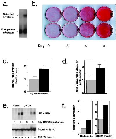
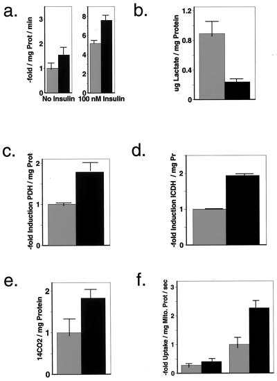
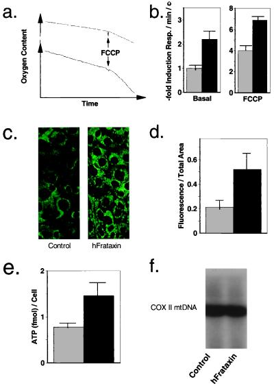
Friedreich's ataxia (FA) is an autosomal recessive disease caused by decreased expression of the mitochondrial protein frataxin. The biological function of frataxin is unclear. The homologue of frataxin in yeast, YFH1, is required for cellular respiration and was suggested to regulate mitochondrial iron homeostasis. Patients suffering from FA exhibit decreased ATP production in skeletal muscle. We now demonstrate that overexpression of frataxin in mammalian cells causes a Ca(2+)-induced up-regulation of tricarboxylic acid cycle flux and respiration, which, in turn, leads to an increased mitochondrial membrane potential (delta psi(m)) and results in an elevated cellular ATP content. Thus, frataxin appears to be a key activator of mitochondrial energy conversion and oxidative phosphorylation.


Figures

Figure 1
Figure 1
Overexpression of frataxin promotes energy storage in adipocytes. (a) A Northern blot of total RNA from 3T3-L1 fibroblasts retrovirally transfected with vehicle (empty pBabe Puro, left lane) and human frataxin (right lane). The blot was probed with a 3′ fragment of a mouse frataxin cDNA. The human frataxin RNA migrates above the endogenous mouse frataxin RNA because of retroviral sequences flanking the transgenic human frataxin. (b) 3T3-L1-cells overexpressing human frataxin (upper row) compared with cells carrying the empty vector (lower row) during differentiation from fibroblasts (day 0, outer left column) to adipocytes (day 9, outer right column). Cells were formalin-fixed on the culture dish and stained with Oil Red O, a fat specific dye. (c) A quantification of triglyceride content in differentiated 3T3-L1 adipocytes (derived from b) using the GPO trinder method. The light bars represent cells transfected with the vehicle (100% equals 291 ± 22.9 μg/mg protein), the dark bars reflect cells overexpressing transgenic human frataxin. (d) The kinetic rate of triglyceride synthesis in cells overexpressing frataxin (dark bars) compared with control cells (light bars). The rate in control cells (100%) was 0.31 ± 0.067 nmol/h/organic soluble material/mg protein. (e) A Northern blot of total RNA extracted from 3T3L1 fibroblasts (day 0 of differentiation) and adipocytes (day 9 of differentiation) overexpressing human frataxin (left lanes) or empty vehicle (right lanes). The blot was first probed for aP2, a downstream regulator in late adipocyte differentiation, then stripped and reprobed with a tubulin probe to estimate the amount of RNA loaded. (f) The quantification of the signals from e by using imagequant software. The light bars represent cells transfected with the vehicle, the dark bars reflect cells overexpressing human frataxin. The left pair of bars compares the amount of RNA in serum-starved cells, whereas the right pair was derived from cells stimulated with insulin at a concentration of 100 nM over a time period of 8 h.
Figure 2
Figure 2
Frataxin induces oxidative glucose metabolism by elevating mitochondrial calcium content. (a) Results of glucose transport assays performed with 3T3-L1-adipocytes expressing vehicle (light bars) or human frataxin (dark bars). Uptake of 3H-labeled 2-deoxy-glucose was measured by scintillation counting in the absence (left pair) and in the presence of insulin (100 nM, 60 min, right pair). The radioactivity in the cell lysates is proportional to the amount of glucose taken up normalized for protein content (100% equals 0.60 ± 0.08 nmol/min/mg protein). (b) Measurement of lactate within cell lysates normalized for protein content. The amount of this tricarbon intermediate and end product of anaerobic glycolysis is depicted in light bars for the vehicle transfected cells (0.89 ± 0.082 μg/mg protein) and dark bars for frataxin overexpressing cells. (c) Photometric quantification of pyruvate dehydrogenase activity in control cells (light bars) and frataxin overexpressing cells (dark bars) normalized for protein content, indicating an increased acetyl-CoA synthesis. (d) Activity of isocitrate dehydrogenase normalized for protein content in an set of cells identical to the above, indicating an up-regulated TCA cycle activity. (e) Production of radiolabeled 14CO2 subsequent to supplementing the cells with 14C-glucose, reflecting increased glucose oxidation. (f) Uptake of 45Ca2+ into isolated mitochondria in presence (left pair of bars) and absence (right pair of bars, light bar represents control [100%], which equals 0.026 ± 0.004 amol/mg mitochondrial protein/s) of ruthenium red, a specific blocker of the mitochondrial Ca2+ uniporter.
Figure 3
Figure 3
Frataxin increases OXPHOS. (a) A typical signal recorded from the oxygen electrode chamber to measure cellular respiration. The upper graph reflects uptake of the control cells, whereas the lower graph was recorded by using frataxin overexpressing cells. (b) The calculated mitochondrial respiration in adipocytes overexpressing human frataxin (dark bars) or cells transfected with the empty vector (light bars) based on the slopes of the graphs in a. The left pair reflects basal oxygen consumption in these cells (100% equals 1.60 ± 0.11 nmol/min/106 cells), indicating increased respiration in cells overexpressing frataxin. The right pair reflects consumption after uncoupling with FCCP, thus reflecting an increased maximal mitochondrial capacity in the frataxin overexpressing cells. (c) A microphotograph (original enlargement 60-fold) of rhodamine 123-stained cells overexpressing frataxin (right) and mock-transfected cells (left), depicting increased fluorescence in overexpressing cells, indicating an elevated mitochondrial membrane potential. (d) Computerized quantification of rhodamine 123 fluorescence in cells overexpressing human frataxin (dark bars) or cells transfected with the empty vector (light bars). (e) Luciferase-based quantification of cellular ATP content in cells overexpressing human frataxin (dark bars) or cells transfected with the empty vector (light bars), indicating increased OXPHOS. (f) A Southern blot of total DNA (10 μg per lane) extracted from differentiated 3T3-L1-adipocytes expressing the empty vector (left lane) or human frataxin (right lane) using a probe for cytochrome c oxidase subunit II, the latter is exclusively encoded in the mitochondrial DNA and thus reflects the amount of mitochondrial DNA relative to total DNA loaded.

References

    1. Friedreich N. Virchows Arch Pathol Anat Physiol Klin Med. 1863;26:391–419.
    1. Finocchiaro G, Baio G, Micossi P, Pozza G, di Donato S. Neurology. 1988;38:1292–1296. - PubMed
    1. Tolis G, Mehta A, Andermann E, Harvey C, Barbeau A. Can J Neurol Sci. 1980;7:397–400. - PubMed
    1. Khan R J, Andermann E, Fantus I G. Metabolism. 1986;35:1017–1023. - PubMed
    1. Hebinck J, Hardt C, Schoels L, Vorgerd M, Briedigkeit L, Kahn C R, Ristow M. Diabetes. 2000;49:1604–1607. - PubMed

Publication types