

Accuracy of 131I tumor quantification in radioimmunotherapy using SPECT imaging with an ultra-high-energy collimator: Monte Carlo study
- PMID: 11038009
- PMCID: PMC2812013
Accuracy of 131I tumor quantification in radioimmunotherapy using SPECT imaging with an ultra-high-energy collimator: Monte Carlo study
Abstract
Accuracy of 131I tumor quantification after radioimmunotherapy (RIT) was investigated for SPECT imaging with an ultra-high-energy (UHE) collimator designed for imaging 511-keV photons.
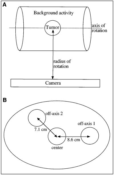
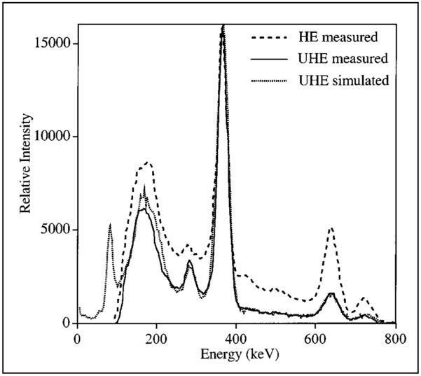
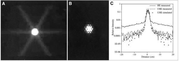
Methods: First, measurements and Monte Carlo simulations were carried out to compare the UHE collimator with a conventionally used, high-energy collimator. On the basis of this comparison, the UHE collimator was selected for this investigation, which was carried out by simulation of spherical tumors in a phantom. Reconstruction was by an expectation-maximization algorithm that included scatter and attenuation correction. Keeping the tumor activity constant, simulations were carried out to assess how volume-of-interest (VOI) counts vary with background activity, radius of rotation (ROR), tumor location, and size. The constant calibration factor for quantification was determined from VOI counts corresponding to a 3.63-cm-radius sphere of known activity. Tight VOIs corresponding to the physical size of the spheres or tumors were used.
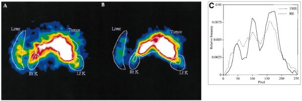
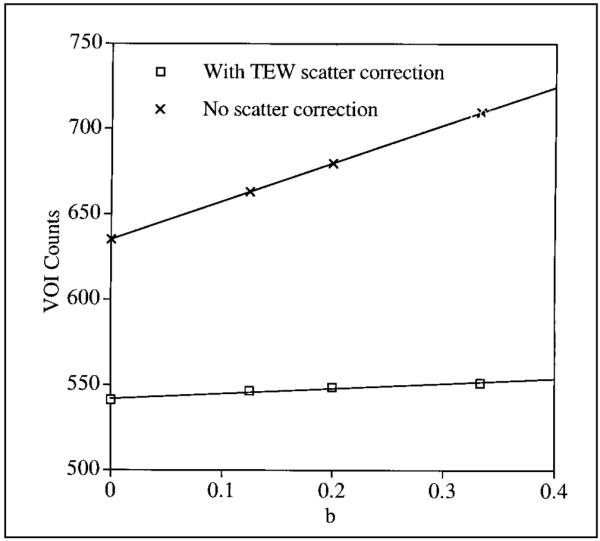
Results: Use of the UHE collimator resulted in a large reduction in 131I penetration, which is especially significant in RIT where background uptake is high. With the UHE collimator, typical patient images showed an improvement in contrast. Considering the desired geometric events, sensitivity was reduced, but only by a factor of 1.6. Simulation results for a 3.63-cm-radius tumor showed that VOI counts vary with background, location, and ROR by less than 3.2%, 3%, and 5.3%, respectively. The variation with tumor size was more significant and was a function of the background. Good quantification accuracy (<6.5% error) was achieved when tumor size was the same as the sphere size used in the calibration, irrespective of the other parameters. For smaller tumors, activities were underestimated by up to -15% for the 2.88-cm-radius sphere, -23% for the 2.29-cm-radius sphere, and -47% for the 1.68-cm-radius sphere.
Conclusion: Reasonable accuracy can be achieved for VOI quantification of 131I using SPECT with an UHE collimator and a constant calibration factor. Difference in tumor size relative to the size of the calibration sphere had the biggest effect on accuracy, and recovery coefficients are needed to improve quantification of small tumors.
Figures
Similar articles
-
Monte Carlo evaluation of object shape effects in iodine-131 SPET tumor activity quantification.Eur J Nucl Med. 2001 Jul;28(7):900-6. doi: 10.1007/s002590100551. Eur J Nucl Med. 2001. PMID: 11504087 Free PMC article.
-
Characterization of scatter and penetration using Monte Carlo simulation in 131I imaging.J Nucl Med. 2000 Jan;41(1):123-30. J Nucl Med. 2000. PMID: 10647615 Free PMC article.
-
Evaluation of Iodine-123 and Iodine-131 SPECT activity quantification: a Monte Carlo study.EJNMMI Phys. 2021 Aug 19;8(1):61. doi: 10.1186/s40658-021-00407-1. EJNMMI Phys. 2021. PMID: 34410539 Free PMC article.
-
Evaluation of quantitative 123I and 131I SPECT with Monte Carlo-based down-scatter compensation.Nucl Med Commun. 2018 Dec;39(12):1097-1102. doi: 10.1097/MNM.0000000000000920. Nucl Med Commun. 2018. PMID: 30222722
-
Quantitative imaging of iodine-131 distributions in brain tumors with pinhole SPECT: a phantom study.J Nucl Med. 1998 May;39(5):856-64. J Nucl Med. 1998. PMID: 9591589
Cited by
-
Simultaneous SPECT imaging of multi-targets to assist in identifying hepatic lesions.Sci Rep. 2016 Jul 5;6:28812. doi: 10.1038/srep28812. Sci Rep. 2016. PMID: 27377130 Free PMC article.
-
EANM Dosimetry Committee series on standard operational procedures for internal dosimetry for 131I mIBG treatment of neuroendocrine tumours.EJNMMI Phys. 2020 Mar 6;7(1):15. doi: 10.1186/s40658-020-0282-7. EJNMMI Phys. 2020. PMID: 32144574 Free PMC article.
-
3-D Monte Carlo-Based Scatter Compensation in Quantitative I-131 SPECT Reconstruction.IEEE Trans Nucl Sci. 2006;53(1):181. doi: 10.1109/TNS.2005.862956. IEEE Trans Nucl Sci. 2006. PMID: 20104252 Free PMC article.
-
Accurate dosimetry in 131I radionuclide therapy using patient-specific, 3-dimensional methods for SPECT reconstruction and absorbed dose calculation.J Nucl Med. 2005 May;46(5):840-9. J Nucl Med. 2005. PMID: 15872359 Free PMC article.
-
Monte Carlo evaluation of object shape effects in iodine-131 SPET tumor activity quantification.Eur J Nucl Med. 2001 Jul;28(7):900-6. doi: 10.1007/s002590100551. Eur J Nucl Med. 2001. PMID: 11504087 Free PMC article.
References
-
- Wahl RL, Zasadny KR, McFarlane D, et al. Iodine-131 anti-B1 antibody for B-cell lymphoma: an update on the Michigan phase I experience. J Nucl Med. 1998;39(suppl 8):21S–27S. - PubMed
-
- Kaminski MS, Zasadny KR, Francis IR, et al. Iodine-131-anti B1 radioimmuno-therapy for B-cell lymphoma. J Clin Oncol. 1996;14:1974–1981. - PubMed
-
- DeNardo GL, Lamborn KR, Goldstein DS, Kroger LA, DeNardo SJ. Increased survival associated with radiolabeled lym-1 therapy for non-Hodgkin’s lymphoma and chronic lymphocytic leukemia. Cancer. 1997;80:2706–2711. - PubMed
-
- Press O, Early J, Appelbaum F, et al. Phase II trial of I-131 B1 (anti-CD20) antibody therapy with autologous stem cell transplantation for relapsed B-cell lymphomas. Lancet. 1995;346:336–340. - PubMed
-
- Koral KF, Zasadny KR, Kessler ML, et al. CT-SPECT fusion plus conjugate views for determining dosimetry in iodine-131-monoclonal antibody therapy of lymphoma patients. J Nucl Med. 1994;35:1714–1720. - PubMed
Publication types
MeSH terms
Substances
Grants and funding
LinkOut - more resources
Full Text Sources