. 2000 Oct 16;151(2):209-20.
doi: 10.1083/jcb.151.2.209.

Ectodomain shedding of epidermal growth factor receptor ligands is required for keratinocyte migration in cutaneous wound healing

Affiliations

Ectodomain shedding of epidermal growth factor receptor ligands is required for keratinocyte migration in cutaneous wound healing

S Tokumaru et al. J Cell Biol. .

Abstract

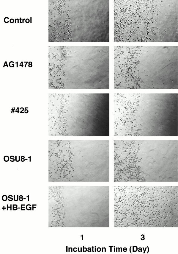
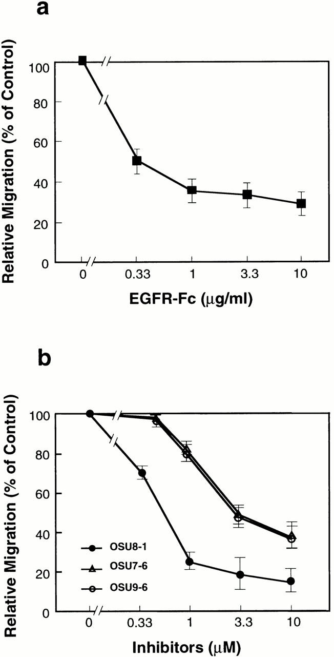
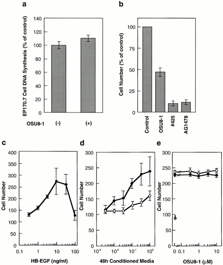
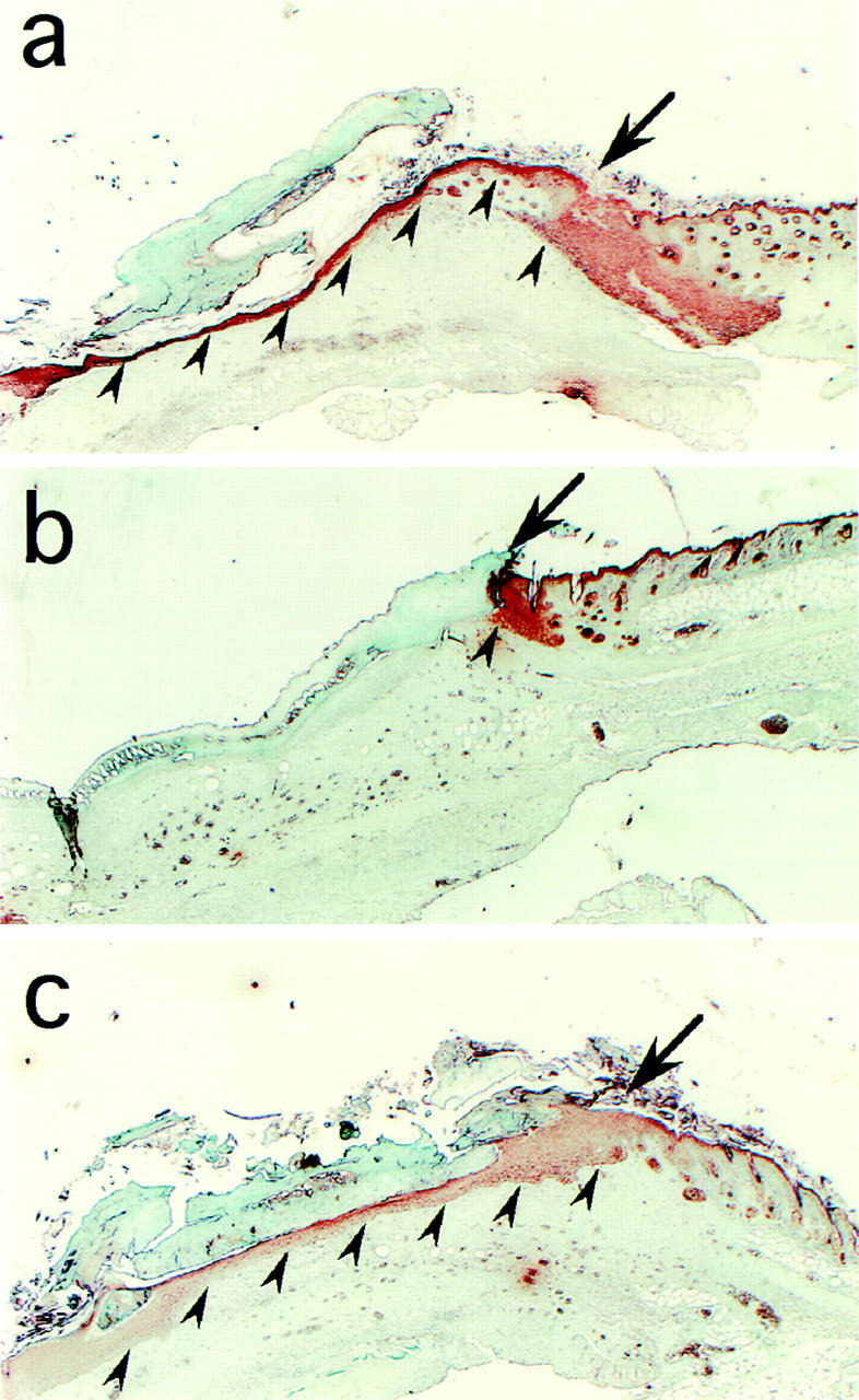
Keratinocyte proliferation and migration are essential to cutaneous wound healing and are, in part, mediated in an autocrine fashion by epidermal growth factor receptor (EGFR)-ligand interactions. EGFR ligands are initially synthesized as membrane-anchored forms, but can be processed and shed as soluble forms. We provide evidence here that wound stimuli induce keratinocyte shedding of EGFR ligands in vitro, particularly the ligand heparin-binding EGF-like growth factor (HB-EGF). The resulting soluble ligands stimulated transient activation of EGFR. OSU8-1, an inhibitor of EGFR ligand shedding, abrogated the wound-induced activation of EGFR and caused suppression of keratinocyte migration in vitro. Soluble EGFR-immunoglobulin G-Fcgamma fusion protein, which is able to neutralize all EGFR ligands, also suppressed keratinocyte migration in vitro. The application of OSU8-1 to wound sites in mice greatly retarded reepithelialization as the result of a failure in keratinocyte migration, but this effect could be overcome if recombinant soluble HB-EGF was added along with OSU8-1. These findings indicate that the shedding of EGFR ligands represents a critical event in keratinocyte migration, and suggest their possible use as an effective clinical treatment in the early phases of wound healing.


Figures

Figure 1
Figure 1
Expression of AP-tagged EGFR ligands. (a) Structure of the AP-tag parental expression plasmid, pSS-AlPh. (b) A schematic diagram of the region within the expression plasmids derived from pss-AlPh that encode the fusions of AP-tag and EGFR ligands. The coding region for the AP-tag HB-EGF fusion was constructed such that, in the encoded fusion, the AP sequence (488 amino acids) was inserted at Ala84 of the HB-EGF precursor. In the AP-tag AR and AP-tag TGF-α fusion proteins, AR (residues 102–252) and TGF-α (residues 45–160), respectively, were substituted for HB-EGF (residues 85–208). (c) Expression of AP-tag HB-EGF, AP-tag AR, and AP-tag TGF-α on the surface of transfected CHO cells. Biotinylated AP-tag HB-EGF, AP-tag AR, and AP-tag TGF-α were immunoprecipitated with human placental AP antibody and analyzed by SDS-PAGE and Western blotting. Biotinylated proteins on the Western blot membranes were detected by HRP-conjugated avidin and ECL. (d) AP activity in the conditioned media of transfectants expressing AP-tag HB-EGF, AP-tag AR, and AP-tag TGF-α. Cells were treated with or without 60 nM TPA for 30 min at 37°C. AP activity was measured as described in Materials and Methods. Each bar is the average of triplicate values.
Figure 2
Figure 2
Effect of OSU8-1 on TPA-inducible shedding of EGFR ligands. (a) AP activities in the conditioned media of CHO transfectants expressing AP-tag HB-EGF, AP-tag AR, and AP-tag TGF-α treated with or without 60 nM of TPA in the presence or absence of OSU8-1. Cells were preincubated with the indicated concentration of OSU8-1 for 10 min at 37°C. The cells were incubated with fresh media containing the indicated concentrations of TPA and OSU8-1 for 30 min at 37°C. AP activity was measured as described in Materials and Methods. Each bar is the average of triplicate values. (b) CHO transfectants expressing wild-type (non–AP-tag fused) HB-EGF, AR, and TGF-α were biotinylated and preincubated with or without 10 μM OSU8-1 for 10 min at 37°C. Cells were incubated with fresh media containing the indicated concentrations of TPA and OSU8-1 for 30 min at 37°C, followed by cell lysis, immunoprecipitation with antibodies against the shed ligands, SDS-PAGE, Western blotting, and avidin-HRP/ECL detection. The wild-type HB-EGF shows different processing forms (Goishi et al. 1995).
Figure 4
Figure 4
Characterization of EGFR-ligand shedding in keratinocytes. (a) Gene expression of EGFR ligands in wounded keratinocytes. After incubation of keratinocytes for the indicated time after wounding, total RNA was extracted and analyzed by Northern blotting. Sequential hybridization using the same membrane is shown. HB-EGF (2.4 kb), AR (1.4 kb), and TGF-α (4.8 kb) messages were detected. (b) Effect of cycloheximide and HB-EGF neutralizing antibody No. 197 on wound-induced EGFR activation in cultured keratinocytes. Keratinocytes were stimulated by tip-scraping in the presence or absence of cycloheximide (50 μM) or HB-EGF neutralizing antibody No. 197 (10 μg/ml). After incubating for the indicated times, the cells were lysed and subjected to immunoprecipitation with EGFR antibodies, fractionation by SDS-PAGE, and Western blotting. Half of the immunocomplex was used for the detection of tyrosine phosphorylation with phosphotyrosine antibody PY-20, and the other half for detection of EGFR protein with EGFR antibody. Cycloheximide (50 μM) did not abrogate the wound-induced tyrosine phosphorylation of EGFR. On the other hand, antibody No. 197 (10 μg/ml) suppressed wound-induced tyrosine phosphorylation of EGFR by 70%. (c) Quantitative analyses of EGFR ligand shedding. Stable transfectants of HaCat cells expressing nearly equal amounts of AP-tag HB-EGF, AP-tag AR, or AP-Tag TGF-α were incubated with or without 1 μM OSU8-1 for 60 min after wounding. AP activities in the conditioned media were measured. Each bar is the average of triplicate values.
Figure 3
Figure 3
Production of soluble EGFR ligands and activation of EGFR in cultured keratinocytes after wounding. (a) Detection of shed EGFR ligands produced by wounded keratinocytes. The levels of released EGFR ligands in conditioned media collected at the indicated times in the presence or absence of 1 μM OSU8-1 were measured by an EP170.7 cell assay. Four independent experiments were carried out and a typical time course of the level of EGFR ligands is shown. Each bar is the average of quadruplicate values. (b) Tyrosine phosphorylation of EGFR in wounded keratinocytes. Keratinocytes, cultured in 10-cm dishes, were stimulated by tip-scraping and incubated for the indicated times in the presence or absence of 1 μM OSU8-1. Wounded keratinocytes were lysed and subjected to immunoprecipitation with EGFR antibodies followed by SDS-PAGE and Western blotting. Half of the immunocomplex was used for the detection of phosphotyrosine with PY-20 antibody, and the other half for the detection of EGFR protein with EGFR antibody.
Figure 5
Figure 5
Evaluation of OSU8-1 and EGFR inactivation on keratinocyte migration. A portion of the keratinocytes were removed from tissue culture plates by scraping, and the remaining cells were cultured for 3 d in the absence or presence of EGFR neutralizing antibody No. 425 (10 μg/ml), EGFR kinase–specific inhibitor AG1478 (30 nM), OSU8-1 (1 μM), or both OSU8-1 (1 μM) and HB-EGF (20 ng/ml). The same area of each dish was monitored microscopically (40×) at day 0 and day 3.
Figure 6
Figure 6
Dose effects of EGFR-Fc (a) and OSU8-1, OSU7-6, and OSU9-6 (b) on keratinocyte migration after wounding. Each point is the average of quadruplicate measurements. Migration was evaluated by the distance between the furthest migrated cell and the scraped edge.
Figure 7
Figure 7
Quantitative evaluation of the effects of OSU8-1 and EGFR inactivation on keratinocyte growth and migration. (a) Effect of 1 μM OSU8-1 on juxtacrine activity of EGFR ligands in cultured keratinocytes. Juxtacrine activity was measured as described in Materials and Methods. (b) The effect of OSU8-1, antibody No. 425 and AG1478 on the growth of keratinocytes. Keratinocytes were seeded at a density of 105 cells/well in 6-well plates and incubated for 3 d in the absence or presence of OSU8-1 (1 μM), antibody No. 425 (10 μg/ml), or AG1478 (30 nM). (a and b) Values represent the average of triplicate experiments. (c) Stimulation of keratinocyte migration by HB-EGF. The ability of recombinant HB-EGF to stimulate keratinocyte migration was tested using a Boyden chamber. (d) Stimulation of keratinocyte migration by conditioned medium. Serially diluted, 48-h conditioned medium obtained from wounded keratinocytes was added to the bottom wells of Boyden chambers and tested for its ability to stimulate keratinocyte migration. Open circles, conditioned medium generated in the presence of 1 μM OSU8-1; closed circles, conditioned medium obtained without 1 μM OSU8-1. (e) Effect of OSU8-1 on the migration response of keratinocytes induced by conditioned medium from wounded keratinocyte cultures. The indicated concentrations of OSU8-1 were added to the top (open circles) and the bottom (closed circles) wells of a Boyden chamber containing wounded keratinocyte conditioned medium in the bottom wells. An open triangle shows migrated cell number obtained using fresh medium in the bottom wells as a control. (c–e) Each point is the average of quadruplicate measurements.
Figure 8
Figure 8
Effect of OSU8-1 on wound healing in vivo. Histology of excised mouse skin on day 6 after wounding, without or with 10 mM OSU8-1 treatment. Keratinocytes were stained with antikeratin/cytokeratin antibody. (a) A typical tissue section from the control wounds (n = 15). Keratinocytes are observed to have migrated from the edge of the wound, spreading under the crust and covering the wound site. Arrowheads indicate the base of the keratinocyte layer. The arrow marks the edge of the wound. (b) A typical tissue section from an OSU8-1–treated wound (n = 10). Both keratinocyte migration and the regeneration of the epidermis were greatly inhibited. (c) A typical tissue section from a wound treated with a cocktail of OSU8-1 and HB-EGF (5 μg/ml; n = 5). Both keratinocyte migration and the regeneration of the epidermis were completely restored.

Similar articles

Cited by

References

    1. Black R.A., Rauch C.T., Kozlosky C.J., Peschon J.J., Slack J.L., Wolfson M.F., Castner B.J., Stocking K.L., Reddy P., Srinivasan S. A metalloproteinase disintegrin that releases tumor-necrosis factor-alpha from cells. Nature. 1997;385:729–733. - PubMed
    1. Blotnick S., Peoples G.E., Freeman M.R., Eberlein T.J., Klagsbrun M. T lymphocytes synthesize and export heparin-binding epidermal growth factor-like growth factor and basic fibroblast growth factor, mitogens for vascular cells and fibroblastsdifferential production and release by CD4+ and CD8+ T cells. Proc. Natl. Acad. Sci. USA. 1994;91:2890–2894. - PMC - PubMed
    1. Boyden S. The chemotactic effect of mixtures of antibodies and antigens on polymorphonuclear leukocytes. J. Exp. Med. 1962;115:435–466. - PMC - PubMed
    1. Brachmann R., Lindquist P.B., Nagashima M., Kohr W., Lipari T., Napier M., Derynck R. Transmembrane TGF-α precursors activate EGF/TGF-α receptors. Cell. 1989;56:691–700. - PubMed
    1. Chen C.A., Okayama H. Calcium phosphate-mediated gene transfera highly efficient transfection system for stably transforming cells with plasmid DNA. Biotechniques. 1988;6:632–638. - PubMed

MeSH terms