. 2000 Oct 16;151(2):235-48.
doi: 10.1083/jcb.151.2.235.

Indications for a novel muscular dystrophy pathway. gamma-filamin, the muscle-specific filamin isoform, interacts with myotilin

Affiliations

Indications for a novel muscular dystrophy pathway. gamma-filamin, the muscle-specific filamin isoform, interacts with myotilin

P F van der Ven et al. J Cell Biol. .

Abstract

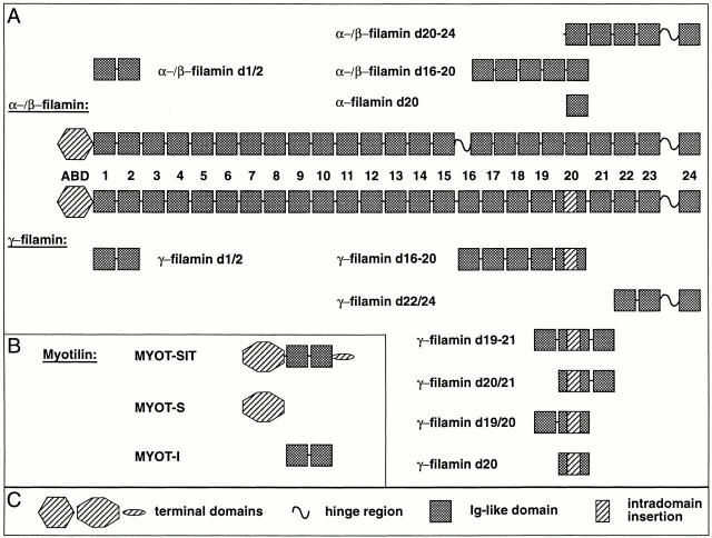
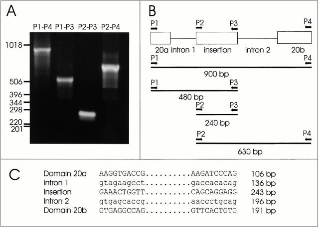
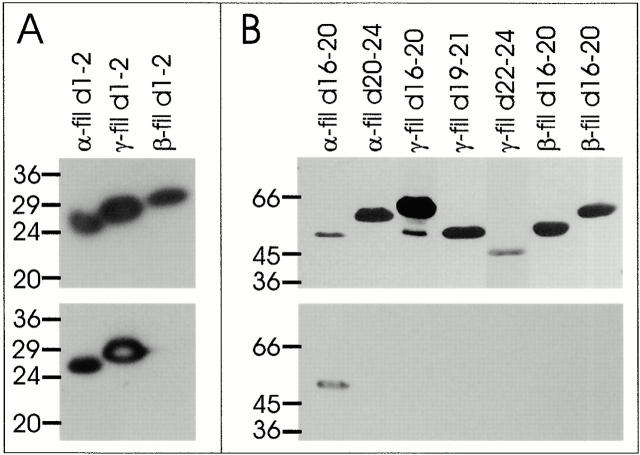
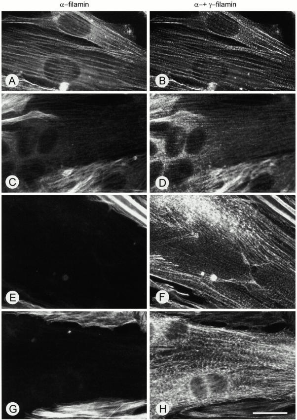
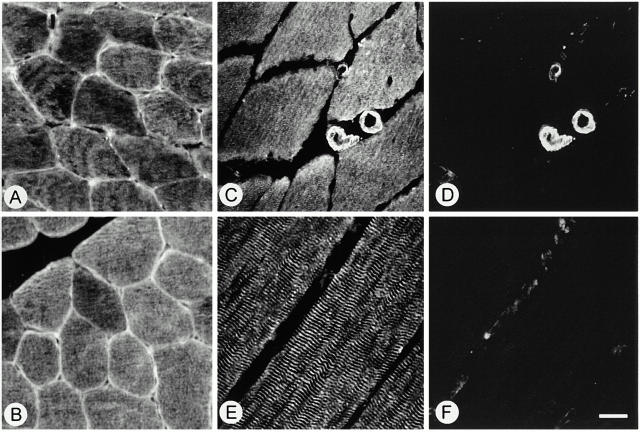
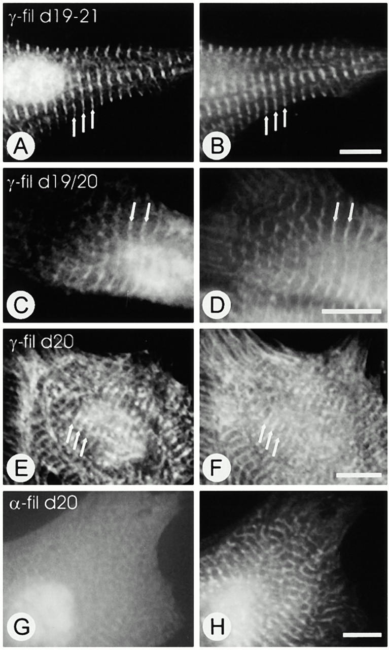
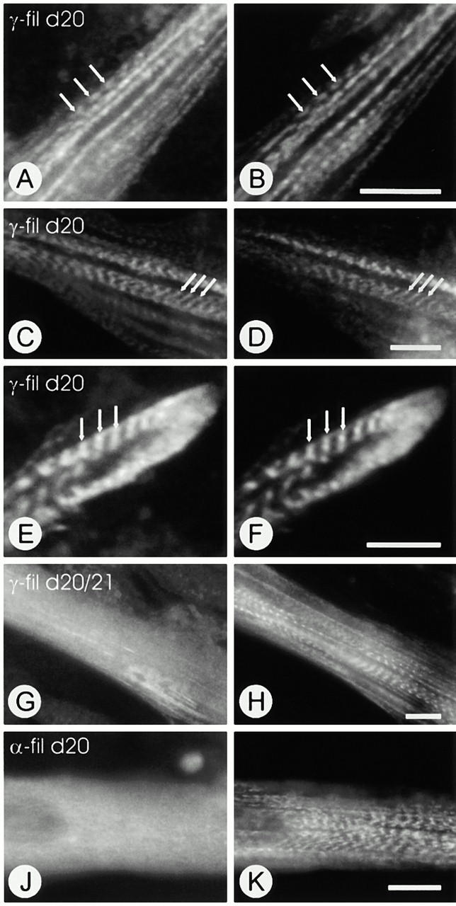
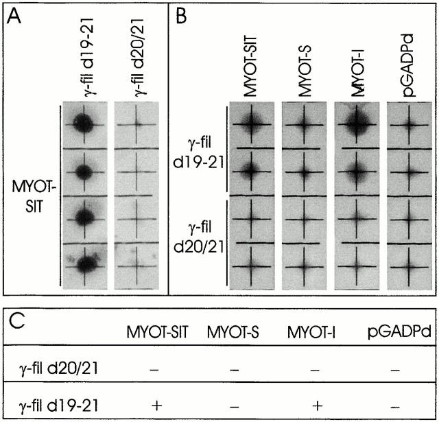
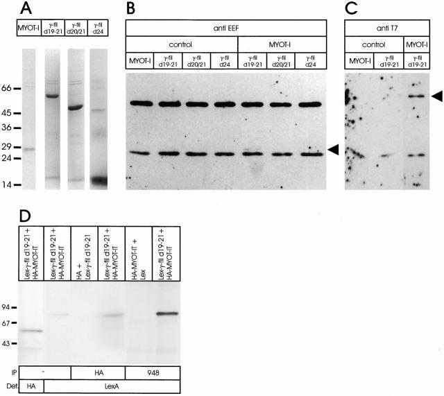
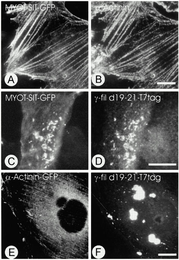
gamma-Filamin, also called ABP-L, is a filamin isoform that is specifically expressed in striated muscles, where it is predominantly localized in myofibrillar Z-discs. A minor fraction of the protein shows subsarcolemmal localization. Although gamma-filamin has the same overall structure as the two other known isoforms, it is the only isoform that carries a unique insertion in its immunoglobulin (Ig)-like domain 20. Sequencing of the genomic region encoding this part of the molecule shows that this insert is encoded by an extra exon. Transient transfections of the insert-bearing domain in skeletal muscle cells and cardiomyocytes show that this single domain is sufficient for targeting to developing and mature Z-discs. The yeast two-hybrid method was used to identify possible binding partners for the insert-bearing Ig-like domain 20 of gamma-filamin. The two Ig-like domains of the recently described alpha-actinin-binding Z-disc protein myotilin were found to interact directly with this filamin domain, indicating that the amino-terminal end of gamma-filamin may be indirectly anchored to alpha-actinin in the Z-disc via myotilin. Since defects in the myotilin gene were recently reported to cause a form of autosomal dominant limb-girdle muscular dystrophy, our findings provide a further contribution to the molecular understanding of this disease.


Figures

Figure 1
Figure 1
Domain organization of the filamins and of myotilin, and recombinant constructs used for transfection assays. A emphasizes the general modular organization of filamins: the actin binding head domain (ABD) is followed by 24 Ig-like modules (identified by numbers between the boxes). Stretches of unique sequence, so-called hinge regions, are found between Ig-like domains 15 and 16, as well as between domains 23 and 24. Muscle γ-filamin, however, lacks this insertion, which might make the molecule less flexible. A conspicuous difference between γ-filamin and the other members of the filamin family is a unique insertion in the middle of Ig-like domain 20. Constructs derived from α- or γ-filamin, which were used for the experiments of this work, are shown schematically above and below the respective mother molecules. For yeast two-hybrid screens and for prokaryotic expression experiments, identical inserts were cloned into the respective vectors (see Materials and Methods). (B) A layout of the structure of myotilin is given. The molecule consists of a serine-rich amino terminal portion (S), followed by two immunoglobulin-like domains (I) and a carboxyterminal “tail” sequence (T). Inserts were cloned into the respective vectors for yeast two-hybrid experiments and for prokaryotic expression. (C) Legend to the domain symbols used in A and B.
Figure 2
Figure 2
Analysis of the structure of the FLNC gene encoding domain 20. (A) PCR products after amplification of human genomic DNA with primers P1 and P4 (P1–P4), P1 and P3 (P1–P3), P2 and P3 (P2–P3), and P2 and P4 (P2–P4). (B) Horizontal lines represent the four PCR products depicted in A, and the derived exon/intron structure of domain 20. Note that the primers used for amplification (P1: GTCACCAACAGCCCCTTCAAG; P2: AAGGTGACCGGCGAGGGCCGC; P3: CACAGTGAACTGAAAGGGGCT; P4: AGCGTCATCCGAGAGGGAGG) all had a tail of nine additional basepairs to allow restriction enzyme–based cloning. Therefore, the actual length of the products was reduced by subtraction of 18 basepairs. (C) Splice junctions and exon and intron sizes of the part of the FLNC gene that encodes domain 20 are shown.
Figure 3
Figure 3
Characterization of the filamin antibodies. Total protein extracts from bacteria expressing the carboxyterminally immunotagged polypeptides indicated above each lane were separated on a polyacrylamide gel and transferred to nitrocellulose. Subsequently, the filters were incubated with YL1/2 (A and B, top), RR90 (A, bottom) or mAb 1680 (B, bottom). The reactivity with the anti–EEF tag antibody indicates that the polypeptides are intact and in the correct reading frame. Note that RR90 recognizes an epitope in the first two immunoglobulin-like domains of α- and γ-filamins, but not in β-filamin. mAb 1680 recognizes an epitope in the polypeptide α-filamin d16–20, and not in β- or γ-filamin, indicating that it is specific for this isoform.
Figure 5
Figure 5
Immunofluorescence localization of filamin isoforms in developing human skeletal muscle cells. The micrographs show cultured human skeletal muscle cells differentiated for 0 (A and B), 2 (C and D), 4 (E and F) or 6 (G and H) d, double stained with an antibody specific for α-filamin (left) and mAb RR90 (right). Note that only in nondifferentiated cells a high level of α-filamin is detected by both antibodies (A and B). After 2 d of differentiation, RR90 strongly stains the young myotubes (D), while the level of α-filamin has strongly decreased (C), indicating a clear predominance of the muscle-specific filamin isoform. In further developed myotubes, α-filamin is not detectable (E and G), and γ-filamin seems to be the only filamin isoform that is expressed at these developmental stages (F and H). Note that, after 6 d of differentiation, γ-filamin is localized in a cross-striated pattern due to its localization in Z-discs. Bar, 20 μm.
Figure 4
Figure 4
Immunolocalization of filamins in sections of human skeletal muscle tissue. Photomicrographs were taken from cryosections of normal human skeletal muscle tissue stained with mAb RR90 (A and B) or double stained with RR90 (C and E) and mAb 1680, an antibody specific for α-filamin (D and F). RR90 detects both sarcoplasmic and sarcolemmal filamins in cross-sectioned myofibers (A and B). In slightly oblique sections, the typical cytoplasmic staining for proteins that show a striated, myofibrillar staining pattern in longitudinal sections is observed (A). Double staining of RR90 (C and E) with mAb 1680 (D and F) shows that blood vessels that are known to contain α-filamin are stained by both antibodies. In contrast, α-filamin cannot be detected at the sarcolemma or in the sarcoplasm of human skeletal muscle fibers, indicating that the filamin isoform detected by RR90 in these cells is exclusively γ-filamin. Bar, 20 μm.
Figure 6
Figure 6
Expression of recombinant filamin fragments in neonatal rat cardiomyocytes. Neonatal rat cardiomyocytes transfected with constructs encoding T7-tagged γ-filamin d19-21 (A and B), γ-filamin d19/20 (C and D), γ-filamin d20 (E and F), or α-filamin d20 (G and H) were fixed and double stained with T7-tag antibody (left) and an antibody specific for sarcomeric α-actinin (right). Note that the recombinant polypeptides γ-filamin d19–21 and γ-filamin d20/21 colocalize precisely with α-actinin in sharp striations, indicating that this part of γ-filamin is targeted to Z-discs (A–D, arrows). The single domain γ-filamin d20 is also targeted to the Z-disc region, although the fluorescent stripes appear less sharp (E and F, arrows). α-filamin d20 is distributed diffusely in the cytoplasm (G) and does not show any specific targeting to Z-discs or myofibrils in general (H). Bars, 10 μm.
Figure 7
Figure 7
Expression of recombinant filamin fragments in differentiating C2C12 mouse myoblasts. C2C12 cells transfected with constructs encoding T7-tagged γ-filamin d20 (A and F), γ-filamin d20/21 (G and H), or α-filamin d20 (J and K) were allowed to differentiate for 2 (A and B) or 4 (C–K) d. Subsequently, they were fixed and double stained with T7-tag antibody (left) and T12 (right), an antibody specific for a Z-disc epitope of titin. Note that γ-filamin d20 exactly colocalizes with Z-disc titin in nascent myofibrils with immature Z-discs (A and B, arrows), as well as in mature myofibrils (C–F) where the recombinant protein and Z-disc epitope of titin are localized in sharp striations (E and F, arrows). Note that, although γ-filamin d20/21 associates with stress fiber–like structures, it is never found at Z-discs (G and H). α-Filamin d20 is distributed diffusely in the cytoplasm (J), and does not show any targeting to immature or mature myofibrils (K). Bars, 20 μm.
Figure 8
Figure 8
Characterization of the interaction of γ-filamin with myotilin. Yeast cells pretransformed with γ-filamin d20/21 or γ-filamin d19–21 in pLexPd were transformed with the empty prey vector (pGADPd) or with the region of the myotilin cDNA encoding its complete polypeptide (MYOT-SIT), its serine rich domain (MYOT-S), or the immunoglobulin domains (MYOT-I) in pGADPd. (A) In four independent colonies double transformed with γ-filamin d19–21 and MYOT-SIT, β-galactosidase activity was restored, indicating an interaction between both polypeptides. This reactivity was absent in all colonies of yeast cells transformed with γ-filamin d20/21 and MYOT-SIT. (B) Two independent yeast colonies cotransformed with γ-filamin d19–21 or γ-filamin d20/21, and truncated myotilin fragments. Note that only in yeast cells cotransformed with γ-filamin d19–21 and MYOT-I or MYOT-SIT β-galactosidase reactivity was restored. These results are summarized in C.
Figure 9
Figure 9
Coimmunoprecipitation of filamin and myotilin fragments. (A) Coomassie blue–stained SDS gel of recombinant protein fragments used for coimmunoprecipitation experiments. The identity of the polypeptides is indicated above each lane. (B and C) T7-tagged MYOT-I and EEF-tagged γ-filamin fragments alone (control) or mixed with MYOT-I (MYOT-I) were incubated with anti–EEF antibody (B) or anti–T7-tag antibody (C), immunoprecipitated as described in Materials and Methods, and subjected to SDS-PAGE and Western blotting. For immunodetection of binding partners of the precipitated polypeptide, anti–T7-tag antibody (B) or anti–EEF antibody (C) were used. Arrowheads indicate positions of proteins that are specifically coprecipitated. Note that MYOT-I is coprecipitated with γ-filamin d19–21, but not with γ-filamin d20/21 or γ-filamin d24 (B), and that γ-filamin d19–21 is coprecipitated with MYOT-I (C). (D) Coimmunoprecipitation from yeast cells double transfected with an empty Lex vector (Lex) or the pLex vector containing γ-filamin d19–21, together with an empty pACT2-HA vector (HA) or this vector with MYOT-IT (HA-MYOT-IT), as indicated above the lanes. In the left two lanes, expression of the proteins was confirmed by Western blotting using an anti–HA or anti–LexA antibody. In the middle two lanes, the complexes immunoprecipitated with the anti–HA antibody were separated and stained with the anti–LexA antibody to detect Lex-γ-filamin d19–21. Note that Lex-γ-filamin d19–21 is only coprecipitated with HA-MYOT-I and not with HA alone. In the right two lanes, complexes immunoprecipitated with the anti–myotilin antibody were separated and stained with the anti–LexA antibody to detect Lex-γ-filamin d19–21. Note that Lex-γ-filamin d19–21 is again coprecipitated with HA-MYOT-I. No signal is obtained if the cells are transfected with the empty LexA vector.
Figure 10
Figure 10
Expression of filamin, myotilin and α-actinin in non-muscle cells. Ptk2 cells were transfected with GFP-tagged MYOT-SIT alone (A and B), GFP-tagged MYOT-SIT together with T7-tagged γ-filamin d19–21 (C and D), or GFP-tagged α-actinin and T7-tagged γ-filamin d19–21 (E and F). The T7-tagged recombinant protein was visualized by staining with the anti–T7-tag antibody (D and F). Endogenous α-actinin was stained with BM-75.2 recognizing all isoforms of α-actinin (B). Full-length myotilin is targeted to stress fibers, where it colocalizes with endogenous α-actinin (A and B). Note that if, in the same cell, γ-filamin d19–21 is expressed, the localization of MYOT-SIT is dramatically changed, and the protein is found in aggregates together with γ-filamin d19–21 (C and D). In contrast, the localization of GFP-tagged α-actinin, which is also targeted to stress fibers, is not changed upon coexpression of γ-filamin d19–21 (E and F), indicating that MYOT-I and not the GFP tag interacts with γ-filamin d19–21. Bars, 10 μm.
Figure 11
Figure 11
Expression of recombinant myotilin fragments in differentiating C2C12 mouse myoblasts. C2C12 cells transfected with constructs encoding T7-tagged immunoglobulin domains of myotilin (MYOT-I; A and F), or full-length myotilin (MYOT-SIT; G and H) were allowed to differentiate for 4 d. Subsequently, they were fixed and double stained with T7-tag antibody (left) and T12 (right), an antibody specific for a Z-disc epitope of titin. Note that MYOT-SIT exactly colocalizes with Z-disc titin in nascent myofibrils with immature Z-discs as well as in mature myofibrils (G and H). MYOT-I associates with stress fiber–like structures (A and B), but it is never found in Z-discs. Moreover, myotubes expressing MYOT-I do not develop myofibrils. Instead, the myofibrillar proteins are colocalized with MYOT-I in filamentous bundles (C and D) or amorphous aggregates (E and F). Bar, 20 μm.

Similar articles

Cited by

References

    1. Abd-el-Basset E.M., Ahmed I., Fedoroff S. Actin and actin-binding proteins in differentiating astroglia in tissue culture. J. Neurosci. Res. 1991;30:1–17. - PubMed
    1. Auerbach D., Bantle S., Keller S., Hinderling V., Leu M., Ehler E., Perriard J.C. Different domains of the M-band protein myomesin are involved in myosin binding and M-band targeting. Mol. Biol. Cell. 1999;10:1297–1308. - PMC - PubMed
    1. Ayscough K.R. In vivo functions of actin-binding proteins. Curr. Opin. Cell Biol. 1998;10:102–111. - PubMed
    1. Barry C.P., Xie J., Lemmon V., Young A.P. Molecular characterization of a multipromoter gene encoding a chicken filamin protein. J. Biol. Chem. 1993;268:25577–25586. - PubMed
    1. Bröcker F., Bardenheuer W., Vieten L., Jülicher K., Werner N., Marguitan G., Michael D., Opalka B., Schütte J. Assignment of human filamin gene FLNB to human chromosome band 3p14.3 and identification of YACs containing the complete FLNB transcribed region. Cytogenet. Cell Genet. 1999;85:267–268. - PubMed

MeSH terms