. 2000 Oct 16;151(2):341-52.
doi: 10.1083/jcb.151.2.341.

The dynamin-related GTPase, Mgm1p, is an intermembrane space protein required for maintenance of fusion competent mitochondria

Affiliations

The dynamin-related GTPase, Mgm1p, is an intermembrane space protein required for maintenance of fusion competent mitochondria

E D Wong et al. J Cell Biol. .

Abstract

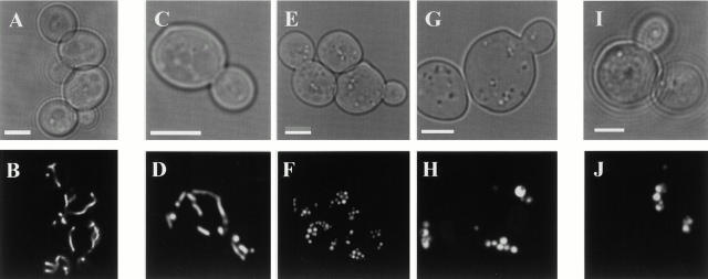
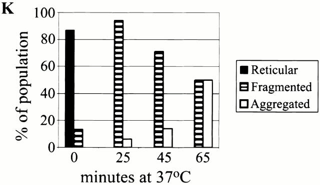
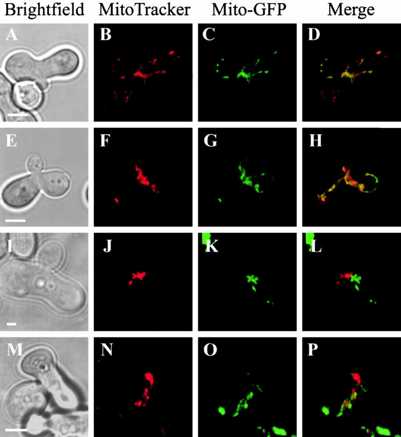
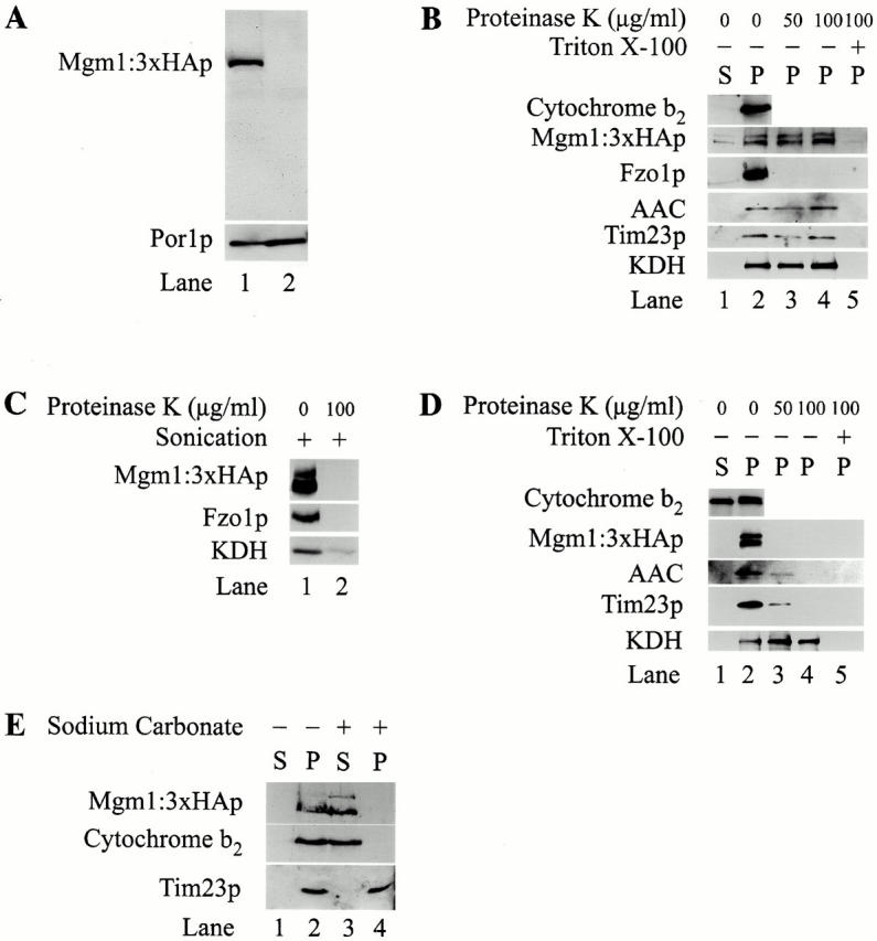
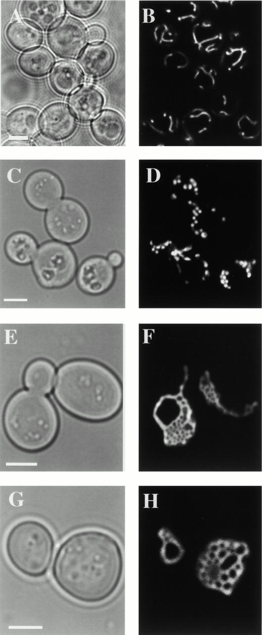
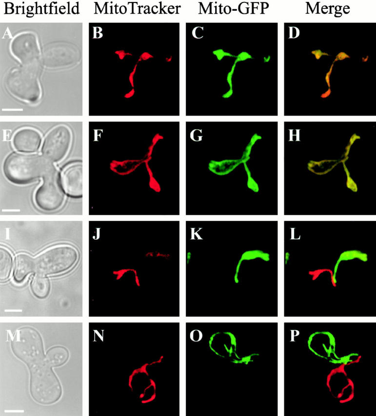
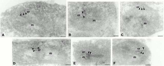
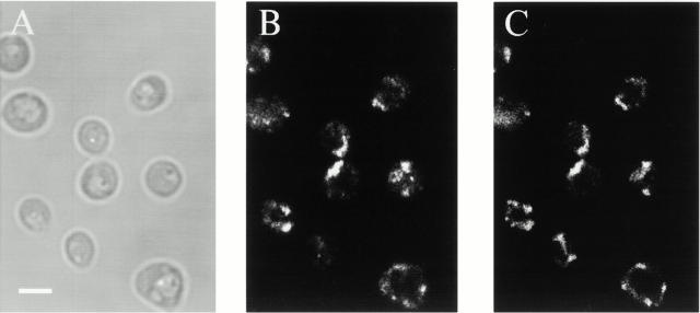
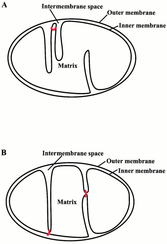
Mutations in the dynamin-related GTPase, Mgm1p, have been shown to cause mitochondrial aggregation and mitochondrial DNA loss in Saccharomyces cerevisiae cells, but Mgm1p's exact role in mitochondrial maintenance is unclear. To study the primary function of MGM1, we characterized new temperature sensitive MGM1 alleles. Examination of mitochondrial morphology in mgm1 cells indicates that fragmentation of mitochondrial reticuli is the primary phenotype associated with loss of MGM1 function, with secondary aggregation of mitochondrial fragments. This mgm1 phenotype is identical to that observed in cells with a conditional mutation in FZO1, which encodes a transmembrane GTPase required for mitochondrial fusion, raising the possibility that Mgm1p is also required for fusion. Consistent with this idea, mitochondrial fusion is blocked in mgm1 cells during mating, and deletion of DNM1, which encodes a dynamin-related GTPase required for mitochondrial fission, blocks mitochondrial fragmentation in mgm1 cells. However, in contrast to fzo1 cells, deletion of DNM1 in mgm1 cells restores mitochondrial fusion during mating. This last observation indicates that despite the phenotypic similarities observed between mgm1 and fzo1 cells, MGM1 does not play a direct role in mitochondrial fusion. Although Mgm1p was recently reported to localize to the mitochondrial outer membrane, our studies indicate that Mgm1p is localized to the mitochondrial intermembrane space. Based on our localization data and Mgm1p's structural homology to dynamin, we postulate that it functions in inner membrane remodeling events. In this context, the observed mgm1 phenotypes suggest that inner and outer membrane fission is coupled and that loss of MGM1 function may stimulate Dnm1p-dependent outer membrane fission, resulting in the formation of mitochondrial fragments that are structurally incompetent for fusion.


Figures

Figure 1
Figure 1
The primary phenotype resulting from loss of MGM1 function is fragmentation of mitochondrial reticuli. Mito-GFP was used to visualize mitochondria in wild-type (A and B), mgm1-5 (C and D), and Δmgm1 (I and J) cells grown overnight at 25°C to log phase, and after shifting to 37°C for 25 min (E and F) or 65 min (G and H). Quantification of mitochondrial morphology phenotypes at 37°C (K). Bars, 2 μm.
Figure 1
Figure 1
The primary phenotype resulting from loss of MGM1 function is fragmentation of mitochondrial reticuli. Mito-GFP was used to visualize mitochondria in wild-type (A and B), mgm1-5 (C and D), and Δmgm1 (I and J) cells grown overnight at 25°C to log phase, and after shifting to 37°C for 25 min (E and F) or 65 min (G and H). Quantification of mitochondrial morphology phenotypes at 37°C (K). Bars, 2 μm.
Figure 2
Figure 2
Mitochondrial fusion is blocked in mgm1-5 cells during mating. Cells of opposite mating type were grown to log phase at 25°C, labeled with either mito-GFP or MitoTracker and mated at 25°C (E–H) or 37°C (A–D, I–P). Mitochondrial fusion was assessed by examining in merged mito-GFP and MitoTracker images of large-budded homozygous zygotes formed from wild-type (A–D), mgm1-5 (E–L), and fzo1-1 (M–P) cells. Bars, 2 μm.
Figure 5
Figure 5
Mgm1p is a mitochondrial intermembrane space protein peripherally associated with the inner membrane. Whole cell extracts of JSY836 and JSY2519 were prepared as described in Materials and Methods and analyzed by SDS-PAGE and Western blotting with indicated antibodies (A). Subcellular fractions of enriched intact mitochondria were isolated, treated with PK after mock-treatment (B), sonication (C), or hypoosmotic shock (D) and analyzed by SDS-PAGE and Western blotting as described. E, Intact mitochondria were treated with 0.1 M sodium carbonate (pH 10.5) as described and fractions were analyzed by SDS-PAGE and Western blotting.
Figure 3
Figure 3
Deletion of DNM1 blocks mitochondrial fragmentation in mgm1-5 cells. Cells were grown to log phase at 25°C and shifted to 37°C for 40 min. Mitochondria were visualized using mito-GFP by fluorescence confocal microscopy in wild-type (A and B), mgm1-5 (C and D), Δdnm1 (E and F), and mgm1-5Δdnm1 (G and H) cells. Bars, 2 μm.
Figure 4
Figure 4
Mitochondrial fusion does not require MGM1 function. Mitochondrial fusion was assessed as described in Fig. 2. Mitochondria in homozygous zygotes formed at 37°C from Δdnm1 (A–D), mgm1-5 Δdnm1 (E–H), fzo1-1 Δdnm1 (I–L), and mgm1-5 fzo1-1 Δdnm1 (M–P) cells were visualized by fluorescence confocal microscopy. Bars, 2 μm.
Figure 7
Figure 7
Mgm1p is associated with the inner membrane. JSY2519 cells were grown in YPD to log phase and processed for cryoimmunoelectron microscopy using anti-HA. Mitochondrial profiles (m) from JSY2519 cells are shown (A–F). Arrows indicate gold particle labeling and critae decorated by gold particles are indicated (cr). Bars, 0.1 μM.
Figure 6
Figure 6
Mgm1:3xHAp localizes to mitochondria in vivo. JSY2519 cells were grown in YPD to log phase, labeled with MitoTracker (B), processed for indirect immunofluorescence with anti-HA (C), and were imaged using fluorescence confocal microscopy as described. Bar, 2 μm.
Figure 8
Figure 8
Models for Mgm1p function in mitochondrial inner membrane remodeling events. Mgm1p oligomeric ring structures are depicted in red. Mgm1p may help form and/or stabilize inner membrane cristae (A and B) or regulate inner membrane fission (B). See Discussion for details.

Comment in

Similar articles

Cited by

References

    1. Beech P.L., Nheu T., Schultz T., Herbert S., Lithgow T., Gilson P.R., McFadden G.I. Mitochondrial FtsZ in a chromophyte alga. Science. 2000;287:1276–1279. - PubMed
    1. Bleazard W., McCaffery J.M., King E.J., Bale S., Mozdy A., Tieu Q., Nunnari J., Shaw J.M. The dynamin-related GTPases, Dnm1, regulates mitochondrial fission in yeast. Nat. Cell Biol. 1999;1:298–304. - PMC - PubMed
    1. Erickson H.P. Dynamin and FtsZmissing links in mitochondrial and bacterial division. J. Cell Biol. 2000;148:1103–1105. - PMC - PubMed
    1. Glick B.S., Brandt A., Cunningham K., Mueller S., Hallberg R.L., Schatz G. Cytochromes c1 and b2 are sorted to the intermembrane space of yeast mitochondria by a stop-transfer mechanism. Cell. 1992;69:809–822. - PubMed
    1. Guan K., Farh L., Marshall T.K., Deschenes R.J. Normal mitochondrial structure and genome maintenance in yeast requires the dynamin-like product of the MGM1 gene. Curr. Genetics. 1993;24:141–148. - PubMed

Publication types

MeSH terms