. 2000 Oct 16;151(2):353-66.
doi: 10.1083/jcb.151.2.353.

Mdv1p is a WD repeat protein that interacts with the dynamin-related GTPase, Dnm1p, to trigger mitochondrial division

Affiliations

Mdv1p is a WD repeat protein that interacts with the dynamin-related GTPase, Dnm1p, to trigger mitochondrial division

Q Tieu et al. J Cell Biol. .

Abstract

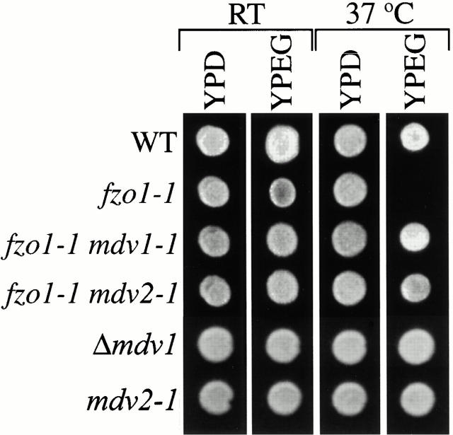
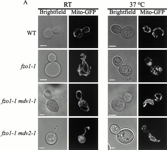
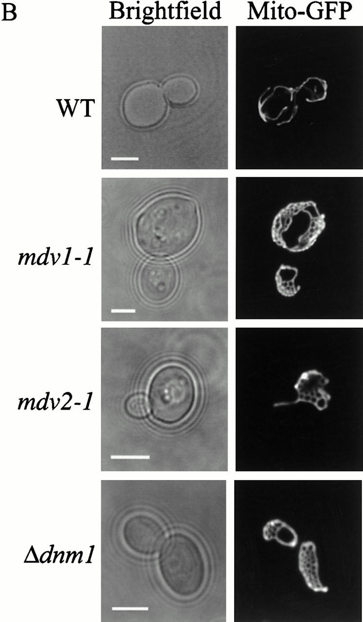
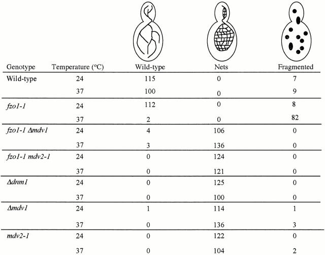
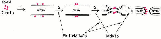
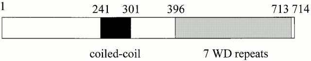
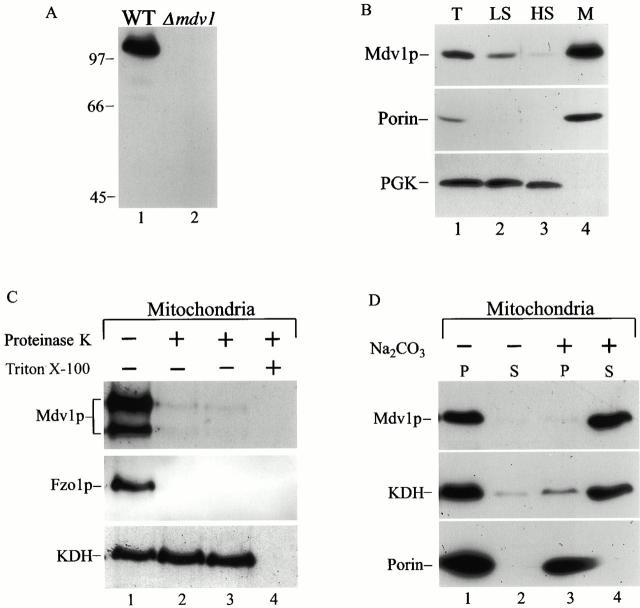
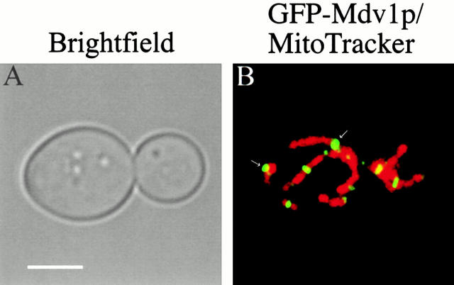
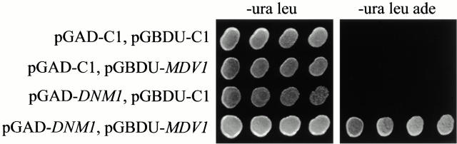
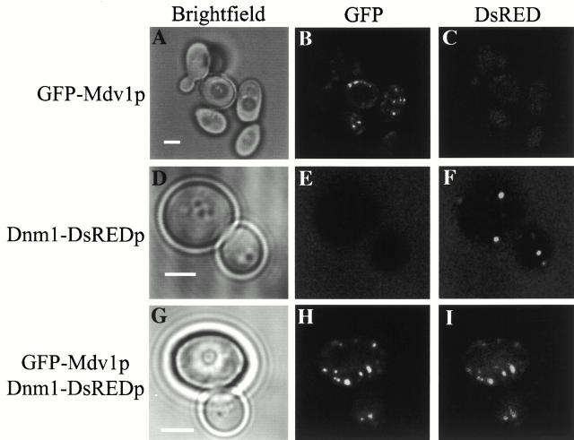
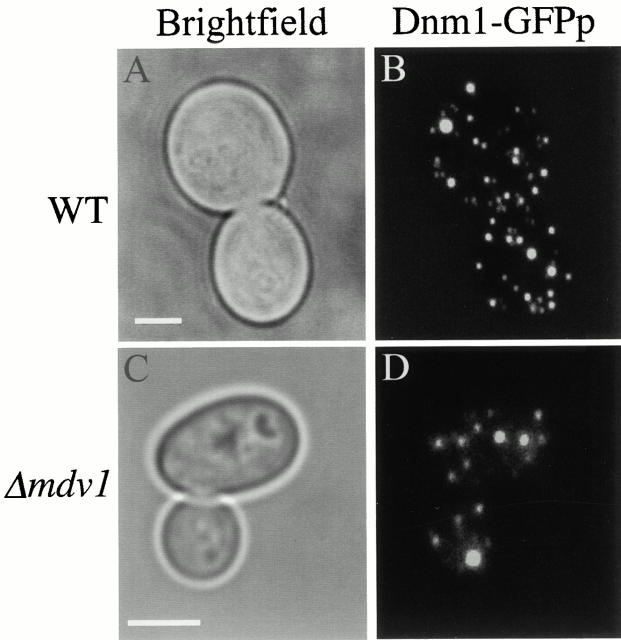
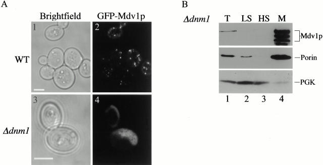
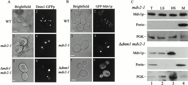
Mitochondrial fission is mediated by the dynamin-related GTPase, Dnm1p, which assembles on the mitochondrial outer membrane into punctate structures associated with sites of membrane constriction and fission. We have identified additional nuclear genes required for mitochondrial fission, termed MDV (for mitochondrial division). MDV1 encodes a predicted soluble protein, containing a coiled-coil motif and seven COOH-terminal WD repeats. Genetic and two-hybrid analyses indicate that Mdv1p interacts with Dnm1p to mediate mitochondrial fission. In addition, Mdv1p colocalizes with Dnm1p in fission-mediating punctate structures on the mitochondrial outer membrane. Whereas localization of Mdv1p to these structures requires Dnm1p, localization of Mdv1p to mitochondrial membranes does not. This indicates that Mdv1p possesses a Dnm1p-independent mitochondrial targeting signal. Dnm1p-independent targeting of Mdv1p to mitochondria requires MDV2. Our data indicate that MDV2 also functions separately to regulate the assembly of Dnm1p into punctate structures. In contrast, Mdv1p is not required for the assembly of Dnm1p, but Dnm1p-containing punctate structures lacking Mdv1p are not able to complete division. Our studies suggest that mitochondrial fission is a multi-step process in which Mdv2p regulates the assembly of Dnm1p into punctate structures and together with Mdv1p functions later during fission to facilitate Dnm1p-dependent mitochondrial membrane constriction and/or division.


Figures

Figure 1
Figure 1
Mutations in MDV genes suppress the loss of mtDNA in fzo1-1 cells. Indicated strains were plated onto YPD and YPG and grown at 25 and 37°C.
Figure 2
Figure 2
Mutations in MDV genes suppress mitochondrial fragmentation in fzo1-1 cells and cause mitochondrial membranes to form net-like structures. Mito-GFP was used to visualize mitochondrial morphology. All strains were grown at 25°C to log phase and imaged at 25°C, or after shifting to 37°C for 1 h. A, Mitochondrial morphology in fzo1-1 mdv double mutants. B, Mitochondrial morphology in mdv single mutants. Bars, 2 μm.
Figure 2
Figure 2
Mutations in MDV genes suppress mitochondrial fragmentation in fzo1-1 cells and cause mitochondrial membranes to form net-like structures. Mito-GFP was used to visualize mitochondrial morphology. All strains were grown at 25°C to log phase and imaged at 25°C, or after shifting to 37°C for 1 h. A, Mitochondrial morphology in fzo1-1 mdv double mutants. B, Mitochondrial morphology in mdv single mutants. Bars, 2 μm.
Figure 3
Figure 3
Quantification of mitochondrial membrane morphology. Mitochondrial membrane was visualized with Mito-GFP.
Figure 12
Figure 12
Schematic model of the mitochondrial fission pathway. Mitochondrial tubules are diagramed in cross-section and red circles represent Dnm1p. Mitochondrial fission is initiated by the recruitment of Dnm1p from the cytosol to the mitochondrial membrane by an unknown regulatory mechanism (1). In a Fis1p-dependent regulated manner, mitochondrial-associated Dnm1p subsequently assembles into higher order structures that possibly form rings around the surface of the mitochondrial outer membrane (2). Via a direct interaction with Dnm1p, Mdv1p either coassembles with Dnm1p during the formation of these higher order structures or associates with Dnm1p in these structures after their formation. Mdv1p functions to recruit either Fis1p or a Fis1p-dependent component to the Mdv1p/Dnm1p structure, thereby catalyzing the potentially rate-limiting steps of membrane constriction (3) and membrane division (4).
Figure 4
Figure 4
Mdv1p contains a novel NH2-terminal domain, a central coiled-coil domain, and a COOH-terminal WD repeat domain. The coiled-coil domain (amino acids 241–303) was identified as described (Lupas et al. 1991). The amino acid regions of the WD repeats are predicted to be the following: WD1 396–427, WD2 439–469, WD3 500–529, WD4 564–592, WD5 604–632, WD6 643–672, WD7 685–713.
Figure 5
Figure 5
Mdv1p is peripherally associated with the mitochondrial outer membrane. All samples were prepared and analyzed by SDS-PAGE and Western blot as described in Materials and Methods. A, Specificity of anti-Mdv1p polyclonal antibodies. Total extract was prepared from JSY1826 (wild-type, lane 1) and JNY552 (Δmdv1, lane 2) cells and analyzed as described in Materials and Methods. B, Fractionation of total JSY1826 (wild-type) cell extract by differential centrifugation. T, Total extract (lane 1); LS, low-speed supernatant (lane 2); HS, high speed supernatant (lane 3); and M, high speed, mitochondrial-enriched pellet (lane 4). C, Topology of Mdv1p. Intact mitochondria were either mock-treated (lane 1), or treated with 50 μg/ml Proteinase-K (lane 2), 100 μg/ml Proteinase-K (lane 3), or 100 μg/ml Proteinase-K with 1% Triton-X 100 (lane 4) for 30 min on ice, and processed as described in Materials and Methods. D, Carbonate extraction of high speed mitochondrial-enriched pellet. Intact mitochondria were either mock-treated (lanes 1 and 2), or treated with 100 mM Na2CO3, pH 10.5 (lanes 3 and 4), subjected to ultracentrifugation at 100,000 g for 20 min, and fractionated into supernatant (S) and pellet (P) fractions. Multiple Mdv1p products observed by Western blotting were variable and likely the result of an in vitro proteolysis artifact.
Figure 6
Figure 6
Mdv1p localizes to punctate structures on mitochondria. JNY556 (GAL1GFP-MDV1) cells were grown in SRaf to log phase and GFP–Mdv1p was expressed by shifting cells into SGal media and incubating at 30°C for 1 h. After expression of GFP–Mdv1p (in green/yellow, see arrows), mitochondria in JNY556 (GAL1GFP-MDV1) cells were labeled with MitoTracker (in red). Cells were visualized directly by confocal microscopy as described in Materials and Methods. Bar, 2 μm.
Figure 7
Figure 7
MDV1 interacts with DNM1 by two-hybrid analysis. Interactions between activating domain (AD) and binding domain (BD) two-hybrid vectors were assessed by growth of indicated transformants on SD media lacking leucine (leu), uracil (ura), and adenine (ade), as described in Materials and Methods.
Figure 8
Figure 8
Mdv1p and Dnm1p colocalize in punctate structures in vivo. GFP–Mdv1p was expressed in JNY556 (GAL1GFP-MDV1) and JNY558 (GAL1GFP-MDV1, pRS315-DNM1-GFP) cells as described in Fig. 6. Cells expressing GFP–Mdv1p (A–C), Dnm1–DsREDp (D–F), or GFP–Mdv1p and Dnm1–DsREDp (G–I) were visualized directly by fluorescence confocal microscopy. Bars, 2 μm.
Figure 9
Figure 9
The assembly of Dnm1p into punctate structures does not required MDV1 function. JNY566 (wild-type, pRS315-DNM1-GFP; A and B) and JNY560 (Δ mdv1, pRS315-DNM1-GFP; C and D) cells were grown in YPG to log phase and Dnm1–GFPp was visualized directly by fluorescence confocal microscopy. Bars, 2 μm.
Figure 10
Figure 10
Mdv1p localization pattern is altered in Δdnm1 cells. A, GFP–Mdv1p was expressed in JNY556 (GAL1GFP-MDV1) and JNY570 (GAL1GFP-MDV1 Δdnm1) cells and visualized directly by fluorescence confocal microscopy as described in Fig. 6. B, Cell extract from JSY1371(Δdnm1; lanes 1–4) was fractionated and analyzed as described in Fig. 5. Bars, 2 μm.
Figure 11
Figure 11
MDV2 regulates the assembly of Dnm1p-containing punctate structures and is required for Mdv1p's association with mitochondrial membranes in the absence of Dnm1p. A, Dnm1–GFPp in JSY1863 (wild-type, pRS315-DNM1-GFP; panel 2), JNY561 (mdv2-1, pRS315-DNM1-GFP; panel 4), and JNY562 (Δmdv1 mdv2-1, pRS315-DNM1-GFP; panel 6) cells. B, GFP–Mdv1p in JNY 556 (GAL1GFP-MDV1; panel 2), JNY571 (GAL1GFP-MDV1;mdv2-1; panel 4), and JNY572 (GAL1GFP-MDV1;mdv2-1, Δdnm1; panel 6) cells. C, JNY567 (mdv2-1; top, lanes 1–5) and JNY569 (Δdnm1; bottom, lanes 1–4) cell extracts were fractionated and analyzed as described in Fig. 5.

Comment in

References

    1. Altschul S.F., Schaffer A.A., Zhang J., Zhang Z., Miller W., Lipman D.J. Gapped BLAST and PSI-BLASTa new generation of protein database search programs. Nucleic Acids Res. 1997;25:3389–3402. - PMC - PubMed
    1. Anderson C.W., Baum P.R., Gesteland R.F. Processing of adenovirus-2 induced proteins. J. Virol. 1973;12:241–252. - PMC - PubMed
    1. Bleazard W., McCaffery J.M., King E.J., Bale S., Mozdy A., Tieu Q., Nunnari J., Shaw J.M. The dynamin-related GTPases, Dnm1, regulates mitochondrial fission in yeast. Nat. Cell Biol. 1999;1:298–304. - PMC - PubMed
    1. Cziepluch C., Kordes E., Pujol A., Jauniax J.-C. Sequencing analysis of a 40.2 kb fragment of yeast chromosome X reveals 19 open reading frames including URA2 (5′ end), TRK1, PBS2, SPT10, GCD14, RPE1, PH086, NCA3, ASF1, CCT7, CZF3, two tRNA genes, three remnant delta elements and a Ty4 transposon. Yeast. 1996;12:1471–1474. - PubMed
    1. Gietz R.D., Schiestl R.H. Transforming yeast with DNA. Methods Mol. Cell. Biol. 1994;5:255–269.

Publication types