. 2000 Oct 16;151(2):367-80.
doi: 10.1083/jcb.151.2.367.

Dnm1p GTPase-mediated mitochondrial fission is a multi-step process requiring the novel integral membrane component Fis1p

Affiliations

Dnm1p GTPase-mediated mitochondrial fission is a multi-step process requiring the novel integral membrane component Fis1p

A D Mozdy et al. J Cell Biol. .

Abstract

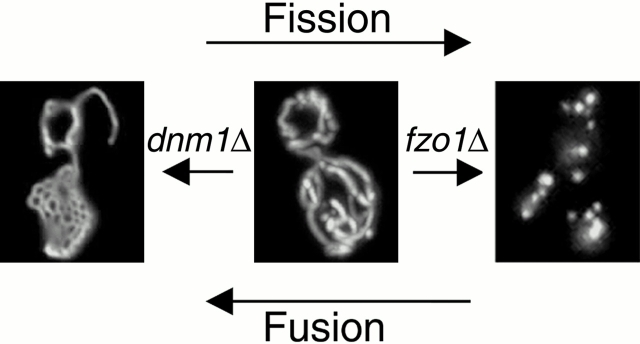
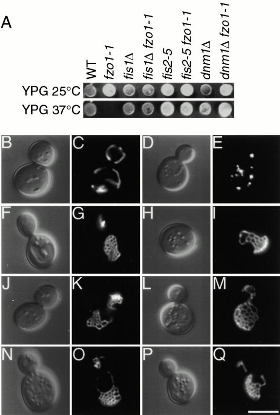
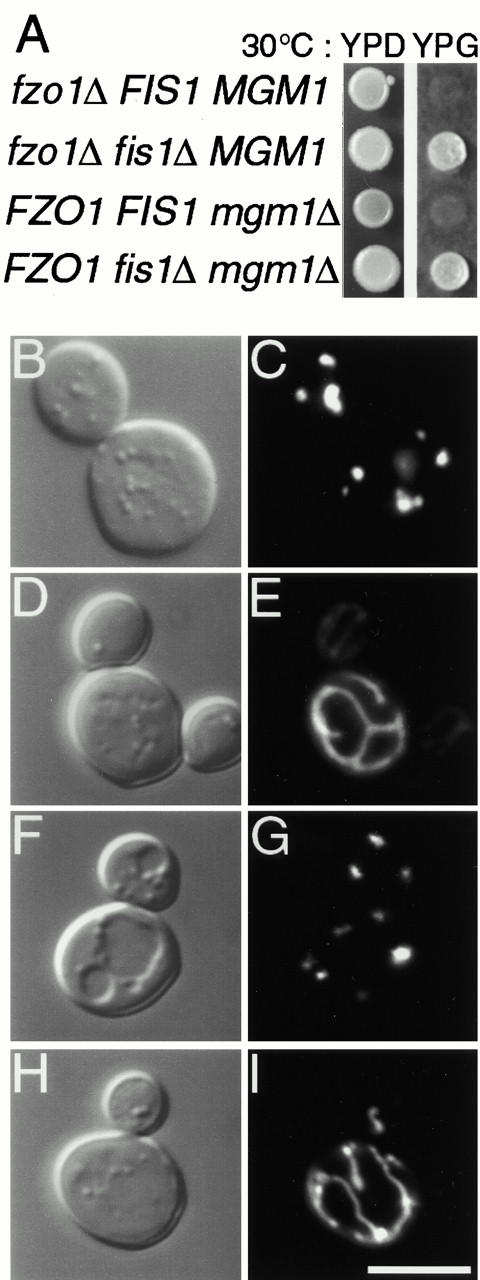
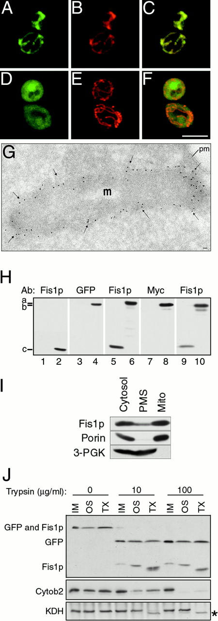
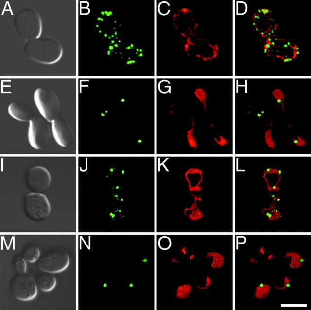
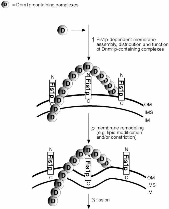
Yeast Dnm1p is a soluble, dynamin-related GTPase that assembles on the outer mitochondrial membrane at sites where organelle division occurs. Although these Dnm1p-containing complexes are thought to trigger constriction and fission, little is known about their composition and assembly, and molecules required for their membrane recruitment have not been isolated. Using a genetic approach, we identified two new genes in the fission pathway, FIS1 and FIS2. FIS1 encodes a novel, outer mitochondrial membrane protein with its amino terminus exposed to the cytoplasm. Fis1p is the first integral membrane protein shown to participate in a eukaryotic membrane fission event. In a related study (Tieu, Q., and J. Nunnari. 2000. J. Cell Biol. 151:353-365), it was shown that the FIS2 gene product (called Mdv1p) colocalizes with Dnm1p on mitochondria. Genetic and morphological evidence indicate that Fis1p, but not Mdv1p, function is required for the proper assembly and distribution of Dnm1p-containing fission complexes on mitochondrial tubules. We propose that mitochondrial fission in yeast is a multi-step process, and that membrane-bound Fis1p is required for the proper assembly, membrane distribution, and function of Dnm1p-containing complexes during fission.


Figures

Figure 1
Figure 1
Dnm1p and Fzo1p act in opposing fission and fusion pathways to maintain the yeast mitochondrial network. Ongoing fission and fusion events maintain a tubular mitochondrial network in wild-type yeast cells (middle). Loss of the Dnm1p GTPase (dnm1Δ) leads to net formation due to unopposed mitochondrial tip fusion (left); loss of the Fzo1p GTPase (fzo1Δ) leads to fragmentation due to unopposed mitochondrial fission (right).
Figure 2
Figure 2
fis1 and fis2 mutations cause mitochondrial net formation and suppress glycerol growth defects and mitochondrial fragmentation in fzo1-1. (A) Wild-type (ADM548), fzo1-1 (ADM547), fis1Δ (ADM549), fis1Δ fzo1-1 (ADM550), fis2-5 (ADM752), fis2-5 fzo1-1 (ADM176), dnm1Δ (ADM379), and dnm1Δ fzo1-1 (ADM378) cells were spotted on YPGlycerol and grown at 25° or 37°C for 5 d. The presence/absence of mtDNA nucleoids in each strain was evaluated by DAPI staining (not shown). (B–Q) Morphology of mito-GFP–labeled (pVT100UGFP) mitochondria in (C) wild-type, (E) fzo1-1, (G) fis1Δ, (I) fis1Δ fzo1-1, (K) fis2-5, (M) fis2-5 fzo1-1, (O) dnm1Δ, and (Q) dnm1Δ fzo1-1 cells grown at 37°C. The corresponding differential interference contrast images are shown in B, D, F, H, J, L, N, and P. Bar, 5 μm.
Figure 3
Figure 3
fis1Δ acts as a bypass suppressor of mitochondrial fragmentation defects. (A) fzo1Δ FIS1 MGM1 (ADM575), fzo1Δ fis1Δ MGM1 (ADM574), FZO1 FIS1 mgm1Δ (ADM764), and FZO1 fis1Δ mgm1Δ (ADM763) strains were spotted on YPDextrose or YPGlycerol and grown at 30°C for 5 d, respectively. Morphology of mito-GFP-labeled (pVT100UGFP) mitochondria in (C) fzo1Δ FIS1 MGM1, (E) fzo1Δ fis1Δ MGM1, (G) FZO1 FIS1 mgm1Δ, and (I) FZO1 fis1Δ mgm1Δ cells grown at 30°C. The corresponding differential interference contrast images are shown in B, D, F, and H. Bar, 5 μm.
Figure 5
Figure 5
Fis1p is a mitochondrial membrane protein with its amino terminus exposed to the cytoplasm. (A–F) Wild-type (ADM551) cells expressing GFP-Fis1p (A–C; pRS415MET25 + rsGFP-FIS1 aa1-155) or GFP-Fis1p lacking the COOH-terminal transmembrane domain (D–F; pRS415MET25 + rsGFP-FIS1 aa1-127) were stained with MitoTracker red CMXRos (B, C, E, and F). Confocal microscopy was used to evaluate the distribution of the GFP fusion proteins (green) and mitochondrial compartments (red) in individual (A, B, D, and E) and merged (C and F) images. Bar, 5μm. (G) The distribution of Myc-Fis1p (black arrows) (ADM551; pRS415MET25 + 9xMYC-FIS1) was determined by immunogold labeling of ultrathin cryosections. 5-nm gold particles were found concentrated at the periphery of the mitochondrial compartment. Percentage distribution of 1,807 gold particles counted: mitochondrial tips 8.5%, mitochondrial sides (excluding constriction sites) 77.6%, mitochondrial constriction sites 12.5%, and cytoplasm 1.4%. m, mitochondria; pm, plasma membrane. Bar, 0.1 μm. (H) Total cell extracts from fis1Δ (lane 1, ADM549), wild-type (lane 2, ADM548), or fis1Δ cells expressing either Fis1p (lanes 3, 5, 7, and 9, ADM549; pRS416 + FIS1), GFP-Fis1p (lanes 4 and 6, ADM549; pRS415MET25 + rsGFP-FIS1) or Myc-Fis1p proteins (lanes 8 and 10, ADM549; pRS415MET25 + 9xMYC-FIS1) were analyzed by Western blotting with anti–Fis1p, –GFP, and –Myc serum as indicated. a, the position of GFP-Fis1p (47 kD); b, Myc-Fis1p (35 kD); and c, native Fis1p (17 kD). (I) Wild-type protein extract was fractionated by differential centrifugation, separated by SDS-PAGE, and analyzed by Western blotting with anti–Fis1p, –porin, and –3-PGK serum. Cytosol, postnuclear cytoplasmic extract; PMS, supernatant depleted of mitochondria; Mito, mitochondrial pellet. (J) Untreated (IM), osmotically shocked (OS), and Triton X-100 solubilized (TX) mitochondrial fractions were treated with 0, 10, or 100 μg/ml Trypsin and analyzed by SDS-PAGE and Western blotting with anti–GFP, –Fis1p, –cytochrome b2, and –KDH serum. *Protease-clipped form of KDH.
Figure 4
Figure 4
Domain structure of the Fis1p Family. Domain structure of eight Fis1p family members with the predicted carboxy-terminal transmembrane domain shown in black (TM). The length in amino acids, percent sequence identity, and percent sequence similarity between the Saccharomyces cerevisiae predicted protein (NCBI RefSeq NP012199) and the predicted Schizosaccharomyces pombe (PIR T39328), Caenorhabiditis elegans (T16053 and T16332), Homo sapiens (NP057152), Arabidopsis thaliana (CAB72179.1), Schistosoma mansoni (AAA99799), and Drosophila melanogaster (AAF45343) orthologs are indicated. The D. melanogaster and S. mansoni amino acid sequences may be incomplete. All sequences can be accessed through the NCBI database (http://www.ncbi.nlm.nih.gov). The Jotun Hein program (MegAlign, DNA*) was used to construct pairwise alignments between the S. cerevisiae protein sequence and each Fis1p ortholog.
Figure 6
Figure 6
Dnm1-GFP localization requires FIS1 but not FIS2 function. (A, E, I, and M) differential interference contrast, (B, F, J, and N) Dnm1-GFP (pRS414-DNM1-rsGFP, PRS416-DNM1-rsGFP, or pHS20), (C, G, K, and O) mito-RFP (pRS416-GAL1 + PrF0ATP9-RFP), and (D, H, L, and P) merged Dnm1-GFP/mito-RFP images of (A–D) wild-type (ADM548), (E–H) fis1Δ (ADM552), (I–L) fis2-5 (ADM752), and (M–P) fis1Δ fis2-5 (ADM742) cells grown at 30°C. Bar, 5 μm.
Figure 7
Figure 7
Model for mitochondrial constriction and fission mediated by Dnm1p-containing complexes. In response to an unknown signal(s), fission complexes containing the Dnm1p GTPase and probably Mdv1p are assembled in a Fis1p-dependent manner on the outer mitochondrial membrane (step 1). Dnm1p-containing fission complexes may assemble into higher-order structures, possibly forming rings or collars around the outer surface of mitochondrial tubules, similar to structures formed by dynamin during endocytosis (rings/collars are visualized as punctate structures in intact yeast cells). Dnm1p-containing complexes could play a role in the mechanical constriction and fission of mitochondrial tubules, or, alternatively, these complexes might recruit or activate additional components that mediate constriction and fission (steps 2 and 3). Although both the inner and outer mitochondrial membranes are shown in cross section, Dnm1p-containing fission complexes are predicted to contact only the outer membrane. OM, outer membrane; IMS, intermembrane space; IM, inner membrane.

Comment in

References

    1. Adari H., Lowy D.R., Willumsen B.M., Der C.J., McCormick F. Guanosine triphosphatase activating protein (GAP) interacts with the p21 ras effector binding domain. Science. 1988;240:518–521. - PubMed
    1. Baudin A., Ozier-Kalogeropoulos O., Denouel A., Lacroute F., Cullin C. A simple and efficient method for direct gene deletion in Saccharomyces cerevisiae . Nucleic Acids Res. 1993;21:3329–3330. - PMC - PubMed
    1. Beech P.L., Gilson P.R. FtsZ and organelle division in Protists. Protist. 2000;151:11–16. - PubMed
    1. Beech P.L., Nheu T., Schultz T., Herbert S., Lithgow T., Gilson P.R., McFadden G.I. Mitochondrial FtsZ in a chromophyte alga. Science. 2000;287:1276–1279. - PubMed
    1. Bleazard W., McCaffery J.M., King E.J., Bale S., Mozdy A., Tieu Q., Nunnari J., Shaw J.M. The dynamin-related GTPase Dnm1 regulates mitochondrial fission in yeast. Nat. Cell Biol. 1999;1:298–304. - PMC - PubMed

Publication types