. 2000 Oct 16;151(2):401-12.
doi: 10.1083/jcb.151.2.401.

Transgenic mice expressing a mutant form of loricrin reveal the molecular basis of the skin diseases, Vohwinkel syndrome and progressive symmetric erythrokeratoderma

Affiliations

Transgenic mice expressing a mutant form of loricrin reveal the molecular basis of the skin diseases, Vohwinkel syndrome and progressive symmetric erythrokeratoderma

Y Suga et al. J Cell Biol. .

Abstract

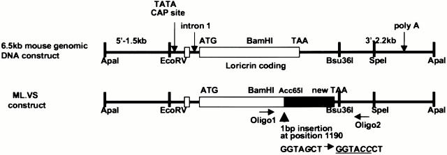
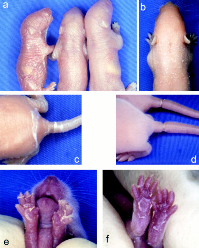
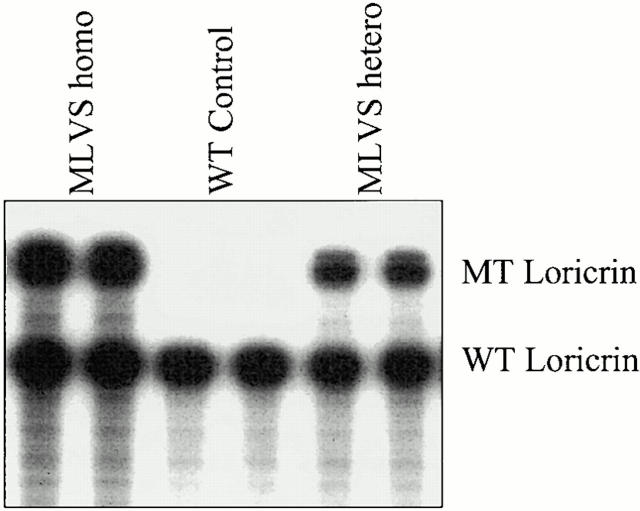
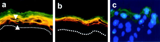
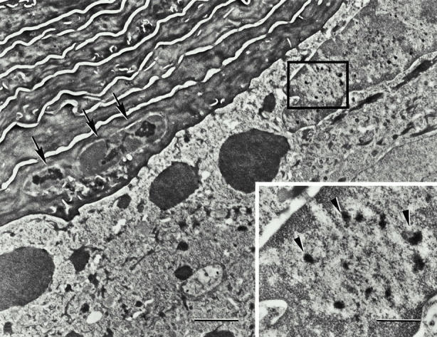
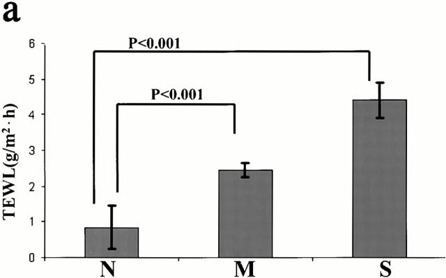
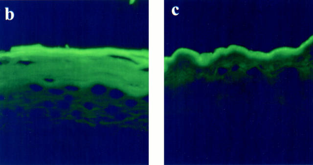
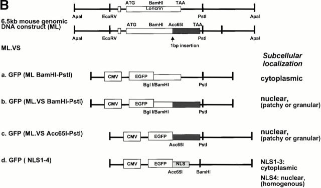
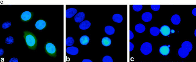
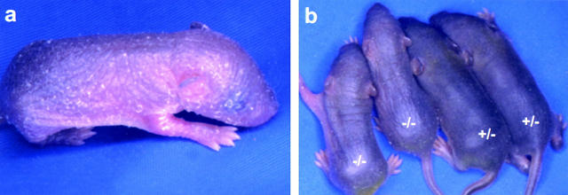
Mutations in the cornified cell envelope protein loricrin have been reported recently in some patients with Vohwinkel syndrome (VS) and progressive symmetric erythrokeratoderma (PSEK). To establish a causative relationship between loricrin mutations and these diseases, we have generated transgenic mice expressing a COOH-terminal truncated form of loricrin that is similar to the protein expressed in VS and PSEK patients. At birth, transgenic mice (ML.VS) exhibited erythrokeratoderma with an epidermal barrier dysfunction. 4 d after birth, high-expressing transgenic animals showed a generalized scaling of the skin, as well as a constricting band encircling the tail and, by day 7, a thickening of the footpads. Histologically, ML. VS transgenic mice also showed retention of nuclei in the stratum corneum, a characteristic feature of VS and PSEK. Immunofluorescence and immunoelectron microscopy showed the mutant loricrin protein in the nucleus and cytoplasm of epidermal keratinocytes, but did not detect the protein in the cornified cell envelope. Transfection experiments indicated that the COOH-terminal domain of the mutant loricrin contains a nuclear localization signal. To determine whether the ML.VS phenotype resulted from dominant-negative interference of the transgene with endogenous loricrin, we mated the ML.VS transgenics with loricrin knockout mice. A severe phenotype was observed in mice that lacked expression of wild-type loricrin. Since loricrin knockout mice are largely asymptomatic (Koch, P.K., P. A. de Viragh, E. Scharer, D. Bundman, M.A. Longley, J. Bickenbach, Y. Kawachi, Y. Suga, Z. Zhou, M. Huber, et al., J. Cell Biol. 151:389-400, this issue), this phenotype may be attributed to expression of the mutant form of loricrin. Thus, deposition of the mutant protein in the nucleus appears to interfere with late stages of epidermal differentiation, resulting in a VS-like phenotype.


Figures

Figure 1
Figure 1
Construction of transgene. Schematic representation of the structures of the ML.VS transgene and the 6.5-kb mouse genomic DNA construct. We introduced a frameshift mutation resulting in a delayed termination of translation (black box) into the second exon (white boxes) of the loricrin gene by inserting a cytosine (C) at position 1190 (arrowhead). An additional Acc65I site was created by the mutation (underlined).
Figure 2
Figure 2
Macroscopic phenotypes of ML.VS transgenic mice. (a) Severely (left) and moderately (middle) affected transgenic neonates, and control (right) littermate F2 pups. Note the erythrokeratoderma with a shiny skin in the transgenic pups. (b) Phenotype of a pup at day 4 after birth. Note the hyperkeratotic, scaly skin, which resembles generalized ichthyosis. (c) Severely affected transgenic mice exhibited hyperkeratotic skin limited to buttocks and tails at day 5 after birth. (d) Hyperkeratotic constricting bands develop at the base of tail at day 7. Note that the distal part of the tail was slightly edematous. (e and f) Front and rear paws of transgenics at day 10. Note the severe scaling of the footpads.
Figure 3
Figure 3
RPAs using RNA from neonatal epidermis. Note the higher expression level of mutant (MT) loricrin transcripts in the severely affected homozygous F2 pups (MLVS homo) compared with the moderately affected heterozygous F2 pups (MLVS hetero). Simultaneous detection of endogenous wild-type (WT) loricrin transcripts provides a loading control.
Figure 4
Figure 4
Immunofluorescence analysis to detect the VS mutant protein. (a and b) Double-immunofluorescent microscopy with LorNAb (FITC) and LorCAb (Texas red) antibodies. LorNAb detects expression of both endogenous and transgenic epitopes, whereas the LorCAb only detects endogenous epitopes. The LorNAb revealed a markedly strong and broad expression in ML.VS transgenics (a) compared with nontransgenics (b). The LorNAb (FITC) also detected immunoreactive granules within the nuclei in the suprabasal layers of ML.VS transgenics (a, arrowheads). Dots mark the dermo–epidermal interface. (c) Nuclear accumulation of the VS mutant protein is more apparent in ML.VS/Lor / neonatal skin (see below) stained with LorNAb and DAPI.
Figure 5
Figure 5
Histological analysis of ML.VS transgenic mice. Hematoxylin and eosin–stained paraffin sections of dorsal skin from transgenic (a) and nontransgenic mice (b). Note the remnants of nuclei present in the stratum corneum of transgenic skin (arrows). (c) Hyperkeratosis and parakeratosis were evident in the cornified cell layers around the constricting band in the transgenic tail. (d) Higher magnification of the cornified layers reveals the retention of nuclei (arrows). (e) Control tail. (f) Hyperkeratosis and parakeratosis were also evident in the transgenic footpad. (g) Higher magnification of f to demonstrate retention of nuclei (arrows).
Figure 6
Figure 6
Ultrastructure of transgenic skin. Electron micrograph of a thin section through the upper epidermis from back skin of a transgenic ML.VS mouse. No gross morphological abnormalities in CE formation are evident in the transgenics, but parakeratotic nuclei are observed in the transitional layer (arrows). Moreover, in the nuclei of granular layer cells, electron-dense granules are visible (arrowheads, bottom right, in the enlargement of boxed area) that are not seen in normal siblings. Bars, 0.5 μm.
Figure 7
Figure 7
Immunoelectron microscopy of transgenic ML.VS mouse skin. Immunogold labeling was performed with LorNAb antibodies and 10-nm gold particles applied to sections of dorsal skin from biopsy samples, prepared according to the method of Tokuyasu 1980. Abnormal granular aggregates observed within parakeratotic corneocyte nuclei (a and b) and granulocyte nuclei (c) labeled positively with LorNAb. The aggregates have a somewhat different visual texture in these preparations compared with Epon embeddings (see Fig. 6). Note that these aggregates (arrows in a and c) do not colocalize with nucleoli. Nu, nucleus; No, nucleolus; Cy, cytoplasm. In contrast, with this antibody, normal skin shows diffuse cytoplasmic staining as well as staining of round L granules in the cytoplasm and nucleus (data not shown, see Steven et al. 1990; Jarnik et al. 1996). Positive labeling of the CE was observed for Lor+/+ transgenics (a), but not for Lor / transgenics (c). These observations imply that for the former mice, the antibody is labeling wild-type loricrin in the cell envelope and the mutant loricrin in the intranuclear aggregates (see Results). In parakeratotic nuclei, we observe characteristic crescent-shaped features of low electron density (a and b, white arrowheads). Their origin is unclear but, to date, we have observed them only in parakeratotic nuclei and in Epon embeddings (data not shown) as well as in Tokuyasu preparations. Bar, 0.5 μm.
Figure 8
Figure 8
Increased BrdU labeling and interfollicular K6 expression in ML.VS transgenic epidermis. BrdU incorporation analysis was used to compare the distribution of S phase nuclei. BrdU labeling (yellow) of transgenic skin (a) shows an increase in mitotic activity versus control (b). Note the additional labeled nuclei in the basal layer in transgenic epidermis. The sections were also double-labeled with K14 (red). Marked interfollicular staining with an antibody to K6 (FITC) is evident in transgenic epidermis (c) compared with control (d). The epidermis was counterstained with antibody to K14 (Texas red). Yellow indicates colocalization at K14 and K6. In both cases, hair follicles stain positive for K6. Note strong interfollicular expression of K6 in transgenic tail from mice at day 7 after birth in addition to the staining of cells in the outer root sheaths.
Figure 9
Figure 9
Defects in the skin barrier function of ML.VS transgenic mice. (a) TEWL was measured in transgenics and normal littermates. Note TEWL was increased more than fourfold in severely affected mice. Error bars represent SEM. Normal (N), n = 20; moderate (M), n = 24; severe (S), n = 12. (b and c) The fluorescent micrographs show the distribution of Lucifer yellow in the skin from severely affected (b) and control mice (c).
Figure 9
Figure 9
Defects in the skin barrier function of ML.VS transgenic mice. (a) TEWL was measured in transgenics and normal littermates. Note TEWL was increased more than fourfold in severely affected mice. Error bars represent SEM. Normal (N), n = 20; moderate (M), n = 24; severe (S), n = 12. (b and c) The fluorescent micrographs show the distribution of Lucifer yellow in the skin from severely affected (b) and control mice (c).
Figure 11
Figure 11
ML.VS loricrin sequence altered GFP localization in HeLa cells. (A) Comparison of wild-type and mutant COOH-terminal loricrin DNA and amino acid sequence resulting from a frameshift mutation and delayed termination of translation. The arrow points to the site of insertion of the point mutation in the ML.VS construct. Some motifs meeting the criteria of NLS in the mutant peptide are shown in bold type and underlined (NLS1–β3). The potential bipartite NLS is boxed, with the relevant basic residues in bold type (NLS4). (B) Diagram illustrating the subcloning of sequences from normal and mutant loricrin COOH terminus into the pEGFP-C vector. BamHI–PstI fragments from wild-type (a) and mutant loricrin constructs (b), respectively, Acc65I–PstI fragment (c), and four nuclear targeting motif candidates (d) from mutant loricrin were fused to the GFP protein. The mutant COOH terminus is represented by a black box. (C) Subcellular localization of GFP. GFP plasmids including the various sequences described above were transfected into HeLa cells. The subcellular localization of the expressed GFP was analyzed 24 h after transfection. GFP fluorescence was detected homogeneously in the cytoplasm of cells transfected with wild-type loricrin COOH terminus (a). However, fluorescence was detected in a patchy or granular pattern in the nuclei of cells transfected with mutant loricrin COOH terminus plasmid GFP(ML.VS.BamHI-PstI) (b) or GFP(ML.VS.Acc65I-PstI) (data not shown). GFP(NLS1), GFP(NLS2), and GFP(NLS3) constructs showed homogeneous distribution in the cytoplasm (data not shown). Only the NLS4 (bipartite NLS) construct was distributed homogeneously in the nuclei (c). Nuclei were visualized by staining with DAPI.
Figure 11
Figure 11
ML.VS loricrin sequence altered GFP localization in HeLa cells. (A) Comparison of wild-type and mutant COOH-terminal loricrin DNA and amino acid sequence resulting from a frameshift mutation and delayed termination of translation. The arrow points to the site of insertion of the point mutation in the ML.VS construct. Some motifs meeting the criteria of NLS in the mutant peptide are shown in bold type and underlined (NLS1–β3). The potential bipartite NLS is boxed, with the relevant basic residues in bold type (NLS4). (B) Diagram illustrating the subcloning of sequences from normal and mutant loricrin COOH terminus into the pEGFP-C vector. BamHI–PstI fragments from wild-type (a) and mutant loricrin constructs (b), respectively, Acc65I–PstI fragment (c), and four nuclear targeting motif candidates (d) from mutant loricrin were fused to the GFP protein. The mutant COOH terminus is represented by a black box. (C) Subcellular localization of GFP. GFP plasmids including the various sequences described above were transfected into HeLa cells. The subcellular localization of the expressed GFP was analyzed 24 h after transfection. GFP fluorescence was detected homogeneously in the cytoplasm of cells transfected with wild-type loricrin COOH terminus (a). However, fluorescence was detected in a patchy or granular pattern in the nuclei of cells transfected with mutant loricrin COOH terminus plasmid GFP(ML.VS.BamHI-PstI) (b) or GFP(ML.VS.Acc65I-PstI) (data not shown). GFP(NLS1), GFP(NLS2), and GFP(NLS3) constructs showed homogeneous distribution in the cytoplasm (data not shown). Only the NLS4 (bipartite NLS) construct was distributed homogeneously in the nuclei (c). Nuclei were visualized by staining with DAPI.
Figure 10
Figure 10
Macroscopic phenotype of ML.VS in the absence of endogenous loricrin (ML.VS Lor /). (a) Newborn transgenics without endogenous loricrin expression seem to have a more scaly and dry skin phenotype than those with endogenous loricrin expression. (b) ML.VS transgenics that are heterozygous (ML.VS Lor+/−) or homozygous (ML.VS Lor /) for the loss of the loricrin gene at day 6. Note that the phenotype of ML.VS Lor / mice was more severe than that of ML.VS Lor+/− mice.

Comment in

References

    1. Armstrong D.K.B., McKenna K.E., Hughes A.E. A novel insertional mutation in loricrin in Vohwinkel's keratoderma. J. Invest. Dermatol. 1998;111:702–704. - PubMed
    1. Boulikas T. Nuclear localization signals (NLS) Crit. Rev. Eukaryot. Gene Expr. 1993;3:193–227. - PubMed
    1. Camisa C., Rossana C. Variant of keratoderma hereditaria mutilans (Vohwinkel's syndrome). Treatment with orally administered isotretinoin. Arch. Dermatol. 1984;120:1323–1328. - PubMed
    1. Dingwall C., Laskey R.A. Nuclear targeting sequence—a consensus? Trends. Biochem. Sci. 1991;16:478–481. - PubMed
    1. DiSepio D., Jones A., Longley M.A., Bundman D., Rothnagel J.A., Roop D.R. The proximal promoter of the mouse loricrin gene contains a functional AP-1 element and directs keratinocyte-specific but not differentiation-specific expression. J. Biol. Chem. 1995;270:10792–10799. - PubMed

Publication types