. 2000 Oct 16;151(2):425-37.
doi: 10.1083/jcb.151.2.425.

The transmembrane domain of influenza hemagglutinin exhibits a stringent length requirement to support the hemifusion to fusion transition

Affiliations

The transmembrane domain of influenza hemagglutinin exhibits a stringent length requirement to support the hemifusion to fusion transition

R T Armstrong et al. J Cell Biol. .

Abstract

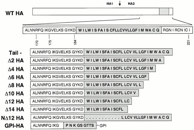
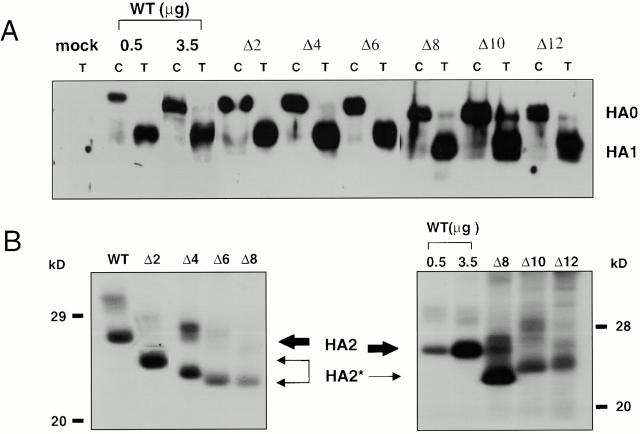
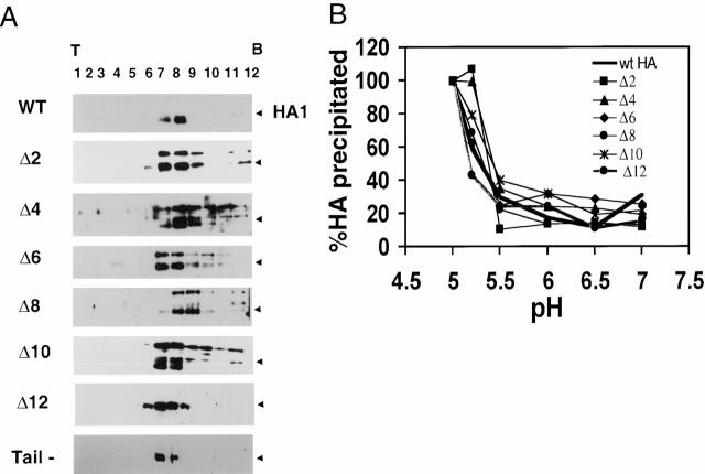
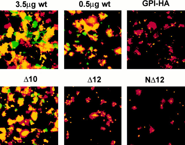
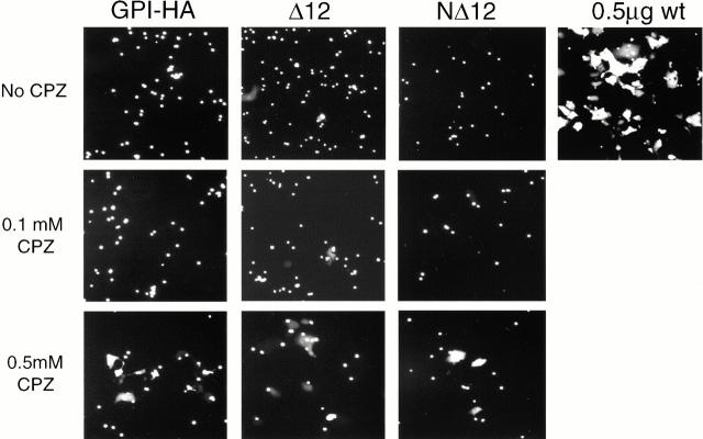
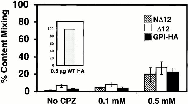
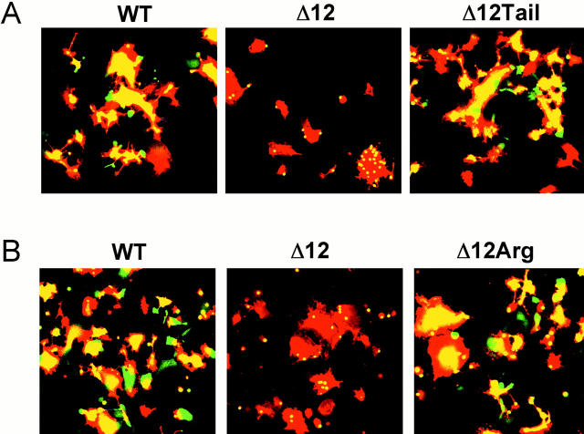
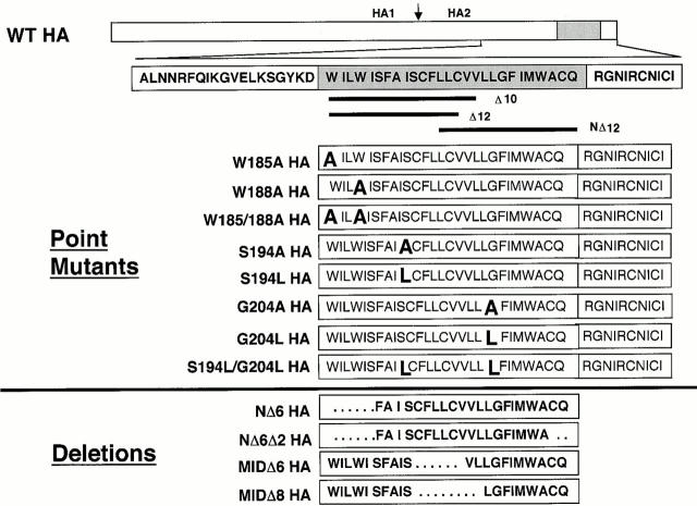
Glycosylphosphatidylinositol-anchored influenza hemagglutinin (GPI-HA) mediates hemifusion, whereas chimeras with foreign transmembrane (TM) domains mediate full fusion. A possible explanation for these observations is that the TM domain must be a critical length in order for HA to promote full fusion. To test this hypothesis, we analyzed biochemical properties and fusion phenotypes of HA with alterations in its 27-amino acid TM domain. Our mutants included sequential 2-amino acid (Delta2-Delta14) and an 11-amino acid deletion from the COOH-terminal end, deletions of 6 or 8 amino acids from the NH(2)-terminal and middle regions, and a deletion of 12 amino acids from the NH(2)-terminal end of the TM domain. We also made several point mutations in the TM domain. All of the mutants except Delta14 were expressed at the cell surface and displayed biochemical properties virtually identical to wild-type HA. All the mutants that were expressed at the cell surface promoted full fusion, with the notable exception of deletions of >10 amino acids. A mutant in which 11 amino acids were deleted was severely impaired in promoting full fusion. Mutants in which 12 amino acids were deleted (from either end) mediated only hemifusion. Hence, a TM domain of 17 amino acids is needed to efficiently promote full fusion. Addition of either the hydrophilic HA cytoplasmic tail sequence or a single arginine to Delta12 HA, the hemifusion mutant that terminates with 15 (hydrophobic) amino acids of the HA TM domain, restored full fusion activity. Our data support a model in which the TM domain must span the bilayer to promote full fusion.


Figures

Figure 1
Figure 1
HA TM domain truncation mutants. Line diagram of the HA gene. The region encompassing the TM domain (gray box) is expanded below. Deletion mutations are shown as sequential removal of two amino acids from the COOH-terminal end of the TM domain (mutants Δ2–14) starting with the tail construct. NΔ12 HA lacks the NH2-terminal 12 amino acids of the TM domain as well as the cytoplasmic tail. GPI-HA has been aligned with the correct ectodomain sequences of the TM truncation mutants.
Figure 2
Figure 2
Processing of HA truncation mutants. (A) Chymotrypsin (C) or trypsin (T) treated CV-1 cells were biotinylated, lysed, immunoprecipitated with the Site A antibody, resolved by SDS-PAGE, transferred to nitrocellulose, and visualized with streptavidin-HRP. Like WT HA, all mutant HAs exhibit efficient processing. (B) CV-1 cells were metabolically labeled, treated with trypsin, lysed, immunoprecipitated with an HA-specific mAb, treated with 1 U of N-Glycosidase F for 2 h at 37°C, and resolved on 15% SDS-PAGE. The gels were dried and exposed to film. Δ2–12 HA exhibit a mobility shift indicative of a truncated HA2 subunit (HA2*).
Figure 3
Figure 3
Biochemical analyses. (A) Sucrose gradient analysis. Transfected CV-1 cells were trypsin treated, lysed, run on 3–30% continuous sucrose gradients, and fractionated as described in Materials and Methods. The samples were precipitated with Con A–agarose, resolved by 10% SDS-PAGE, and analyzed for HA protein by Western blotting. Processed forms of Δ2–12 HA migrate to a similar position on sucrose gradients as the WT HA trimer (black arrowhead). (B) Conformational change assay. Transfected CV-1 cells were metabolically labeled, trypsin treated, incubated at indicated pH values for 10 min at 37°C, reneutralized, and lysed. Cell lysates were then immunoprecipitated with the C-HA1 antibody, which recognizes only the low pH conformation of HA, resolved by 10% SDS-PAGE, and quantitated by PhosphorImager® analysis. The amount of HA precipitated when the total HA precipitated at pH 5 is considered as 100%. Δ2–12 HA change conformation with a pH dependence similar to WT HA. The results presented for WT HA and Δ2–Δ10 are from a typical experiment. The values given for Δ12 HA are the average from three independent experiments.
Figure 4
Figure 4
Quantitation of lipid mixing and content mixing. CV-1 cells expressing WT or mutant HAs were prepared for fusion as indicated in Materials and Methods, except that fusion was triggered at pH 5.0 for 2 min at 37°C and reneutralized. The amount of cDNA used per transfection is indicated. Lipid and content mixing events were averaged from 4–12 random fields (mean ± SEM). Percent lipid dye transfer (hatched bars) was determined by dividing the number of cells receiving lipid dye by the number of cells with bound RBCs in each field. Percent content dye transfer (black bars) was determined by dividing the number of cells receiving content dye by the number of cells with bound RBCs in each field. Relative surface expression of HA in fluorescence units (FU), was obtained by FACS® analyses, and is presented as the mean fluorescence intensity per cell normalized to that of 3.5 μg WT HA cDNA.
Figure 5
Figure 5
Fusion activity: lipid and content mixing. CV-1 cells were transfected with the indicated amounts of WT HA cDNA, 5.0 μg GPI-HA cDNA, or 7.5 μg mutant HA cDNA. The cells were prepared as described in the legend to Fig. 4 and observed (within 15–30 min) by fluorescence microscopy. Δ10 HA (17–amino acid TM domain) mediates full fusion, whereas Δ12 HA and NΔ12 HA (15–amino acid TM domain) are arrested at hemifusion, as is GPI-HA.
Figure 7
Figure 7
Effect of CPZ on content mixing. CV-1 cells expressing GPI-HA, NΔ12, and Δ12 HA cDNA were processed for fusion with CF-labeled RBCs in the absence or presence of the indicated amount of CPZ, as described in the legend to Fig. 6. Only 0.5 mM CPZ is able to promote significant content dye transfer (see Fig. 6).
Figure 6
Figure 6
CPZ induces transfer of aqueous dye (CF) from RBCs to hemifused NΔ12, Δ12, and GPI-HA–expressing cells. CV-1 cells expressing WT and mutant HAs were prepared as described in the legend to Fig. 4, except that fusion was triggered for 5 min at pH 5.0 and 37°C before reneutralization. After triggering fusion, these cells were exposed to either 0.1 or 0.5 mM CPZ for 1 min at room temperature. The CPZ solution was replaced with PBS+ and the cells were observed as above. In control experiments, virtually all WT HA–expressing cells (0.5 μg WT HA, inset graph) were stained with CF in the absence of CPZ. Percent content mixing was determined by dividing the number of cells receiving CF by the number of cells with bound RBCs. Error bars show the SEM for four to five independent experiments (mean ± SEM). Only 0.5 mM CPZ promoted significant content dye transfer between labeled RBCs and cells expressing NΔ12 HA, Δ12 HA, and GPI-HA.
Figure 8
Figure 8
Effect of carbonate extraction and cholesterol depletion on HA TM mutants. (A) Carbonate extraction. Microsomal membranes were prepared, adjusted to pH 11.0, and HA in the supernatant and pellet fractions was prepared, immunoprecipitated with an anti-HA mAb, and detected as described in Materials and Methods. Processed HA (WT and mutant) was only found in the pellet fraction, indicating that it is not extracted by high pH. (B) Triton X-100 insolubility. CV-1 cells expressing WT or mutant HAs were incubated in the absence (−) or the presence (+) of the cholesterol-depleting reagent MβCD (20 mM) for 30 min at 37°C. HA was prepared and divided into insoluble and soluble fractions and detected as described in Materials and Methods. The percentage of HA found in the insoluble fraction in the absence or presence of MβCD was determined. Cholesterol depletion by treatment with MβCD increases the Triton X-100 solubility of WT HA and GPI-HA, but not of Δ12 HA (n = 4). (C) Effect of cholesterol depletion on fusion. CV-1 cells expressing WT or mutant HAs were prepared as described in the legend to Fig. 4, depleted of cholesterol as described in B, bound to labeled RBCs, and triggered for fusion. Cholesterol depletion by treatment with MβCD does not affect the fusion phenotype of WT HA, GPI-HA, Δ12 HA, or NΔ12 HA (see Fig. 5).
Figure 10
Figure 10
Addition of the cytoplasmic tail or a single arginine to Δ12 HA restores fusion. CV-1 cells transfected with 0.5 μg WT, 7.5 μg Δ12, 5.0 μg Δ12Tail, and 5.0 μg Δ12Arg were prepared for fusion as described in Materials and Methods. Fusion was triggered at pH 5.0 for 2 min at 37°C. Images presented in A and B are from separate experiments. Δ12Tail and Δ12Arg were expressed at the cell surface at levels comparable to that using 0.5 μg WT HA cDNA. The COOH-terminal sequences of Δ10, Δ11, Δ12, and Δ12Arg HA mutants are: Δ10WILWISFAISCFLLCVV Δ11WILWISFAISCFLLCV Δ12WILWISFAISCFLLC Δ12Arg WILWISFAISCFLLCR.
Figure 9
Figure 9
(A) Model of possible interactions between the Δ12 (and NΔ12) TM domains and membranes: (1) The truncated TM domains of Δ12 and NΔ12 may span a thinned bilayer; (2a) the TM domain may project into, but not span, the bilayer in a perpendicular orientation relative to the membrane surface; (2b) the TM domain may project obliquely into the bilayer; (2c) the TM domain may be anchored at the surface parallel to the lipid bilayer. HA is presented as a monomer for clarification. The TM domain is depicted as an α-helix, but it may adopt other structures. (B) Models of HA and SNARE-mediated hemifusion to fusion transition. Multimers of HA trimers promote hemifusion (mixing of the outer, but not inner leaflets of the lipid bilayer). Subsequent interactions between the fusion peptides (white) and the TM domains (dark gray), either alone or in concert, with the hemifusion diaphragm may promote full fusion. In the case of the SNAREs, the TM domains of the t- (gray) and v-SNARE (black) may perform analogous functions.
Figure 11
Figure 11
Additional TM domain deletion and point mutants. Line diagram of the HA gene. In detail is the region surrounding the HA TM domain (gray box). Point mutations were made in the context of full-length HA and are indicated in large font. Deletion mutations were made in the context of tail HA and are indicated as spaces. Δ10, Δ12, and NΔ12 HA are included as a reference.

Comment in

Similar articles

Cited by

References

    1. Blumenthal R., Sarkar D.P., Durell S., Howard D.E., Morris S.J. Dilation of the influenza hemagglutinin fusion pore revealed by kinetics of individual cell–cell fusion events. J. Cell Biol. 1996;135:63–71. - PMC - PubMed
    1. Bullough P.A., Hughson F.M., Skehel J.J., Wiley D.C. Structure of influenza haemagglutinin at the pH of membrane fusion. Nature. 1994;371:37–43. - PubMed
    1. Carr C.M., Kim P.S. A spring-loaded mechanism for the conformational change of influenza hemagglutinin. Cell. 1993;73:823–832. - PubMed
    1. Chen J., Skehel J.J., Wiley D.C. N- and C-terminal residues combine in the fusion-pH influenza hemagglutinin HA(2) subunit to form an N cap that terminates the triple-stranded coiled coil. Proc. Natl. Acad. Sci. USA. 1999;96:8967–8972. - PMC - PubMed
    1. Chernomordik L.V., Melikyan G.B., Chizmadzhev Y.A. Biomembrane fusiona new concept derived from model studies using two interacting planar lipid bilayers. Biochim. Biophys. Acta. 1987;906:309–352. - PubMed

Publication types

MeSH terms