

Comment
doi: 10.1083/jcb.151.2.f9.
HIV-1 membrane fusion: targets of opportunity
Affiliations
- PMID: 11038194
- PMCID: PMC2192632
- DOI: 10.1083/jcb.151.2.f9
Item in Clipboard
Comment
HIV-1 membrane fusion: targets of opportunity
J Cell Biol.
.
No abstract available
Figures
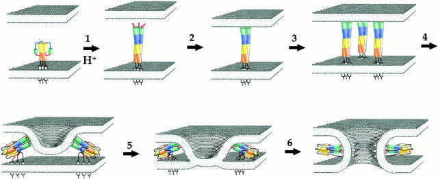
Model for HA-induced membrane fusion. The HA trimer consists of both HA1 (not shown for clarity) and HA2 subunits. The fusion peptides are hidden in the native conformation, being tucked into the trimer interface. Upon acid activation, the fusion peptides are exposed and directed towards the target membrane by a loop to helix transition (blue), thus extending the triple-stranded coiled-coil (1). Insertion of the fusion peptide into the target membrane (2) links the viral and target membranes. Lateral aggregation of several HA trimers (3) is likely needed for a fusion pore to form. Tilting of the HA trimers may be accomplished by folding of the base of triple stranded coiled-coil upon itself, forming a six-helix bundle, as has been seen for other viral fusion proteins (4). This may bring the membranes in close enough proximity such that hemifusion occurs (5). If the HA trimer is GPI-anchored or if the transmembrane domain is not of sufficient length to span the bilayer, the fusion process does not extend past this point. However, if the TM domain is well anchored in the viral membrane, fusion occurs, and, judging by the work by Melikyan et al. 2000(this issue) may be coincident with the completion of the six-helix bundle that brings the TM domain and fusion peptides into close proximity (6).
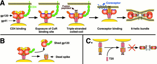
A, Model for HIV-1 Env membrane fusion. Binding of CD4 to the gp120 subunit of Env induces exposure of a conserved region in gp120 implicated in coreceptor binding (purple; Rizzuto et al. 1998). In addition, CD4 binding appears to trigger exposure of the triple-stranded coiled-coil, and presumably exposure of the fusion peptide, although coreceptor binding could increase the efficiency and kinetics of this process. It is not known if the more COOH-terminal helical region in each gp41 subunit (red) interact with each other as drawn, but it is known that the extodomain of gp41 in general plays an important role in mediating Env oligomerization. Binding to coreceptor could bring Env in closer proximity to the target membrane, enabling the fusion peptide to insert in the bilayer, or it could impact formation of the six-helix bundle, the transition to which leads to membrane fusion. Note that in the six-helix bundle, the NH2-terminal helices form the core of the helix, with the COOH-terminal helices packing in the grooves on the outside of the structure. It is not known if gp120 remains associated with gp41 throughout the fusion process. B, Formation of dead spikes. Binding of soluble CD4 to Env can induce shedding of gp120 from gp41, and can even induce formation of the six-helix bundle. A similar process is likely to occur at the cell surface. Such modified Env proteins are not fusogenic, but may serve as immunologic decoys. C, Inhibition of fusion by T20. T20 is a small peptide based on the COOH-terminal helical region in gp41. It binds to the grooves on the outside of the triple-stranded coiled-coil formed by the NH2-terminal helices. Therefore, it prevents transition to the six-helix bundle and membrane fusion. Only gp41 is depicted for clarity.
Comment on
-
Evidence that the transition of HIV-1 gp41 into a six-helix bundle, not the bundle configuration, induces membrane fusion.J Cell Biol. 2000 Oct 16;151(2):413-23. doi: 10.1083/jcb.151.2.413. J Cell Biol. 2000. PMID: 11038187 Free PMC article.
-
The transmembrane domain of influenza hemagglutinin exhibits a stringent length requirement to support the hemifusion to fusion transition.J Cell Biol. 2000 Oct 16;151(2):425-37. doi: 10.1083/jcb.151.2.425. J Cell Biol. 2000. PMID: 11038188 Free PMC article.
References
-
- Bullough P.A., Hughson F.M., Skehel J.J., Wiley D.C. Structure of influenza hemagglutinin at the pH of membrane fusion. Nature. 1994;371:37–43. - PubMed
-
- Chan D.C., Fass D., Berger J.M., Kim P.S. Core structure of gp41 from the HIV envelope glycoprotein. Cell. 1997;89:263–273. - PubMed
-
- Chan D.C., Kim P.S. HIV entry and its inhibition. Cell. 1998;93:681–684. - PubMed
Publication types
MeSH terms
Substances
Grants and funding
LinkOut - more resources
Full Text Sources
Other Literature Sources
