

doi: 10.1073/pnas.94.10.5467.
Expression of early nodulin genes in alfalfa mycorrhizae indicates that signal transduction pathways used in forming arbuscular mycorrhizae and Rhizobium-induced nodules may be conserved
Affiliations
- PMID: 11038545
- PMCID: PMC24702
- DOI: 10.1073/pnas.94.10.5467
Item in Clipboard
Expression of early nodulin genes in alfalfa mycorrhizae indicates that signal transduction pathways used in forming arbuscular mycorrhizae and Rhizobium-induced nodules may be conserved
Proc Natl Acad Sci U S A.
.
Abstract
Transcripts for two genes expressed early in alfalfa nodule development (MsENOD40 and MsENOD2) are found in mycorrhizal roots, but not in noncolonized roots or in roots infected with the fungal pathogen Rhizoctonia solani. These same two early nodulin genes are expressed in uninoculated roots upon application of the cytokinin 6-benzylaminopurine. Correlated with the expression of the two early nodulin genes, we found that mycorrhizal roots contain higher levels of trans-zeatin riboside than nonmycorrhizal roots. These data suggest that there may be conservation of signal transduction pathways between the two symbioses-nitrogen-fixing nodules and phosphate-acquiring mycorrhizae.
Figures
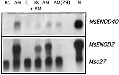
Early nodulin (MsENOD40 and MsENOD2) genes are expressed in roots colonized by AM fungi, but not in those infected by a pathogen only. Rs, R. solani; C, uninoculated control; Rs + AM, inoculated with both R. solani and AM fungi; AM(29), mycorrhizae 29 days after inoculation; N, nitrogen-fixing nodules.
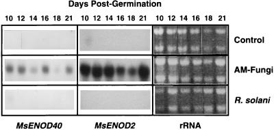
RNA transfer blot showing MsENOD40 and MsENOD2 expression in control, mycorrhizal, and R. solani-infected roots 10–21 days post germination. Approximately 10 μg of RNA was loaded per lane. There were some fluctuations in the amount of RNA loaded in several lanes, as can be seen from the ethidium bromide-stained rRNA bands (rRNA lanes).
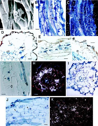
(A) Scanning electron micrograph of an arbuscule in an alfalfa root. (Bar = 5 μm.) (B) Bright-field micrograph of a mycorrhizal root sectioned longitudinally. Arrows indicate arbuscules. The pericycle (p) and stele (s) are indicated. (Bar = 22 μm.) (C) Dark-field micrograph of B illustrating the presence of MsENOD40 transcripts (shown here as white dots representing silver grains) in root cortical cells surround the arbuscules (a) and in the pericycle (arrow), but not in the central part of the stele (arrowhead); 35S-labeled probe. Exposed for 6 weeks. (D) Bright-field micrograph of a transverse section of a young mycorrhizal root; arbuscules are not fully developed. Blue color indicating the presence of MsENOD40 transcript is detected in the pericycle (open arrow), the inner cortical cells (small arrows), and an epidermal cell (arrow); DIG-labeled probe. (Bar = 22 μm.) (E) Off-median longitudinal section through the inner cortex of a root. Arbuscules (small arrows) are in the inner cortical cells, and the blue color indicating the presence of MsENOD40 mRNAs is present in these cells; DIG-labeled probe. (Bar = 44 μm.) (F) Longitudinal section. Blue color is found in the epidermal cells (open arrows) as well as the pericycle (p) and stele (s); DIG-labeled probe. (Bar = 22 μm.) (G) Longitudinal section of a mycorrhizal root taken with Nomarski optics. Blue color indicating MsENOD40 transcript localization is found in the stele and pericycle and in two infected cells (arrowhead). Note that the fully developed arbuscule (open arrow) does not show the blue color; DIG-labeled probe. (Bar = 22 μm.) (H) Dark-field micrograph of a transverse section of mycorrhizal root. Silver grains indicating MsENOD2 mRNAs are clustered over the inner cortical cells, which contain arbuscules (arrows); 35S-labeled probe. Exposed for 3 weeks. (Bar = 22 μm.) (I) Bright-field micrograph of H. Arbuscules (stained purple, arrowheads) are within inner cortical cells. (J) Bright-field micrograph of an off-median longitudinally sectioned mycorrhizal root. The arrowheads indicate the arbuscules. (Bar = 22 μm.) (K) Dark-field view of J. Silver grains indicating MsENOD2 mRNAs are clustered over the arbuscules (arrows); 35S-labeled probe. Exposed for 3 weeks.
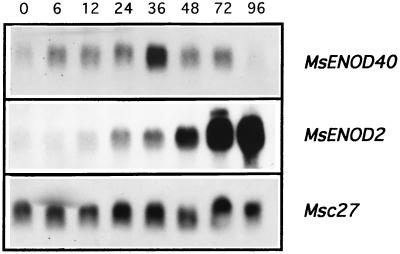
BAP at 10−6 M induces MsENOD40 or MsENOD2 gene expression in uninoculated alfalfa roots harvested at the indicated time points (h). Msc27 was used to standardize RNA levels.
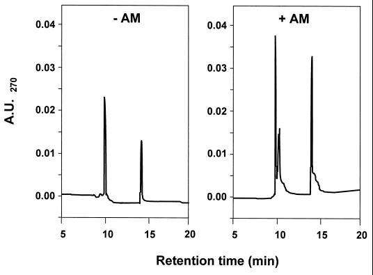
HPLC chromatogram of IAC-purified extract of nonmycorrhizal (− AM) and mycorrhizal (+ AM) alfalfa roots. Samples (0.5 g dry weight) from 18-day-old plants were subjected to the extraction protocol described in the text. Peaks with retention times of 10 and 14 min correspond to the cytokinin standards, zeatin glucoside, and ZR, respectively.
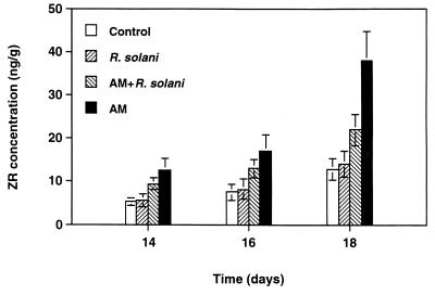
Concentration of ZR (ng/g) over time in noncolonized roots, roots with AM, roots infected with R. solani, and roots with both R. solani and AM fungi.
References
-
- LaRue T A, Weeden N F. In: Proceedings of the 1st European Nitrogen Fixation Conference. Kiss G B, Endre G, editors. Szeged, Hungary: Officina; 1994. pp. 147–151.
-
- Bradbury S M, Peterson R L, Bowley S R. New Phytol. 1993;124:665–673. - PubMed
-
- Duc E, Trouvelot A, Gianinazzi-Pearson V, Gianinazzi S. Plant Sci. 1989;60:215–222.
LinkOut - more resources
Full Text Sources
