. 1997 Jun 24;94(13):6868-73.
doi: 10.1073/pnas.94.13.6868.

RecA-like proteins are components of early meiotic nodules in lily

Affiliations

RecA-like proteins are components of early meiotic nodules in lily

L K Anderson et al. Proc Natl Acad Sci U S A. .

Abstract

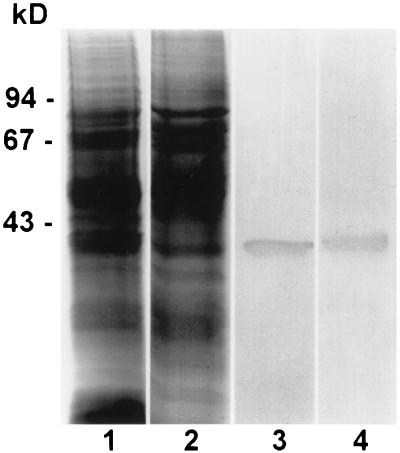
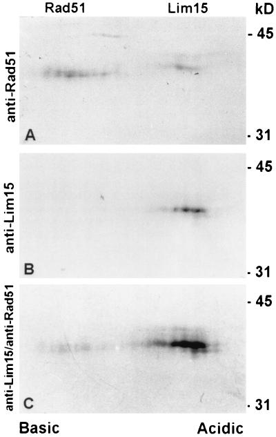
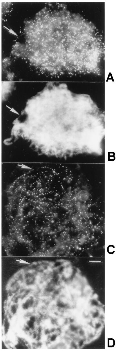
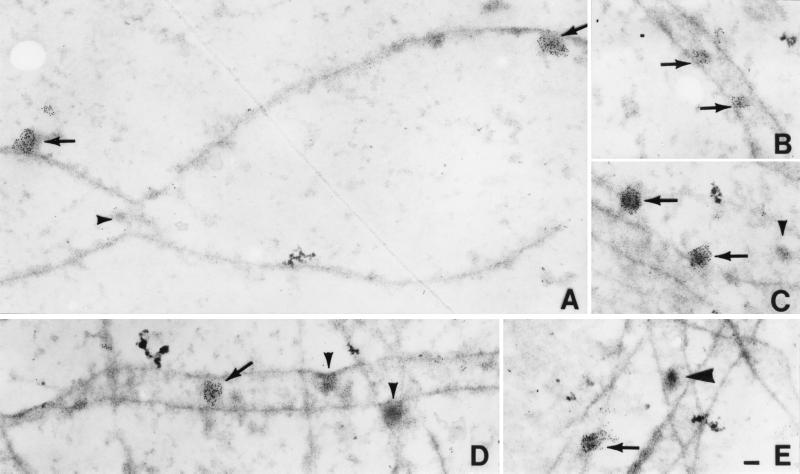
Early meiotic nodules (also called recombination nodules) are proteinaceous structures about 100 nm in diameter that are associated with forming synaptonemal complexes (SCs) during early prophase I of meiosis. Although their function is unknown, early nodules may be involved in searching for DNA homology before synaptic initiation. Two potential components of early nodules are Rad51 and Dmc1 proteins. These proteins are important for meiotic recombination in eukaryotes and are homologous to RecA, the major protein that catalyzes homologous pairing and DNA strand exchange in prokaryotes. In addition, Rad51 has been localized by immunofluorescence in abundant foci that may correspond to early nodules in yeast, lily, and mouse. In yeast and lily, Dmc1 and Lim15, the lily homolog of Dmc1, colocalize with Rad51. Here, using electron microscopic immunogold localization to spreads of zygotene and early pachytene SCs from lily, we confirm that RecA-like proteins are components of early nodules. The antibody used was generated to full-length tomato Rad51 protein and binds to both Rad51 and Lim15 in immunoblots of lily primary microsporocyte proteins. The labeled early nodules are heterogeneous in size and are associated with both axial elements and SCs. There are two classes of early nodules, those that are densely labeled with gold and those that are not labeled at all. This result may be due to technical limitations associated with using spread preparations or to differences in the nodules themselves. The presence of Rad51 and/or Lim15 proteins in early nodules supports the hypothesis that early nodules are involved in recombination-related events during meiosis.


Figures

Figure 1
Figure 1
Extracts of total protein from lily primary microsporocytes at zygonema-pachynema (lanes 1 and 3) and tomato anthers containing primary microsporocytes at zygonema-pachynema (lanes 2 and 4) separated in one dimension and stained with Coomassie blue (lanes 1 and 2) or immunostained with affinity-purified polyclonal antibodies to full-length tomato Rad51 protein (lanes 3 and 4). In each immunoblot, the antibodies bind to a single protein band of approximately 40 kDa. From DNA sequence analysis, the predicted mass of tomato Rad51 protein is about 37 kDa (28). Molecular size standards are indicated on the left in kilodaltons.
Figure 2
Figure 2
Total protein from lily primary microsporocytes separated in two dimensions, blotted, and immunostained with affinity-purified antibodies to Rad51 (A) or antibodies to the nonconserved N terminus of Lim15 (B). (C) The blot from B was subsequently immunostained with affinity-purified antibodies to Rad51. In A, affinity-purified antibodies to Rad51 protein bind to two proteins that migrate at 40 kDa and 41 kDa, which are separated by less than 1 pH unit. In the second blot (B and C), antibodies to Lim15 bind to the more acidic protein and subsequent staining of the same blot with antibodies to Rad51 not only intensifies the stain of the Lim15 protein, but also reveals the more basic Rad51 protein. Based on these results and the predicted molecular masses and pIs for Rad51 (37 kDa; pI = 5.9) and Lim15 (38 kDa; pI = 5.5), the protein on the left corresponds to Rad51 and the protein on the right corresponds to Lim15. Molecular size standards are indicated on the right in kilodaltons.
Figure 3
Figure 3
Squashed lily primary microsporocytes at zygonema (A and B) and early pachynema (C and D). (A and C) Immunofluorescent images using affinity-purified antibodies to Rad51 protein and secondary antibodies conjugated to fluorescein isothiocyanate. (B and D) DAPI images of the corresponding nuclei. Arrows in A and B show that individual foci follow unsynapsed chromosomes, and arrows in C and D show that foci also follow synapsed chromosomes. The squashes are not completely flat, and many Rad51 foci are not in the plane of focus. (Bar = 10 μm.)
Figure 4
Figure 4
Immunogold labeling of spread SCs from lily using affinity-purified antibodies to Rad51 protein and secondary antibodies conjugated to 5-nm gold particles. (A) Portion of two converging axial elements from a mid- to late zygotene nucleus. Two early nodules associated with one axial element are labeled (arrows), but a small nodule, present at the site of convergence, is not labeled (arrowhead). (B) Portion of an SC from a zygotene nucleus with two small labeled early nodules (arrows). (C) An axial element and an SC from a zygotene nucleus with two early nodules that are abundantly labeled (arrows) and a smaller nodule that is not labeled (arrowhead). (D) Portion of a zygotene nucleus with one SC that extends horizontally across the figure and several more or less vertically oriented axial elements. One early nodule is labeled (arrow) and two other early nodules are not labeled (arrowhead). Notice that both labeled and unlabeled nodules are variable in size (compare Fig. 4 AD). (E) Portions of four SCs from an early pachytene nucleus. One early nodule is labeled (arrow), and one late nodule is not labeled (large arrowhead). The latter nodule was identified as a late nodule because of the following combination of characteristics: association with the central region of the SC, the dense stain, and ellipsoidal shape (50 × 100 nm). (Bar = 100 nm.)

References

    1. Moses M. Annu Rev Genet. 1968;2:363–412.
    1. Gillies C B. CRC Crit Rev Plant Sci. 1984;2:81–116.
    1. von Wettstein D, Rasmussen S W, Holm P B. Annu Rev Genet. 1984;18:331–413. - PubMed
    1. Carpenter A T C. Proc Natl Acad Sci USA. 1975;72:3186–3189. - PMC - PubMed
    1. Carpenter A T C. In: Genetic Recombination. Kucherlapati R, Smith G R, editors. Washington, DC: Am. Soc. Microbiol.; 1988. pp. 529–548.