

Review
doi: 10.1136/heart.84.suppl_2.ii2.
Three dimensional colour Doppler echocardiography for the characterisation and quantification of cardiac flow events
Affiliations
- PMID: 11040028
- PMCID: PMC1766541
- DOI: 10.1136/heart.84.suppl_2.ii2
Item in Clipboard
Review
Three dimensional colour Doppler echocardiography for the characterisation and quantification of cardiac flow events
Heart.
2000 Nov.
No abstract available
Figures
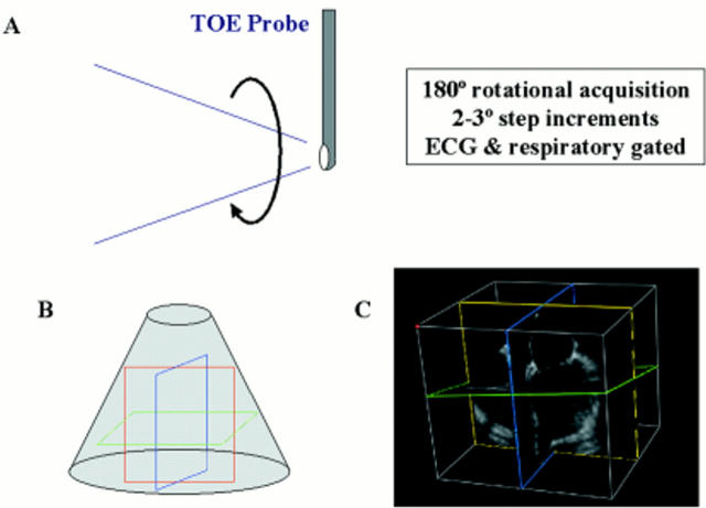
(A) Acquisition of multiple 2D images by rotation of multiplane transoesophageal probe through 180°. (B) Assimilation of multiple 2D imaging planes to generate a 3D volume of data. Coloured lines show typical cutplanes within the dataset. (C) Example of 3D dataset with demonstration of cutplanes.
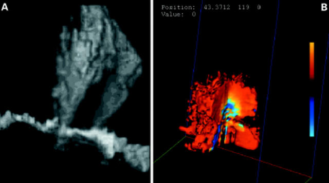
(A) Grey scale reconstruction of twin regurgitant jets extending upwards into the cavity of the left atrium. (B) Digital 3D colour Doppler rendering of a flow convergence region (in vitro acquisition).
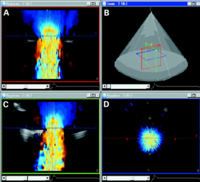
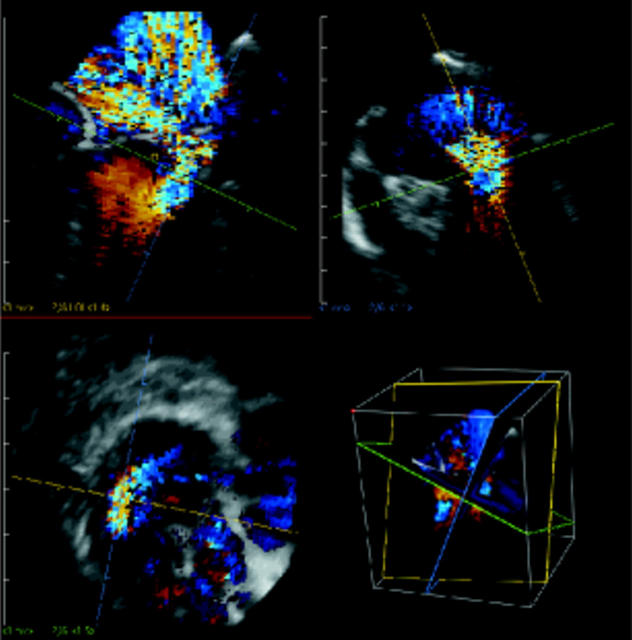
Localisation of the vena contracta in the 3D dataset (in vitro acquisition). Colour Doppler flow data have been acquired parallel to flow. (A) and (C) show direction of flow from top to bottom in the two. (D) A perpendicular (blue cutplane) cut through the narrowest portion of the regurgitant jet, the vena contracta.
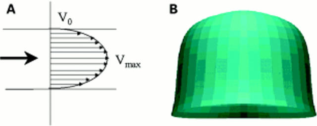
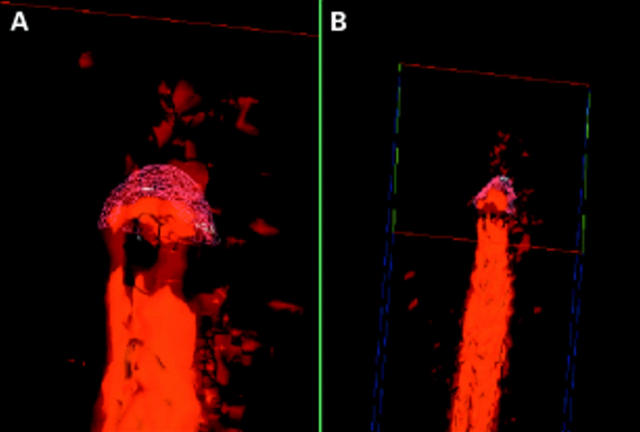
(A) Example of laminar flow pattern, such as might be encountered in the ventricular outflow tracts or the great vessels. (B) 3D reconstruction of a laminar flow profile (in vitro acquisition).
Identification of the vena contracta in a case of anterior mitral valve leaflet prolapse. In spite of the eccentricity of the jet, the cutplanes can still be manipulated to produce a cross section of the vena contracta (bottom left panel). It is then a simple process to measure its area. This should give an approximation of the regurgitant orifice area.
Calculation of the flow convergence region surface area in the 3D dataset. (A) is a magnified view of (B). A wire model framework has been fitted to the surface of the flow convergence region. Surface area computations are made using this model. Note that no geometric assumptions are involved in the measurement of the surface area.
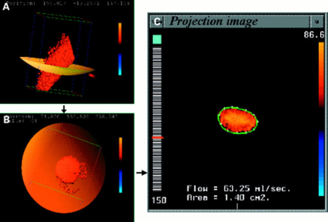
Computation of instantaneous flow rate from a digital 3D Doppler dataset, acquired from an in vitro tube model of laminar flow. The colour flow cross section is identified within the dataset (A), (B). The computer performs a spatial integration of the individual velocity vectors contained within the flow cross-section (C) to generate an instantaneous flow rate.
Similar articles
-
Three-dimensional echocardiography of colour Doppler flow.Arch Cardiovasc Dis. 2010 May;103(5):333-9. doi: 10.1016/j.acvd.2010.01.007. Epub 2010 Jun 23. Arch Cardiovasc Dis. 2010. PMID: 20619244 Review.
-
Current status of three-dimensional color flow Doppler.Cardiol Clin. 2007 May;25(2):297-303. doi: 10.1016/j.ccl.2007.06.006. Cardiol Clin. 2007. PMID: 17765109 Review.
-
Evaluation of a new 3-dimensional color Doppler flow method to quantify flow across the mitral valve and in the left ventricular outflow tract: an in vitro study.J Ultrasound Med. 2014 Feb;33(2):265-71. doi: 10.7863/ultra.33.2.265. J Ultrasound Med. 2014. PMID: 24449729
-
Anterior mitral valve perforation in the absence of acute infection: Diagnosis by two-dimensional and three-dimensional transesophageal echocardiography.Echocardiography. 2017 Dec;34(12):1953-1955. doi: 10.1111/echo.13734. Epub 2017 Oct 26. Echocardiography. 2017. PMID: 29071745
-
Real-time three-dimensional color Doppler echocardiography overcomes the inaccuracies of spectral Doppler for stroke volume calculation.J Am Soc Echocardiogr. 2006 Nov;19(11):1403-10. doi: 10.1016/j.echo.2006.05.010. J Am Soc Echocardiogr. 2006. PMID: 17098150 Review.
Cited by
-
Echocardiographic assessment after surgical repair of tetralogy of fallot.Front Pediatr. 2015 Feb 2;3:3. doi: 10.3389/fped.2015.00003. eCollection 2015. Front Pediatr. 2015. PMID: 25699243 Free PMC article. Review.
-
Quantitative Doppler-echocardiographic determination of regurgitant volume in patients with aortic insufficiency.Open Cardiovasc Med J. 2008 Mar 4;2:12-9. doi: 10.2174/1874192400802010012. Open Cardiovasc Med J. 2008. PMID: 19590613 Free PMC article.
References
Publication types
MeSH terms
LinkOut - more resources
Full Text Sources