. 2000 Oct 15;14(20):2587-95.
doi: 10.1101/gad.834100.

Dmrt1, a gene related to worm and fly sexual regulators, is required for mammalian testis differentiation

Affiliations

Dmrt1, a gene related to worm and fly sexual regulators, is required for mammalian testis differentiation

C S Raymond et al. Genes Dev. .

Abstract

The only molecular similarity in sex determination found so far among phyla is between the Drosophila doublesex (dsx) and Caenorhabditis elegans mab-3 genes. dsx and mab-3 contain a zinc finger-like DNA-binding motif called the DM domain, perform several related regulatory functions, and are at least partially interchangeable in vivo. A DM domain gene called Dmrt1 has been implicated in male gonad development in a variety of vertebrates, on the basis of embryonic expression and chromosomal location. Such evidence is highly suggestive of a conserved role(s) for Dmrt1 in vertebrate sexual development, but there has been no functional analysis of this gene in any species. Here we show that murine Dmrt1 is essential for postnatal testis differentiation, with mutant phenotypes similar to those caused by human chromosome 9p deletions that remove the gene. As in the case of 9p deletions, Dmrt1 is dispensable for ovary development in the mouse. Thus, as in invertebrates, a DM domain gene regulates vertebrate male development.


Figures

Figure 1
Figure 1
Strategy for producing the Dmrt1 deletion mutant. (A) Diagram of Dmrt1 wild-type and mutant alleles. Homologous recombination of the targeting vector with the wild-type Dmrt1 allele (Dmrt1+) resulted in the loxP-flanked (floxed) allele Dmrt1neo. This allele also contains within the first intron a neomycin-resistance cassette (Pgk–neo) flanked by Flp recombinase recognition sites (frt sites). Mice heterozygous for the Dmrt1neo allele were mated with transgenic mice expressing Cre recombinase under the control of a β-actin promoter (Lewandoski et al. 1997), resulting in deletion of the sequences between the two loxP sites, including the major transcriptional start, the first exon (containing the DM domain), Pgk–neo, and part of the first intron. The resulting deletion allele is called Dmrt1. (B) Genotyping of Dmrt1 alleles. PCR of genomic DNA from wild-type Dmrt1+/+, heterozygous Dmrt1+/− and homozygous Dmrt1−/− animals is shown. For each animal, PCR reactions were run to test for each allele (CR92/CR99 to detect Dmrt1+, and KOS1/KOS3 to detect Dmrt1), and the products were mixed and resolved by gel electrophoresis. (C) RT–PCR of Dmrt1 mRNA. RT–PCR of E13.5 testis mRNA was performed using primers from exons 3 and 4, 3′ to the region deleted by the Dmrt1 mutation (CR132/CR133; see Materials and Methods). Hprt mRNA is a positive control.
Figure 2
Figure 2
Dmrt1 mutant testes are severely dysmorphic. (A) Adult testis size is reduced in Dmrt1−/− testes (two large testes at left) relative to Dmrt1+/− (four small testes at right). Testes are from 10-month-old adults. (B–D) Sections of Dmrt1+/− and Dmrt1−/− testes stained with hematoxylin and eosin. (B) Adult (16 weeks postpartum) Dmrt1+/− testis, showing normal morphology. (C) Dmrt1−/− testis at 6 wk post partum. Seminiferous tubules are present and contain immature Sertoli cells, but are devoid of germ cells. There are numerous Leydig cells. (D) Example of adult (16 weeks postpartum) Dmrt1−/− testis. Small numbers of seminiferous tubules are separated by moderate numbers of interstitial cells, some undergoing vacuolation and fatty degeneration. There is extensive infiltration by cells that often contain light brown pigment (ceroid/lipofuscin). These are considered to be of macrophage and/or interstitial cell origin. Degenerating seminiferous tubules are attenuated and contain variable numbers of immature Sertoli cells, foamy macrophages, and as yet unidentified large eosinophillic cells, but are devoid of identifiable spermatogonia and spermatids.
Figure 3
Figure 3
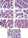
Germ cell death and Sertoli cell over-proliferation in Dmrt1 mutant testes. Sections of Dmrt1+/− and Dmrt1−/− testes of littermates stained with hematoxylin and eosin. (A) P1 Dmrt1+/− testis, showing seminiferous tubules with Sertoli cells (small dark blue nuclei) around periphery, and germ cells (larger round red nuclei; arrowheads) in center. (B) Section of P1 Dmrt1−/− testis, with normal morphology. Germ cells are present in normal numbers (arrowheads). (C) P10 Dmrt1+/− testis. Germ cells have migrated to the periphery (arrowheads). Meiosis has begun and differentiating germ cells (round dark blue nuclei) are visible in some tubules. (D) P10 Dmrt1−/− testis. Sertoli cells have begun to over-proliferate. Germ cells are nearly absent, and the few remaining germ cells have failed to migrate to the periphery of the seminiferous tubules (a few germ cells are visible in this section; two are indicated by arrowheads). No meiosis is detectable. (E) P14 Dmrt1+/− testis, showing meiotic germ cells (round dark blue nuclei) within seminiferous tubules. (F) P14 Dmrt1−/− testis. Immature Sertoli cells are present and have overproliferated, whereas germ cells are absent. (G) P14 c-kitW/W-v mutant testis. Germ cells are absent, as in Dmrt1−/− testis, but differentiated Sertoli cells are present in normal numbers.
Figure 4
Figure 4
Dmrt1 protein expression. Immunofluorescence of testis sections stained with anti-Dmrt1 antibody. (A) E18.5 testis. Expression is detectable in Sertoli cells and not in germ cells. (B) P1 testis. Expression is detectable Sertoli cells (S) and in some germ cells (g). (C) P4 testis. Expression is detectable in Sertoli cells and becoming strong in most germ cells (g). (D) P7 testis. Expression continues in Sertoli and has become strong in all germ cells. (E) P10 testis. Meiosis has begun. Expression is detectable in Sertoli cells and undifferentiated germ cells (spermatogonia) only. (F) P14 testis. All Sertoli cells and all undifferentiated germ cells express Dmrt1. (G) Sixteen-week-old adult testis. All Sertoli cells (S) express Dmrt1, but spermatogonia (g) express Dmrt1 only in regions early in the spermatogenic cycle (bottom tubule but not the top tubule, which is later in the cycle). (H) P14 Dmrt1−/− testis. No Dmrt1 expression is detectable. Brightly autofluorescent interstitial cells in this and other panels are blood cells.
Figure 5
Figure 5
Gata-4 expression persists in Dmrt1−/− mutant Sertoli cells. Immunofluorescence of testis sections stained with Gata-4 antibody. Genotype and developmental stage are indicated. (A) P7 Dmrt1+/− testis. Sertoli cells strongly express Gata-4 and germ cells do not. Many germ cells have migrated to periphery of seminiferous tubules (arrowheads). (B) P7 Dmrt1−/− testis, Gata-4 expression is normal in Sertoli cells. Most germ cells have failed to migrate to the periphery of the seminiferous tubules and remain in the center (arrowheads). (C) P10 Dmrt1+/− testis. Sertoli cells continue to express high levels of Gata-4. All germ cells are at periphery of seminiferous tubules (arrowheads). (D) P10 Dmrt1−/− testis. Sertoli cells express high levels of Gata-4 and have begun to overproliferate, and germ cells are nearly absent. (E) P14 Dmrt1+/− testis. Gata-4 expression has decreased in Sertoli cells. (F) P14 Dmrt1−/− testis. Sertoli cells continue to express high levels of Gata-4 and to proliferate. Germ cells are absent. (G) P14 c-kitW/W-v mutant testis. Sertoli cells have normal morphology (see Fig. 2G) but express high levels of Gata-4, indicating that germ cells are required for down-regulation of Gata-4 in Sertoli cells.
Figure 6
Figure 6
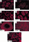
Gata-1 expression is delayed and reduced in Dmrt1 mutant Sertoli cells. Immunofluorescence of testis sections stained with Gata-1 antibody. (A) P10 Dmrt1+/− testis. Most Sertoli cells have begun to express Gata-1. (B) P10 Dmrt1−/− testis. Gata-1 expression is lower than in heterozygous littermate shown in A. (C) P14 Dmrt1+/− testis. Most Sertoli cells express high levels of Gata-1. (D) P14 Dmrt1−/− testis. Most Sertoli cells express some Gata-1, but generally at lower levels than in heterozygous littermate shown in C. (E) Adult (16 weeks post partum) Dmrt1+/− testis. Gata-1 expression is strong in Sertoli cells (brightly staining cells spaced around periphery of seminiferous tubule) and also is present in meiotic germ cells. (F) Six-week postnatal Dmrt1−/− testis. Gata-1 expression has not been maintained, and Sertoli cells express low levels. (Most staining is nonspecific background.) (G) Adult c-kitW/W-v mutant testis. Sertoli cells express high levels of Gata-1, demonstrating that the germ line is not necessary for high-level Gata-1 expression.

References

    1. Baker BS, Ridge K. Sex and the single cell: On the action of major loci affecting sex determination in Drosophila melanogaster. Genetics. 1980;94:383–423. - PMC - PubMed
    1. Bennett CP, Docherty Z, Robb SA, Ramani P, Hawkins JR, Grant D. Deletion 9p and sex reversal. J Med Genet. 1993;30:518–520. - PMC - PubMed
    1. Bitgood MJ, Shen L, McMahon AP. Sertoli cell signaling by Desert hedgehog regulates the male germline. Curr Biol. 1996;6:298–304. - PubMed
    1. Calvari V, Bertini V, De Grandi A, Peverali G, Zuffardi O, Ferguson-Smith M, Knudtzon J, Camerino G, Borsani G, Guioli S. A new submicroscopic deletion that refines the 9p region for sex reversal. Genomics. 2000;65:203–212. - PubMed
    1. Coschigano KT, Wensink PC. Sex-specific transcriptional regulation by male and female doublesex proteins of Drosophila. Genes & Dev. 1993;7:42–54. - PubMed

Publication types

MeSH terms