. 2000 Oct 15;14(20):2610-22.
doi: 10.1101/gad.181700.

Raf induces TGFbeta production while blocking its apoptotic but not invasive responses: a mechanism leading to increased malignancy in epithelial cells

Affiliations

Raf induces TGFbeta production while blocking its apoptotic but not invasive responses: a mechanism leading to increased malignancy in epithelial cells

K Lehmann et al. Genes Dev. .

Abstract

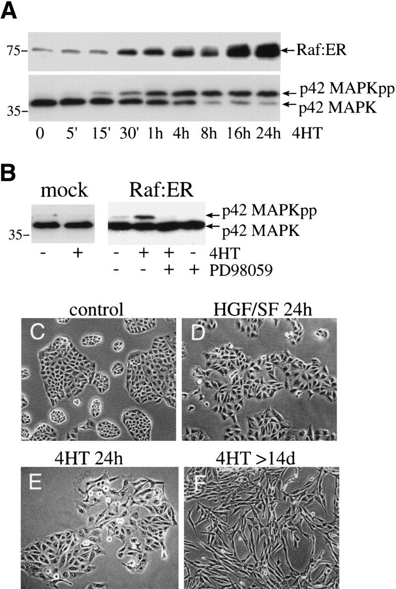
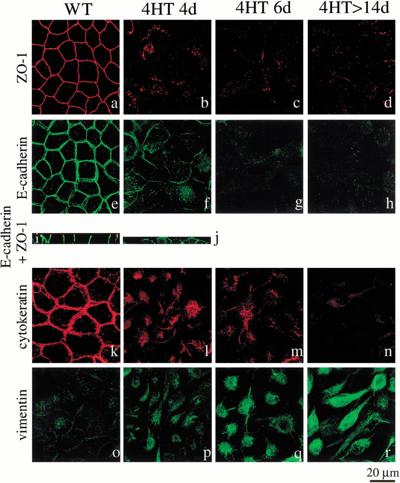
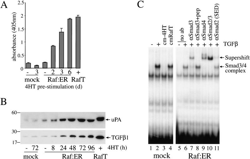
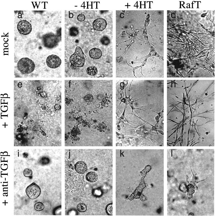
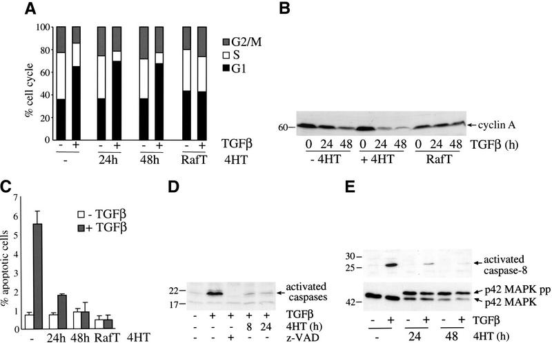
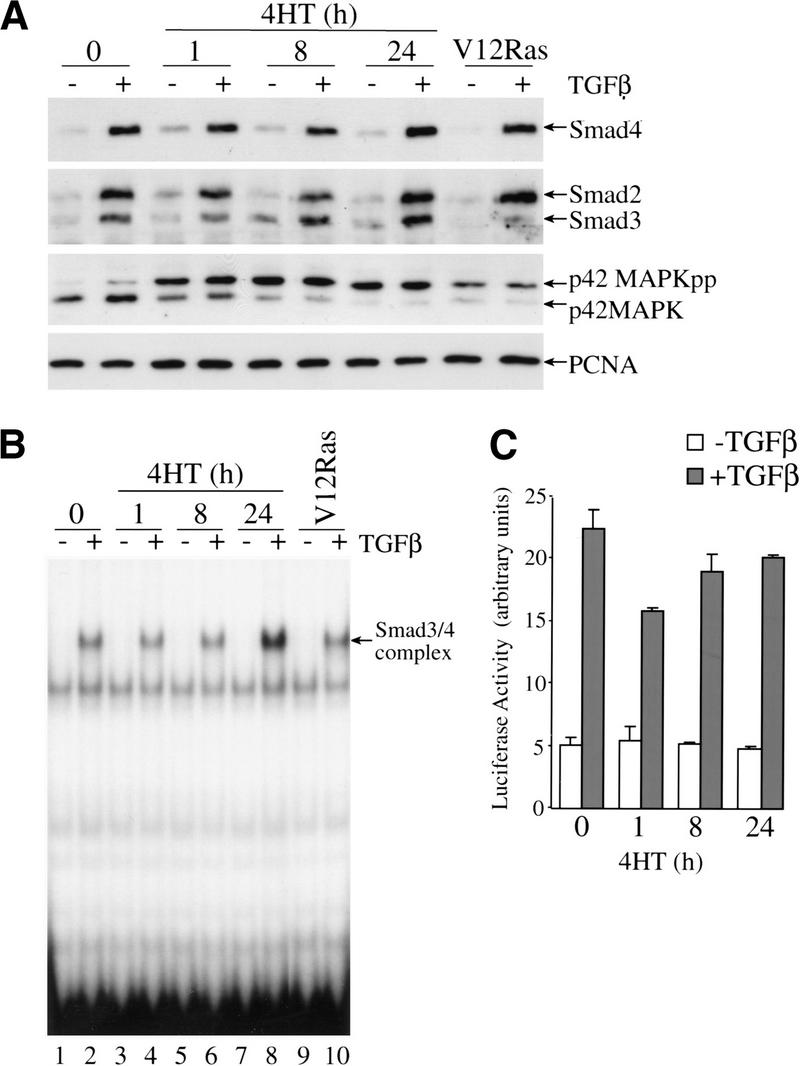
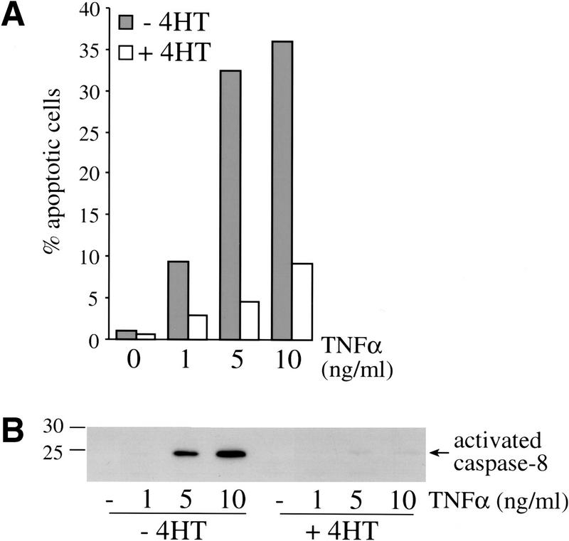
c-Raf-1 is a major effector of Ras proteins, responsible for activation of the ERK MAP kinase pathway and a critical regulator of both normal growth and oncogenic transformation. Using an inducible form of Raf in MDCK cells, we have shown that sustained activation of Raf alone is able to induce the transition from an epithelial to a mesenchymal phenotype. Raf promoted invasive growth in collagen gels, a characteristic of malignant cells; this was dependent on the operation of an autocrine loop involving TGFbeta, whose secretion was induced by Raf. TGFbeta induced growth inhibition and apoptosis in normal MDCK cells: Activation of Raf led to inhibition of the ability of TGFbeta to induce apoptosis but not growth retardation. ERK has been reported previously to inhibit TGFbeta signaling via phosphorylation of the linker region of Smads, which prevents their translocation to the nucleus. However, we found no evidence in this system that ERK can significantly influence the function of Smad2, Smad3, and Smad4 at the level of nuclear translocation, DNA binding, or transcriptional activation. Instead, strong activation of Raf caused a broad protection of these cells from various apoptotic stimuli, allowing them to respond to TGFbeta with increased invasiveness while avoiding cell death. The Raf-MAP kinase pathway thus synergizes with TGFbeta in promoting malignancy but does not directly impair TGFbeta-induced Smad signaling.


Figures

Figure 1
Figure 1
Activation of Raf–ER leads to rapid phosphorylation of p42MAPK and induction of morphological changes in MDCK cells. (A) MDCK cells expressing Raf–ER were serum starved in DMEM +0.5% BSA for 24 h before treatment with 100 nM 4-hydroxy-tamoxifen (4HT) for the indicated times. Cytoplasmic lysates were assayed for the expression of Raf–ER fusion protein using an antiestrogen receptor antibody and the mobility shift of p42MAPK to the phosphorylated form. (B) Control MDCK cells carrying empty vector (mock) and MDCK Raf–ER cells were serum starved for 24 h and either untreated or treated with 100 nM 4HT for 1 h. The MEK inhibitor PD98059 (30 μM) was applied 20 min before 4HT treatment, and p42MAPK activity was assayed by immunoblotting.(C) MDCK Raf–ER cells were either untreated or (E) treated with 100 nM 4HT for 24 h or (F) >14 d or (D) with 10 ng/ml HGF/SF for 24 h and examined by phase contrast microscopy.
Figure 2
Figure 2
Activation of Raf–ER leads to repression of epithelial marker proteins and induction of expression of a mesenchymal marker protein. MDCK Raf–ER cells either untreated (a,e,i,k,o) or treated with 100 nM 4HT for 4 d (b,f,j,l,p), 6 d (c,g,m,q) or after sustained Raf activation (>14 d; d,h,n,r) were immunostained with antibodies recognizing E-cadherin, ZO-1, cytokeratin, and vimentin and examined by confocal laser scanning microscopy. Confocal images show three-dimensional stacks of horizontal sections (ah,kr) or of vertical sections (i,j).
Figure 3
Figure 3
Activation of Raf in MDCK cells induces secretion of TGFβ. (A) MDCK Raf–ER cells, control MDCK cells, and MDCK Raf–ER cells transformed by long-term culture in 4HT (Raf T) were cultivated in the presence or absence of 100 nM 4HT in DMEM +2% FCS for 24 h. Cells were cultured in DMEM +0.5% BSA for a further 24 h before collecting the supernatants. Cell culture supernatants were examined for TGFβ1 levels by ELISA. The values shown were normalized for cell number. (B) MDCK Raf–ER cells, control MDCK cells, and Raf-transformed MDCK Raf–ER cells (RafT) were cultured in DMEM +2% FCS and stimulated with 100 nM 4HT for the indicated times. The cell culture supernatant was concentrated by ultrafiltration and equal aliquots were analyzed by 12% SDS-PAGE under nonreducing conditions and immunoblotted for TGFβ and Urokinase-type Plasminogen Activator (uPA). (C) MDCK control cells were either untreated or treated for 1 h with 2 ng/mL TGFβ, with conditioned medium of 4HT untreated MDCK Raf–ER cells (cm-4HT) or with conditioned medium of MDCK Raf–ER cells transformed by long-term exposure to 4HT (cmRafT). Nuclear extracts were assayed for the binding of activated Smad3/4 complexes to a 32P-labeled c-jun oligonucleotide probe by EMSA (lanes 1–4). Untreated MDCK Raf–ER cells were stimulated with 2 ng/mL TGFβ for 1 h, and nuclear extracts were assayed in supershifts for the presence of Smads in complexes bound to the c-jun probe with the following antibodies: anti-Smad3, anti-Smad4, anti-Smad2, cross-reacting with Smad3 (Transduction Laboratories), and anti-Smad2 (SED). Peptide competition was performed with anti-Smad3 (Smad3 +pep).
Figure 4
Figure 4
Activation of Raf in MDCK cells leads to formation of an invasive phenotype in collagen gels dependent on autocrine TGFβ effects. Wild-type MDCK cells (WT; a,e,i), MDCK Raf–ER cells either untreated (b,f,j) or treated (c,g,k) with 200 nM 4HT for 6 d and MDCK Raf–ER cells transformed by long-term 4HT stimulation (RafT; d,h,l) were grown in type I collagen matrices in the absence (ad) or presence of 5 ng/mL TGFβ (eh) or neutralizing TGFβ antibodies (il) for a further 6 d under serum-free conditions. Structures were photographed at 20× magnification.
Figure 5
Figure 5
Short-term activation of Raf does not prevent TGFβ-induced cell cycle arrest but blocks TGFβ-induced apoptosis. (A) MDCK Raf–ER cells either untreated or treated with 100 nM 4HT for 24 or 48 h and MDCK Raf–ER cells transformed by long-term exposure to 4HT (RafT) were stimulated with TGFβ (7.5 ng/mL) for 24 h. The medium was changed to DMEM +2% FCS 24 h before TGFβ treatment. The cell cycle distribution was assayed by flow cytometry following propidium iodide staining. (B) MDCK Raf–ER cells either untreated or pretreated with 100 nM 4HT and MDCK Raf–ER cells transformed by long-term treatment with 4HT (RafT) were stimulated with 7.5 ng/mL TGFβ for the indicated times. Cells were cultured in DMEM +2% FCS for 24 h before TGFβ stimulation and assayed for cyclin A expression by Western blotting. (C) MDCK Raf–ER cells transformed by long-term exposure to 4HT (RafT) and MDCK Raf–ER cells either untreated or pretreated with 100 nM 4HT for the indicated periods of time were stimulated with 7.5 ng/mL TGFβ for 24 h. The percentage of apoptotic cells was determined by Hoechst staining. Data shown are the means ± standard deviations of three independent experiments performed in duplicate. (D) Effect of activated Raf on TGFβ-induced caspase activation. MDCK Raf–ER cells either untreated or pretreated with 100 nM 4HT for 8 h or 24 h were stimulated with 7.5 ng/mL TGFβ for 24 h. To inhibit caspase activation cells were pretreated with 100 μM z-VAD for 20 min. Cytosolic lysates were incubated with ZEK(bio)D-aomk peptide for 5 min at 37°C and assayed by Western blotting. (E) Activation of caspase-8 and p42MAPK was detected in MDCK Raf–ER cells either untreated or pretreated with 100 nM 4HT for 24 or 48 h. After stimulation with TGFβ (7.5 ng/mL) for 24 h, total cell lysates were analyzed by Western blotting.
Figure 6
Figure 6
TGFβ-induced nuclear translocation, DNA-binding, and transcriptional activity of Smad3/4 complexes is not affected by Raf activation. MDCK Raf–ER cells either untreated or pretreated with 100 nM 4HT for the indicated time periods and V12Ras MDCK cells were stimulated with TGFβ (2 ng/mL) for 1 h. (A) Nuclear translocation of Smad4, Smad2, Smad3, and activation of p42MAPK were detected in nuclear extracts by Western blotting. Expression of PCNA, a nuclear protein was assayed to show equal loading of nuclear extracts. (B) Activated Smad3/4 binding to a 32P labeled c-jun probe was examined in nuclear extracts by EMSA. (C) MDCK Raf–ER cells were transiently transfected with ARE-Luc reporter, pEF-XFast1, and pEF-lacZ. After treatment with 100 nM 4HT for the indicated periods of time, cells were stimulated with 2 ng/mL TGFβ for 6 h and activation of the ARE-Luc reporter was measured. Luciferase activity was normalized to the activity of the cotransfected β-galactosidase control plasmid. Data shown are the means ±average deviations of duplicates from one out of three representative experiments.
Figure 7
Figure 7
Raf inhibits apoptosis induced by TNFα plus cycloheximide. MDCK Raf–ER cells were either untreated or treated with 100 nM 4HT for 24 h and stimulated with the indicated concentrations of TNFα and cycloheximide (2 μg/mL) for 4 h. (A) The percentage of apoptotic cells was assayed by Hoecht staining. Data shown represent at least three independent experiments performed in duplicates. (B) Activation of caspase-8 was detected by Western blotting, showing the processed and activated p20 subunit of caspase-8.

Similar articles

Cited by

References

    1. Akhurst RJ, Balmain A. Genetic events and the role of TGFβ in epithelial tumor progression. J Pathol. 1999;187:82–90. - PubMed
    1. Birchmeier W, Weidner KM, Behrens J. Molecular mechanisms leading to loss of differentiation and gain of invasiveness in epithelial cells. J Cell Sci (Suppl.) 1993;17:159–164. - PubMed
    1. Birchmeier C, Meyer D, Riethmacher D. Factors controlling growth, motility, and morphogenesis of normal and malignant epithelial cells. Int Rev Cytol. 1995;160:221–266. - PubMed
    1. Bosch E, Cherwinski H, Peterson D, McMahon M. Mutations of critical amino acids affect the biological and biochemical properties of oncogenic A-Raf and Raf-1. Oncogene. 1997;15:1021–1033. - PubMed
    1. Boyer B, Tucker GC, Valles AM, Franke WW, Thiery JP. Rearrangements of desmosomal and cytoskeletal proteins during the transition from epithelial to fibroblastoid organization in cultured rat bladder carcinoma cells. J Cell Biol. 1989;109:1495–1509. - PMC - PubMed

MeSH terms