. 2000 Nov 1;192(1):125-31.
doi: 10.1111/j.1574-6968.2000.tb09370.x.

Characterisation of the bovine enteric calici-like virus, Newbury agent 1

Affiliations

Characterisation of the bovine enteric calici-like virus, Newbury agent 1

A M Dastjerdi et al. FEMS Microbiol Lett. .

Abstract

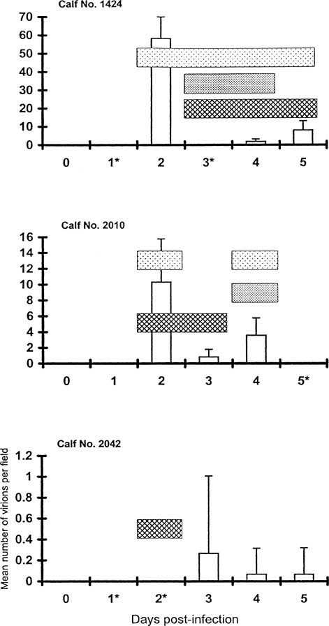
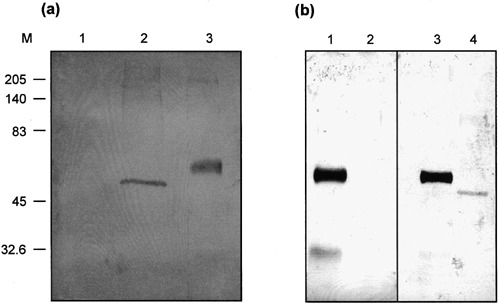
The bovine enteric calici-like virus, Newbury agent 1 (NA1) was characterised to determine if it is a member of the Caliciviridae and to establish its antigenic relationship to the established bovine enteric calicivirus Newbury agent 2 (NA2). Solid phase immune electron microscopy (SPIEM) allowed quantification of NA1 virions and identification of faecal samples with optimal virus levels. NA1 particles were 36.6 nm in diameter, had an indefinite surface structure resembling that of human small round structured viruses (SRSVs), and a buoyant density of 1.34 g ml(-1). A single capsid protein of 49.4 kDa was detected by Western blotting in purified NA1 preparations prepared from post-infection but not pre-infection faecal samples and with post- but not pre-infection sera. NA1 was antigenically unrelated to the bovine enteric calicivirus NA2 by SPIEM. These properties were consistent with classification of NA1 within the Caliciviridae but demonstrated heterogeneity in the capsid composition of bovine enteric caliciviruses.


Figures

Figure 1
Figure 1
Clinical signs and faecal NA1 excretion by SPIEM (white box) in three gnotobiotic calves inoculated with NA1. Faecal colour changes (box with dots), increased rectal temperatures (box with dashed lines) and diminished appetite (box with lines) are indicated. Absence of a parameter indicates it was not present. Asterisks indicate that a faecal sample could not be collected on that day. The diminished appetite and abnormal faecal colour lasted till day 7 p.i. for calf 1424 (not shown). Error bars represent standard deviations of the mean particle counts per field.
Figure 2
Figure 2
NA1 virus particles trapped by SPIEM from the day 2 faecal sample of gnotobiotic calf 1424 using a 1:20 dilution of the NA1 immunoglobulin concentrate. Bar indicates 100 nm. Inset shows surface structure of an NA1 particle and spiked outer edge; bar indicates 50 nm. Stained with 2% PTA at pH 6.0.
Figure 3
Figure 3
Mean number of NA1 particles (•) trapped per field by SPIEM in fractions from a representative CsCl density gradient centrifuged to equilibrium. Buoyant density (◯) determined by refractometry.
Figure 4
Figure 4
(a) Western blotting analysis of fractions from day 0 (track 1) and day 2 (track 2) faecal samples of calf 1424 purified by CsCl centrifugation stained with the post‐inoculation NA1 serum. A mean of 130.5 NA1 particles per field were present by SPIEM in the fraction from the day 2 sample; no particles were detected in the fraction from the day 0 sample. Track 3: bovine IgG as a control for electroblotting and immunostaining. M, molecular mass markers (kDa). (b) Western blotting analysis of fractions from the day 2 faecal sample of calf 1424 stained with the pre‐ (track 2) and the post‐inoculation NA1 serum (track 4). Tracks 1 and 3: bovine IgG as a control for electroblotting and immunostaining. M, molecular mass markers (kDa).
Figure 5
Figure 5
(a) Molecular mass estimation of the capsid protein of NA1 particles purified by CsCl density gradient centrifugation. M, molecular masses of biotinylated standard proteins (kDa). Track 1: bovine IgG as a control for blotting and staining with the anti‐bovine conjugate; track 2: biotinylated standard proteins; track 3: biotinylated standard proteins plus the NA1 capsid protein; track 4: the NA1 capsid protein; arrow indicates the NA1 protein. (b) Molecular mass estimation of the capsid protein of FCV grown in CRFK cells using the same electrophoresis conditions as those used for NA1. Arrow indicates the FCV capsid protein. M, molecular masses of biotinylated standard proteins (kDa). Track 1: preparation from uninfected CRFK cells; track 2: biotinylated standard proteins plus the preparation from FCV‐infected cells; track 3: FCV‐infected cells; track 4: rabbit IgG (the FCV NADC rabbit antiserum) used as a control for blotting and immunostaining with the anti‐rabbit conjugate.

Similar articles

Cited by

References

    1. Glass, R.I. , Noel, J. , Ando, T. , Fankhauser, R. , Belliot, G. , Mounts, A. , Parashar, U.D. , Breese, J.S. , Monroe, S.S. (2000) The epidemiology of enteric caliciviruses from humans: a reassessment using new diagnostics. J. Infect. Dis. 181 (suppl. 2), S254–S261. - PubMed
    1. Woode, G.N. , Bridger, J.C. (1978) Isolation of small viruses resembling astroviruses and caliciviruses from acute enteritis of calves. J. Med. Microbiol. 11, 441–452. - PubMed
    1. Reynolds, D.J. , Morgan, J.H. , Chanter, N. , Jones, P.W. , Bridger, J.C. , Debney, T.G. , Bunch, K.J. (1986) Microbiology of calf diarrhoea in Southern Britain. Vet. Rec. 119, 34–39. - PubMed
    1. Günther, H. , Otto, P. , Heilmann, P. (1984) Untersuchungen zum Durchfall Junger Kälber. Arch. Exp. Vet. Med. 38, 781–792. - PubMed
    1. Granzow, V.H. , Schirrmeier, H. (1985) Elektronmikroskopischer Direktnachweis von 32‐nm‐Viruspartikeln im Kot Durchfallkranker Kälber (Kurzmitteilung). Monatsh. Vet. Med. 40, 228–229.