

Detection of c-kit mutation Asp 816 to Val in microdissected bone marrow infiltrates in a case of systemic mastocytosis associated with chronic myelomonocytic leukaemia
- PMID: 11040941
- PMCID: PMC1186968
- DOI: 10.1136/mp.53.4.188
Detection of c-kit mutation Asp 816 to Val in microdissected bone marrow infiltrates in a case of systemic mastocytosis associated with chronic myelomonocytic leukaemia
Abstract
Background/aims: The occurrence of myeloid leukaemia in patients with systemic mastocytosis is a well recognised phenomenon. However, the pathophysiological basis of such a coevolution has not been clarified. Recent data have shown that the c-kit mutation Asp 816 to Val is detectable in neoplastic mast cells in most patients with systemic mastocytosis, including those who have associated haematological disorders. The aim of this study was to study clonal disease evolution by analysing bone marrow cells from a patient with systemic mastocytosis and associated chronic myelomonocytic leukaemia (CMML) for the presence of this mutation.
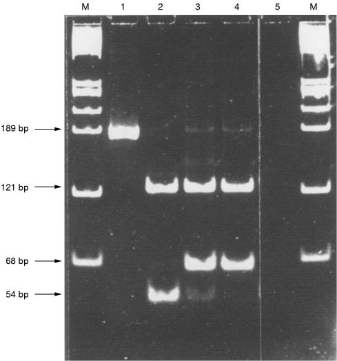
Methods: The DNA of microdissected bone marrow cells from a patient with systemic mastocytosis and associated CMML was analysed for the presence of the c-kit mutation Asp 816 to Val by means of HinfI digestion and direct sequencing of semi-nested polymerase chain reaction (PCR) products.
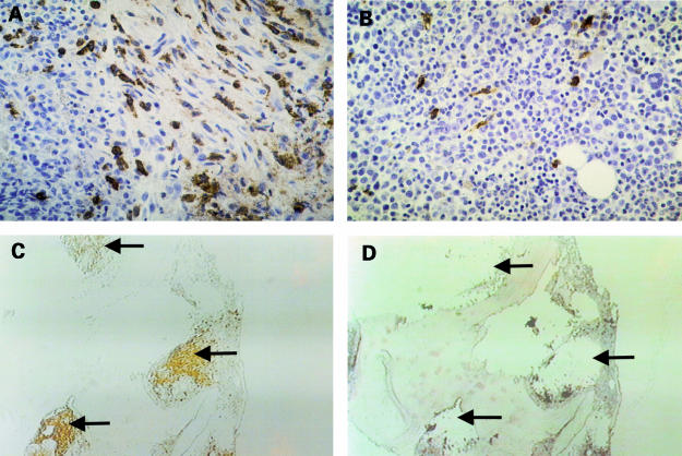
Results: The two neoplasms could easily be identified and discriminated in paraffin wax embedded bone marrow sections by tryptase and chloroacetate esterase staining. A total number of 10 tryptase positive systemic mastocytosis infiltrates and 10 tryptase negative CMML infiltrates were removed by microdissection. As assessed by HinfI digestion and direct sequencing of semi-nested PCR products, the c-kit mutation Asp 816 to Val was detected in five of seven systemic mastocytosis infiltrates and four of six CMML infiltrates. By contrast, no c-kit mutation Asp 816 to Val was found in bone marrow infiltrates in patients with CMML without associated systemic mastocytosis (n = 20).
Conclusion: These data support a monoclonal evolution of systemic mastocytosis and concurrent CMML in the patient studied.
Figures

References
-
- Lennert K, Parwaresch MR. Mast cells and mast cell neoplasia: a review. Histopathology 1979;3:349–65. - PubMed
-
- Horny H-P, Parwaresch MR, Lennert K. Bone marrow findings in systemic mastocytosis. Hum Pathol 1985;16:808–14. - PubMed
-
- Stein DH. Mastocytosis: a review. Pediatr Dermatol 1986;3:365–75. - PubMed
-
- Metcalfe DD. Classification and diagnosis of mastocytosis: current status. J Invest Dermatol 1991;96:2S–4S. - PubMed
-
- Johnson WC, Helwig EB. Solitary mastocytosis (urticaria pigmentosa). Arch Dermatol 1961;84:806–15. - PubMed
Publication types
MeSH terms
Substances
LinkOut - more resources
Full Text Sources
Research Materials