. 2000 Oct;12(10):1799-810.
doi: 10.1105/tpc.12.10.1799.

Separable whorl-specific expression and negative regulation by enhancer elements within the AGAMOUS second intron

Affiliations

Separable whorl-specific expression and negative regulation by enhancer elements within the AGAMOUS second intron

M K Deyholos et al. Plant Cell. 2000 Oct.

Abstract

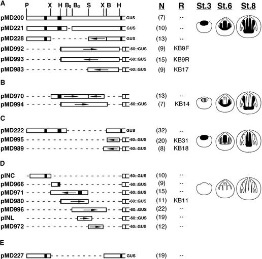
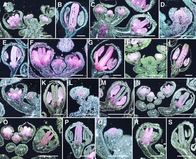
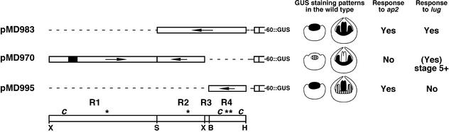
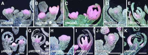
We analyzed the 4-kb intragenic control region of the AGAMOUS (AG) gene to gain insight into the mechanisms controlling its expression during early flower development. We identified three major expression patterns conferred by 19 AG::reporter gene constructs: the normal AG pattern, a stamen-specific pattern, and a predominantly carpel pattern. To determine whether these three expression patterns were under negative control by APETALA2 (AP2) or LEUNIG (LUG), we analyzed beta-glucuronidase staining patterns in Arabidopsis plants homozygous for strong ap2 and lug mutations. Our results indicated that the stamen-specific pattern was independent of AP2 but dependent on LUG; conversely, the carpel-specific pattern was independent of LUG but dependent on AP2. These results lead to a model of control of AG expression such that expression in each of the two inner whorls is under independent positive and negative control.


Figures

Figure 1
Figure 1
Structure of AG::GUS Reporter Gene Constructs. (A) Reporter gene constructs that conferred a normal AG pattern to the reporter gene during early flower development (stages 1 to 9). (B) Reporter gene constructs that conferred a stamen expression pattern to the reporter gene during early flower development (stages 1 to 9). (C) Reporter gene constructs that conferred a carpel expression pattern to the reporter gene during early flower development (stages 1 to 9). (D) Reporter gene constructs that failed to confer reporter gene expression during early flower development (stages 1 to 9). (E) Reporter gene construct that gave variable GUS staining patterns. White bars indicate noncoding sequences; black boxes indicate the first two AG exons. Arrows indicate orientation of fragments; where arrows are omitted, the fragments are in their normal 5′ to 3′ orientation. Letters above pMD200 in (A) indicate restriction sites used for construction of the reporter gene constructs. P indicates PstI, and for convenience we refer to this as nucleotide position 1. X, XbaI, with sites at positions 1272 and 3936; H, HindIII, with sites at positions 1753 and 4762; Bg, BglII, with sites at 2127 and 2455; S, SpeI, with a site at position 3109; B, BamHI, with a site at position 4018. Numbers beneath N (within parentheses) indicate the number of independently transformed lines examined for each construct. Designations beneath R, when present, indicate the construct number containing the same AG DNA used in studies by Busch et al. (1999) and Bomblies et al. (1999). Diagrams at right represent the GUS staining patterns of stage (St.) 3, stage 6, and stage 8 flowers; black areas indicate strong GUS staining, and striped areas indicate weak GUS staining.
Figure 2.
Figure 2.
GUS Staining Patterns in Wild-Type Plants carrying AG::GUS Reporter Gene Constructs. (A) and (B) GUS staining in transgenic plants carrying pMD200. (A) In a longitudinal section through the inflorescence meristem, stage 3, 4, and 8 flowers show GUS staining in the central region of the floral meristem and in the developing stamen and carpel primordia. (B) This stage 12 flower shows GUS staining in the stamens and carpels but not in the sepals or petals. (C) GUS staining in a transgenic plant carrying pMD221. GUS staining in the stage 4, 6, and 10 flowers is restricted to the central region of the floral meristem and the developing stamens and carpels. (D) and (E) GUS staining in transgenic plants carrying pMD992. (D) This stage 4 flower shows GUS staining in the central region of the floral meristem. (E) This stage 10 flower shows GUS staining in the stamens and carpels but not in the sepals and petals. (F) and (G) GUS staining in transgenic plants carrying pMD993. (F) Stage 5, 7, and 8 flowers show GUS staining in the developing stamens and carpels. (G) This stage 9 flower shows GUS staining in the stamens and carpels but not in the sepals and petals. (H) and (I) GUS staining in transgenic plants carrying pMD983. (H) These stage 4 and 7 flowers show GUS staining in the floral meristem and in the stamen and carpel primordia, respectively. No GUS staining was detected in the inflorescence meristem (IM) or the developing sepals. (I) GUS staining in this stage 12 flower is in the stamens and carpels but not in the sepals and petals. (J) and (K) GUS staining in transgenic plants carrying pMD970. (J) The stage 3 flower shows GUS staining in a small domain in the core of the floral meristem; the stage 7 flower has GUS staining in stamen primordia and the receptacle but not in the carpel or sepal primordia. (K) This stage 9 flower shows GUS staining in the developing stamens but not in the sepals or carpels. (L) and (M) GUS staining in transgenic plants carrying pMD994. (L) GUS staining in this stage 4 flower is restricted to a small domain in the core of the floral meristem. (M) This stage 9 flower shows GUS staining in the stamens but not in the carpels, sepals, or petals. (N) GUS staining in a transgenic plant carrying pMD222. GUS staining in the stage 4 flower is strong throughout the central apical region of the floral meristem. In the two stage 7 flowers, GUS staining is strong only in the carpel primordia; weak GUS staining can be detected in the abaxial proximal regions of the developing stamens. In the stage 9 flower, GUS staining is present in the carpels and receptacle only. (O) and (P) GUS staining in transgenic plants carrying the pMD989 transgene. (O) GUS staining in the stage 4 flower is restricted to the central region of the floral meristem. In the stage 6 flower, GUS staining is present in both stamen and carpel primordia. In the stage 7 flower, GUS staining is strong only in the carpels. In the stage 9 flower, GUS staining is detected only in the carpels. (P) GUS staining in this stage 10 flower is present in the carpels but absent in the sepals and stamens. (Q) to (S) GUS staining in transgenic plants carrying pMD995. (Q) GUS staining is present in the central apical region of the floral meristem of this stage 3 flower. (R) GUS staining is present in the carpel primordia but not in the sepal or stamen primordia of this stage 7 flower. (S) GUS staining is lost from all floral organs by stage 10. Numbers at the base of the flowers indicate their floral stage; IM, the inflorescence meristem. formula image; formula image.
Figure 3.
Figure 3.
Summary of Negative Regulation Experiments and Results. Depicted are the three constructs (pMD983, pMD970, and pMD995) used in experiments examining GUS staining in ap2 and lug mutants; beneath them, the regions R1 to R4 are indicated. R1 begins at 1272 (relative to the upstream PstI site as 1) and extends to 3109 (SpeI site). R2 begins at 3110 and extends to 3936 (XbaI site). R3 begins at 3937 and extends to 4018 (BamHI). R4 extends from 4019 to 4762 (HindIII). The c and the asterisk indicate approximate positions of CArG boxes and LFY binding sequences, respectively. pMD983 contains R2, R3, and R4; pMD970 contains R1 and R2; and pMD995 contains only R4. Cartoons depicting the staining patterns of stage 3 and 6 flowers are presented with each construct. At right are indicated the constructs that showed an expansion of the GUS staining domain to include outer-whorl organs when present in either an ap2 or a lug mutant background. Other symbols and abbreviations are as given in Figure 1.
Figure 4.
Figure 4.
GUS Staining Patterns in Flowers of Transgenic ap2 and lug Plants. (A) to (E) GUS staining in strong ap2 mutants. (A) and (B) GUS staining in ap2-2 plants carrying the pMD995 transgene. (A) Pattern seen in approximately half the plants examined, in which intense GUS staining is restricted to the central region of the floral meristem in stage 4 flowers and is not present in the sepal primordia of these pMD995 ap2-2 plants. (B) Pattern seen in the remaining plants (also ∼50%), in which intense GUS staining is seen throughout the central floral meristem and the sepal primordia of the late stage 4 flower, as well as throughout the apical portion of the early stage 3 floral meristem. (C) GUS staining in ap2-2 plants carrying the pMD983 transgene, in which the stage 4 and 7 flowers have intense GUS staining through all floral organ primordia. (D) and (E) GUS staining in ap2-9 plants carrying the pMD970 transgene. (D) GUS staining in a stage 4 flower is restricted to a small domain in the center of the floral meristem. (E) GUS staining in this stage 7 flower is observed only in a developing stamen and not in any outer-whorl organs. (F) to (K) GUS staining patterns in lug-1 mutants. (F) and (G) GUS staining in lug-1 plants carrying the pMD995 transgene. GUS staining is present in the central floral meristem of the stage 4 flower, in the carpel primordia of the stage 6 flower. In stage 8 flowers, GUS staining was highly reduced (F), and when present, was restricted to carpels (G). (H) and (I) GUS staining in lug-1 plants carrying the pMD983 transgene. GUS staining in an early stage 3 flower is present in sepal primordia as well as the floral meristem (H) but in this stage 8 flower is present in developing carpels and in proximal regions of the sepals (I). (J) and (K) GUS staining in lug-1 plants carrying the pMD970 transgene. GUS staining in this stage 3 floral meristem is restricted to the central core (J), whereas in this stage 8 flower it is present in outer-whorl sepals (K). formula image.

References

    1. Baranowskij, N., Frohberg, C., Prat, S., and Willmitzer, L. (1994). A novel DNA binding protein with homology to Myb oncoproteins containing only one repeat can function as a transcriptional activator. EMBO J. 13, 5383–5392. - PMC - PubMed
    1. Bechtold, N., Ellis, J., and Pelletier, G. (1993). In planta Agrobacterium-mediated gene transfer by infiltration of adult Arabidopsis plants. C. R. Acad. Sci. Paris 316, 1194–1199.
    1. Benfey, P.N., and Chua, N.-H. (1990). The cauliflower mosaic virus 35S promoter: Combinatorial regulation of transcription in plants. Science 250, 959–966. - PubMed
    1. Bomblies, K., Dagenais, N., and Weigel, D. (1999). Redundant enhancers mediate transcriptional repression of AGAMOUS by APETALA2. Dev. Biol. 216, 260–264. - PubMed
    1. Bowman, J.L., Smyth, D.R., and Meyerowitz, E.M. (1989). Genes directing flower development in Arabidopsis. Plant Cell 1, 37–52. - PMC - PubMed

Publication types

Associated data