. 2000 Oct;12(10):1811-22.
doi: 10.1105/tpc.12.10.1811.

Simulation of fungal-mediated cell death by fumonisin B1 and selection of fumonisin B1-resistant (fbr) Arabidopsis mutants

Affiliations

Simulation of fungal-mediated cell death by fumonisin B1 and selection of fumonisin B1-resistant (fbr) Arabidopsis mutants

J M Stone et al. Plant Cell. 2000 Oct.

Abstract

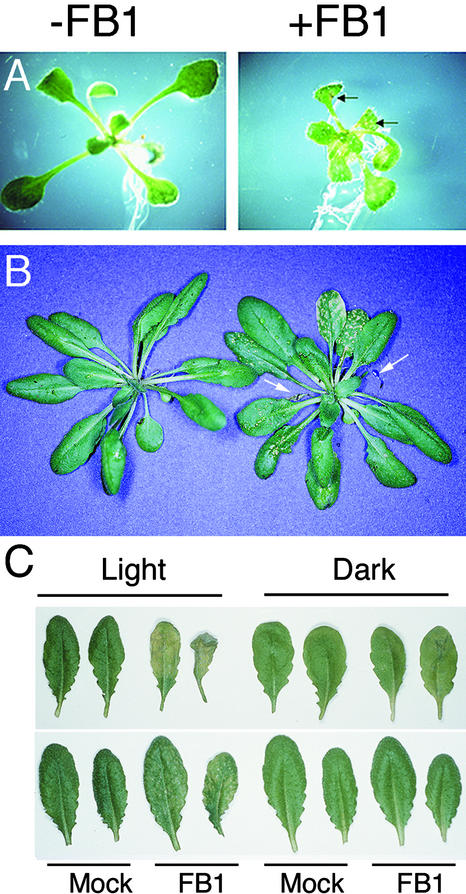
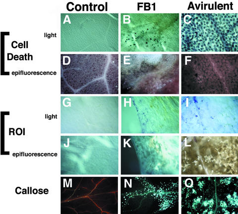
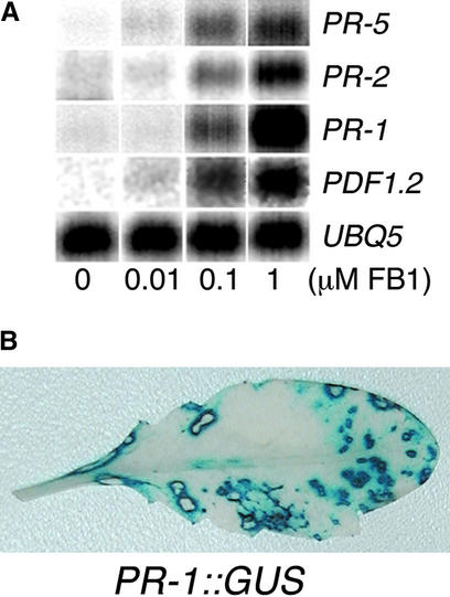
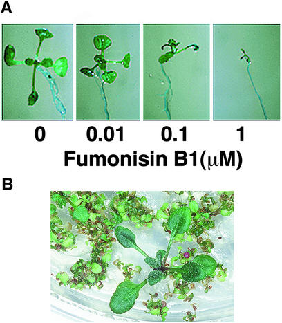
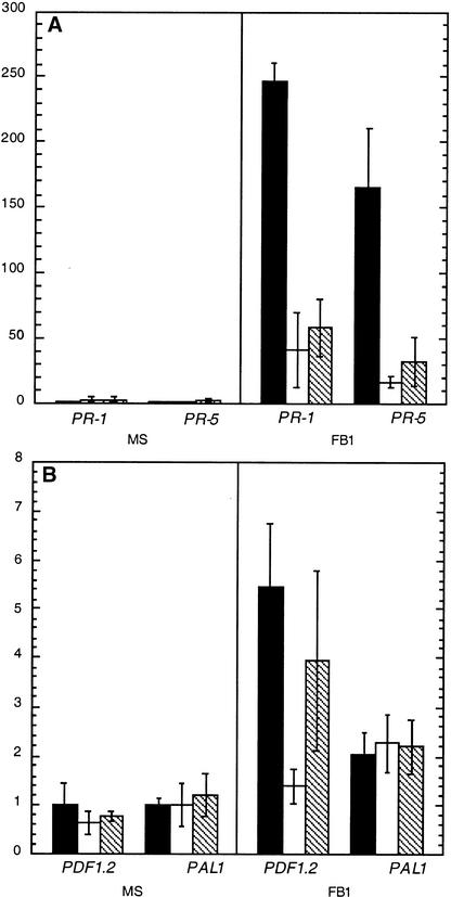
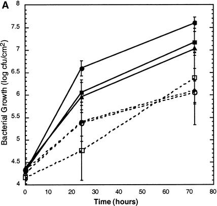
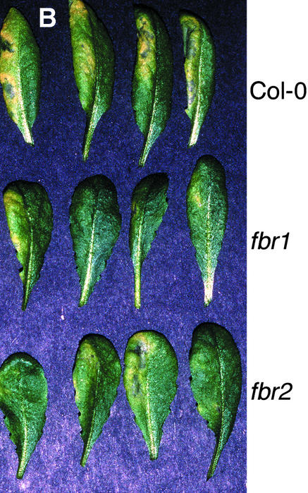
Fumonisin B1 (FB1), a programmed cell death-eliciting toxin produced by the necrotrophic fungal plant pathogen Fusarium moniliforme, was used to simulate pathogen infection in Arabidopsis. Plants infiltrated with 10 microM FB1 and seedlings transferred to agar media containing 1 microM FB1 develop lesions reminiscent of the hypersensitive response, including generation of reactive oxygen intermediates, deposition of phenolic compounds and callose, accumulation of phytoalexin, and expression of pathogenesis-related (PR) genes. Arabidopsis FB1-resistant (fbr) mutants were selected directly by sowing seeds on agar containing 1 microM FB1, on which wild-type seedlings fail to develop. Two mutants chosen for further analyses, fbr1 and fbr2, had altered PR gene expression in response to FB1. fbr1 and fbr2 do not exhibit differential resistance to the avirulent bacterial pathogen Pseudomonas syringae pv maculicola (ES4326) expressing the avirulence gene avrRpt2 but do display enhanced resistance to a virulent isogenic strain that lacks the avirulence gene. Our results demonstrate the utility of FB1 for high-throughput isolation of Arabidopsis defense-related mutants and suggest that pathogen-elicited programmed cell death of host cells may be an important feature of compatible plant-pathogen interactions.


Figures

Figure 1.
Figure 1.
FB1 Induces Lesion Formation in Arabidopsis. (A) Ten-day-old Col-0 Arabidopsis seedlings were transferred to MS plates (−FB1) or MS plates supplemented with 1 μM FB1 (+FB1) and photographed 4 days later. Necrotic lesions on the FB1-treated seedling are indicated by arrows. (B) Two lower leaves of 4-week-old Arabidopsis plants were infiltrated with 10 mM MgSO4 (left) or 10 μM FB1 in 10 mM MgSO4 (right) and photographed 7 days later. Dead FB1-infiltrated leaves are indicated with arrows. (C) Lower leaves of 4-week-old Arabidopsis plants were infiltrated with 10 mM MgSO4 (Mock) or 10 μM FB1 in 10 mM MgSO4 (FB1). Plants were exposed to a 12-hr-light photoperiod (Light) or covered with aluminum foil (Dark) for 3 days. Infiltrated leaves are shown above; systemic, noninfiltrated leaves are shown below.
Figure 2.
Figure 2.
FB1-Induced Lesions Resemble HR Lesions. Transfer of 10-day-old seedlings to plates containing 1 μM FB1 or inoculation of 4-week-old leaves with P. s. maculicola 4326 avrRpt2 induced formation of macroscopic lesions. Comparisons were made by light or epifluorescence microscopic examination of stained leaves. See Methods for details. (A) to (F) Leaves stained with lactophenol–trypan blue, revealing cell death. (G) to (L) Leaves stained with nitroblue tetrazolium, revealing ROIs. (M) to (O) Leaves stained with aniline blue, revealing callose deposition. Control, MgSO4-treated leaves; FB1, FB1-treated leaves; Avirulent, P. s. maculicola ES4326 (avrRpt2)–infected leaves.
Figure 3.
Figure 3.
Treatment of Arabidopsis with FB1 Induces Expression of Defense-Related Genes. (A) Defense gene activation in response to increasing concentrations of FB1. RNA was isolated from seedlings treated for 4 days on agar media with various concentrations of FB1 and analyzed by RNA gel blot analysis. UBQ5 was used as a loading control. (B) Two lower leaves of transgenic Arabidopsis harboring a PR-1 promoter::GUS reporter gene fusion were infiltrated with 10 μM FB1 solution. After 1 week, the plants were histochemically stained for GUS activity. A noninfiltrated leaf that developed lesions is shown. Plants infiltrated with 10 mM MgSO4 showed no detectable GUS activity.
Figure 4.
Figure 4.
FB1 Inhibits Seedling Germination. (A) Arabidopsis Col-0 seeds were germinated on MS agar media supplemented with various concentrations of FB1 as indicated and photographed 10 days later. (B) Mutagenized seeds were sowed directly on MS agar media supplemented with 0.5 μM FB1. An example of selection of a putative fbr mutant is shown.
Figure 5.
Figure 5.
fbr Mutants Display Aberrant Defense Gene Induction in Response to FB1. Wild-type Col-0, fbr1, and fbr2 seedlings were grown axenically for 10 days, then transferred to MS plates (MS) or MS plates supplemented with 1 μM FB1 (FB1). After 4 days of treatment, RNA was isolated and analyzed by RNA gel blot analysis as described in Methods. A representative experiment with triplicate RNA samples is shown. Experiments were repeated four times with similar results, except that PDF1.2 expression in fbr2 seedlings was generally found to be similar to that in fbr1. Data are presented as means ±sem. The relative expression of wild-type Col-0 is shown by black bars, fbr1 by white bars, and fbr2 by hatched bars. (A) RNA gel blot analysis for SA-dependent and systemic acquired resistance–associated pathogenesis-related genes PR-1 and PR-5. (B) RNA gel blot analysis for SA-independent genes PDF1.2 and PAL1.
Figure 6.
Figure 6.
fbr Mutants Display Enhanced Resistance to a Virulent Bacterial Pathogen. (A) Growth of P. s. maculicola ES4326 in wild-type Col-0 (circles), fbr1 (squares), and fbr2 (triangles) plants. Leaves were infiltrated with virulent P. s. maculicola ES4326 (closed symbols) or with the avirulent isogenic strain expressing the avrRpt2 avirulence gene (open symbols). Leaf disks were harvested at 0, 24, and 72 hr after infiltration, and bacterial counts were determined by serial dilution. The data shown are means for six leaves ±sem from a representative experiment; these experiments were repeated three times with fbr2 and five times with fbr1 with similar results. cfu, colony-forming units. (B) Disease symptoms elicited by inoculation with P. s. maculicola ES4326. The left half of each leaf was infiltrated with bacterial suspension (104 cfu/mL), and the leaves were photographed 2 days later.
Figure 6.
Figure 6.
fbr Mutants Display Enhanced Resistance to a Virulent Bacterial Pathogen. (A) Growth of P. s. maculicola ES4326 in wild-type Col-0 (circles), fbr1 (squares), and fbr2 (triangles) plants. Leaves were infiltrated with virulent P. s. maculicola ES4326 (closed symbols) or with the avirulent isogenic strain expressing the avrRpt2 avirulence gene (open symbols). Leaf disks were harvested at 0, 24, and 72 hr after infiltration, and bacterial counts were determined by serial dilution. The data shown are means for six leaves ±sem from a representative experiment; these experiments were repeated three times with fbr2 and five times with fbr1 with similar results. cfu, colony-forming units. (B) Disease symptoms elicited by inoculation with P. s. maculicola ES4326. The left half of each leaf was infiltrated with bacterial suspension (104 cfu/mL), and the leaves were photographed 2 days later.

References

    1. Abbas, H.K., Tanaka, T., Duke, S.O., Porter, J.K., Wray, E.M., Hodges, L., Sessions, A.E., Wang, E., Merrill, A.H., and Riley, R.T. (1994). Fumonisin- and AAL-toxin–induced disruption of sphingolipid metabolism with accumulation of free sphingoid bases. Plant Physiol. 106, 1085–1093. - PMC - PubMed
    1. Allen, L.J., MacGregor, K.B., Koop, R.S., Bruce, D.H., and Karner, J., and Bown, A.W. (1999). The relationship between photosynthesis and a mastoparan-induced hypersensitive response in isolated mesophyll cells. Plant Physiol. 119, 1233–1241. - PMC - PubMed
    1. Aravind, L., Dixit, V.M., and Koonin, E.V. (1999). The domains of death: Evolution of the apoptosis machinery. Trends Biochem. Sci. 24, 47–53. - PubMed
    1. Asai, T., Stone, J.M., Heard, J.E., Kovtun, Y., Yorgey, P., Sheen, J., and Ausubel, F.M. (2000). Fumonisin B1–induced cell death in Arabidopsis protoplasts requires jasmonate-, ethylene-, and salicylate-dependent signaling pathways. Plant Cell 12, in press. - PMC - PubMed
    1. Beers, E.P. (1997). Cell death during plant growth and development. Cell Death Differ. 4, 649–661. - PubMed

Publication types