Comparative Study
. 2000 Oct;12(10):1903-16.
doi: 10.1105/tpc.12.10.1903.

AHM1, a novel type of nuclear matrix-localized, MAR binding protein with a single AT hook and a J domain-homologous region

Affiliations
Comparative Study

AHM1, a novel type of nuclear matrix-localized, MAR binding protein with a single AT hook and a J domain-homologous region

G Morisawa et al. Plant Cell. 2000 Oct.

Abstract

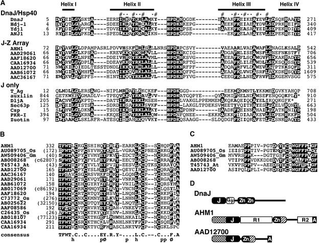
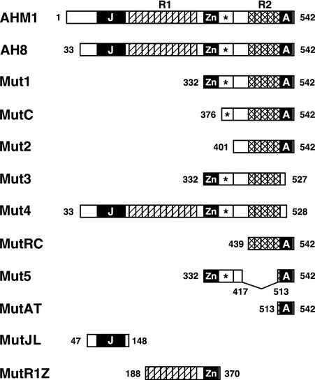
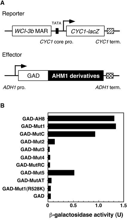
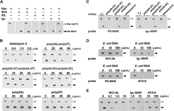
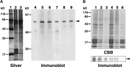
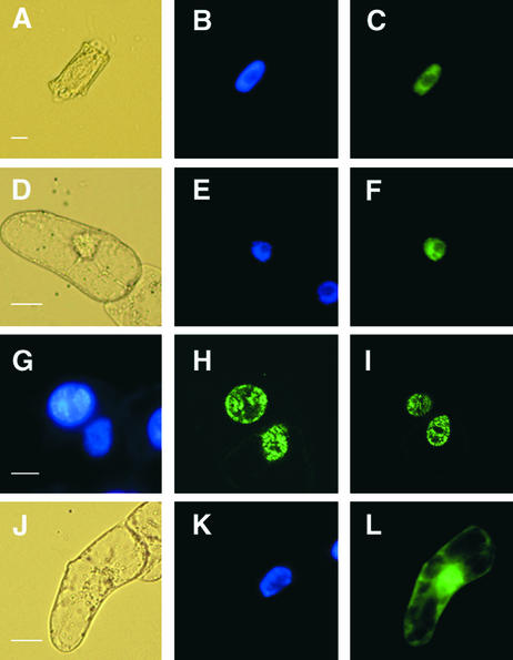
Interactions between the nuclear matrix and special regions of chromosomal DNA called matrix attachment regions (MARs) have been implicated in various nuclear functions. We have identified a novel protein from wheat, AT hook-containing MAR binding protein1 (AHM1), that binds preferentially to MARs. A multidomain protein, AHM1 has the special combination of a J domain-homologous region and a Zn finger-like motif (a J-Z array) and an AT hook. For MAR binding, the AT hook at the C terminus was essential, and an internal portion containing the Zn finger-like motif was additionally required in vivo. AHM1 was found in the nuclear matrix fraction and was localized in the nucleoplasm. AHM1 fused to green fluorescent protein had a speckled distribution pattern inside the nucleus. AHM1 is most likely a nuclear matrix component that functions between intranuclear framework and MARs. J-Z arrays can be found in a group of (hypothetical) proteins in plants, which may share some functions, presumably to recruit specific Hsp70 partners as co-chaperones.


Figures

Figure 1.
Figure 1.
Amino Acid Sequences of AHM1 and AHM2. (A) Amino acid sequence of AHM1 deduced from the sequences of the genomic and cDNA clones. A J domain–like region (positions 72 to 143), a Zn finger–like motif (positions 332 to 366), and a C-terminal basic region (positions 517 to 539) containing an AT hook are boxed. Key amino acids in their respective motifs are highlighted in black. Direct repeats (R1, positions 151 to 315; R2, positions 439 to 516) are indicated with arrows. Internal extra amino acids in the first and second repeats of R2 are indicated with broken lines. A conserved region (positions 367 to 393; see Figure 2C) following the Zn finger–like motif is double underlined. (B) Partial amino acid sequence of AHM2 deduced from the sequence of a cDNA clone. A direct repeat (positions 138 to 221) and an AT hook (positions 220 to 231) are marked as in (A).
Figure 2.
Figure 2.
Comparisons of AHM1-Related Proteins. (A) An alignment of J domains. J domain proteins can be divided into three groups: DnaJ/Hsp40, which includes the canonical DnaJ and closely related homologs; J-Z array, which includes AHM1 and selected Arabidopsis proteins (shown with their GenBank accession numbers) having the Zn finger–like motif shown in (B); and J only, a diverse set of proteins that have no important similarity outside of the J domain. Above the alignment, four α-helical regions resolved in DnaJ (Pellecchia et al., 1996) are shown. Hydrophobic residues participating in the short coiled-coil structure between helices II and III and solvent-accessible residues are indicated with (#) and (•), respectively. Conserved residues (>50% in the alignment) are highlighted, and related residues (hydrophobic or basic) are in boldface. Asterisks denote the ends of proteins. Amino acid positions of the N-terminal residue in the alignment are shown at left. The total length of each protein is shown at right. GenBank accession numbers are as follows: DnaJ, P08622; human Hdj-1, P25685; yeast Ydj1, P25491; Atriplex nummularia ANJ1, P43644; SV40 T antigen (T_Ag), P03070; bovine auxilin, Q27974; E. coli DljA, P31680; yeast Sec63p, P14906; mouse cysteine-string protein (Csp), P54101; bovine double-stranded RNA-induced protein kinase inhibitor (PKR-I), AAA17795; and yeast zuotin, P32527. (B) An alignment of the Zn finger–like motif. Names of hypothetical proteins are denoted by GenBank accession numbers. Expressed sequence tag clones from rice, soybean, and Arabidopsis are indicated with the respective suffixes Os, Gm, and At after their accession numbers. The others were predicted from the Arabidopsis genomic sequence. Conserved residues are highlighted. The consensus sequence deduced is shown at the bottom; h, p, and Ø denote hydrophobic, polar, and aromatic residues, respectively. Sequences of hypothetical proteins that do not have GenBank accession numbers are indicated with nucleotide positions shown in parentheses. c denotes translation of the complementary strand. (C) An alignment of the conserved region immediately adjoining the Zn finger–like motif. The proteins aligned are the same as the first six proteins shown in (B). No amino acids are present between the sequences shown in (B) and (C) (see Figure 1A for AHM1). Conserved and related residues are highlighted as in (A). (D) Topological comparison of DnaJ, AHM1, and an Arabidopsis protein (AAD12700). Homologous regions of AHM1 and AAD12700, adjacent to the N terminus of the J domainlike region and adjacent to the C terminus of the Zn fingerlike motif, are indicated with the same hatch patterns. A, AT hook; Gly/Phe-rich linker region; J, J domain–like region; G/F, Zn, Zn finger–like motif.
Figure 3.
Figure 3.
Schematic Representation of AHM1 and Its Derivatives. The molecular organization of AHM1 is shown schematically at the top. Shown below are AHM1 derivatives expressed in E. coli as a His-tagged protein and/or in yeast as a fusion with the Gal4p activation domain. Amino acid positions at the junctions and ends are indicated. The J domain–like region (J), the Zn finger–like motif (Zn), and the C-terminal basic region containing an AT hook (A) are indicated by black boxes; each unit of direct repeats (R1 and R2) is indicated by a hatched or cross-hatched box. The conserved region immediately following the Zn finger–like motif is indicated by an asterisk.
Figure 4.
Figure 4.
MAR Binding Activity of AHM1 in Vitro. (A) Dissection of AHM1. His-tagged AHM1 derivatives were purified and separated on an SDS–12% polyacrylamide gel and then visualized with Coomassie blue staining (CBB) or subjected to a filter binding assay with WCI-3b MAR as a probe (MAR binding). Positions of molecular mass markers are shown at left in kilodaltons. The positions of His-Mut3 and His-Mut4 are marked with arrowheads. (B) Importance of the AT hook in MAR binding. Total extracts of E. coli expressing His-Mut1 and His-Mut1(R528K) were treated as in (A). IPTG denotes the extract derived from uninduced E. coli cells harboring pET-Mut1. The position of His-Mut1 is indicated by the arrowhead at right.
Figure 5.
Figure 5.
MAR Binding Activity of AHM1 in Yeast. (A) Schematic representation of the effector and reporter genes. The CYC1-lacZ reporter gene carries the WCI-3b MAR upstream of the CYC1 minimum promoter (CYC1 core pro.). The effectors used are the activation domain of Gal4p (GAD) alone or GAD fused with AHM1 derivatives shown in Figure 3. The effector genes are under the control of the ADH1 promoter (ADH1 pro.). Terminators (term.) are shown by a hatched or cross-hatched box. (B) β-Galactosidase activity in yeast cells carrying the reporter construct and an indicated effector construct. U, specific activity of β-galactosidase expressed as nanomoles catalyzed per minute per microgram of protein.
Figure 6.
Figure 6.
DNA Binding Specificity of AHM1. (A) MAR binding activity of AHM1. His-tagged HBP1a(17) (His-1a[17]) and His-Mut1, 0.1 μg each, were electrophoresed side by side and subjected to the filter binding assay with the 32P-labeled, 31-bp-long oligonucleotide Hex31, which contained Hex (CCACGTCA), a high-affinity binding site for HBP-1a(17), with or without WCI-3b MAR (WCI), the MAR-1 of Arabidopsis PS locus (PS), or Igκ MAR (Igκ) as a probe. The probes included in the reaction are indicated with (+). The length of MARs used and their AT content (in parentheses) are as follows: WCI-3b MAR, 0.88 kb (81%); PS MAR, 1.3 kb (70%); and Igκ MAR, 0.44 kb (72%). (B) Effects of competitors. The MAR binding assay was performed as in (A) in the presence of an increasing amount of distamycin A, poly(dA)·poly(dT), poly(dA-dT)·poly(dA-dT), poly(dI-dC)·poly(dI-dC), poly(dA), or poly(dT). The probes used were a mixture of labeled Hex31 and WCI-3b MAR. (C) Binding specificity of AHM1 for PS MAR and Igκ MAR. The MAR binding assay was performed as in (A). The probes used were a mixture of Hex31 and either PS MAR or Igκ MAR. Competitors included were 40 μg/mL poly(dA-dT)·poly(dA-dT), 40 μg/mL poly(dI-dC)·poly(dI-dC), or 1 μM distamycin A. (D) Specific binding of AHM1 to MARs. The binding assay was performed with WCI-3b MAR, Igκ MAR, PS MAR, or AT410 as a probe in the presence of an increasing amount of E. coli DNA. AT410 is a 0.41-kb-long, AT-rich non-MAR fragment (63% AT richness). Only His-Mut1 (0.3 μg) was included in this experiment because the binding of His-1a(17) to Hex31 was inhibited by excess E. coli DNA. (E) Competition of MAR binding of AHM1 by MARs. The probes used were a mixture of labeled Hex31 and WCI-3b MAR. The MAR binding assay was performed with 0.05 μg each of His-1a(17) and His-Mut1 in the presence of 20 ng/mL E. coli DNA plus an increasing amount of WCI-3b MAR, Igκ MAR, or AT410. The positions of His-1a(17) and His-Mut1 are shown with open and closed arrowheads, respectively.
Figure 7.
Figure 7.
Immunoblot Analysis of AHM1. (A) Immunodetection of AHM1. Wheat nuclear proteins and nuclear matrix proteins were extracted 40 hr after imbibition. Proteins were separated on an SDS–12% polyacrylamide gel for silver staining (lanes 1 to 3) or on an SDS–10% polyacrylamide gel for immunoblotting (lanes 4 to 9). Lane 1, molecular mass markers given at left in kilodaltons; lanes 2, 5, and 8, total nuclear proteins; lanes 3, 6, and 9, lithium diiodosalicylate (LIS)–extracted nuclear matrix proteins; and lanes 4 and 7, affinity-purified His-AH8. The amounts of proteins loaded were derived from 1 and 3 A260 units of nuclei for silver staining and immunoblot, respectively. Anti-AHM1 antibodies were affinity-purified with His-MutJL (lanes 4 to 6) or His-MutR1Z (lanes 7 to 9) and used to detect AHM1. An arrowhead indicates the position of AHM1. (B) AHM1 in the nuclear matrix fraction. Nuclei were prepared 5 days (lanes 1 to 3) or 40 hr (lanes 4 to 6) after imbibition. Proteins were separated on an SDS–12% polyacrylamide gel for Coomassie blue staining (top, CBB) or on an SDS–10% polyacrylamide gel for immunoblotting with the IgG fraction of the anti-AHM1 antibody (bottom, Immunoblot). Lane 1, total nuclear proteins; lanes 2 and 3, LIS-extracted nuclear matrix proteins prepared with (lane 2) or without (lane 3) heat stabilization; lane 4, nuclear proteins after DNase I treatment; lane 5, nuclear matrix proteins prepared from DNase I–treated nuclei by the high-salt extraction method; and lane 6, nuclear proteins solubilized during the high-salt treatment. The amounts of proteins loaded for immunoblot were equivalent to 6 A260 units (lanes 1 to 3) or 4 A260 units (lanes 4 to 6) of nuclei; for CBB staining, they were equivalent to 2 A260 units (lanes 1 to 3) or 1.3 A260 units (lanes 4 to 6) of nuclei. An arrowhead indicates the position of AHM1. Positions of molecular mass markers are indicated at left in kilodaltons.
Figure 8.
Figure 8.
Subcellular Localization of AHM1. (A) to (C) Nuclear localization of AHM1 in a root apical cell of wheat. AHM1 was detected by using an anti-AHM1 antibody (affinity-purified with His-MutJL) and an FITC-conjugated secondary antibody. (A) Phase-contrast image; (B) DAPI staining; (C) FITC-derived fluorescent emission. (D) to (F) Localization of the G3GFP–AH8 fusion protein in a tobacco BY2 cell. (D) Phase-contrast image; (E) DAPI staining; (F) GFP-derived fluorescent emission. (G) to (I) Subnuclear distribution of GFP–AH8 in two adjacent BY2 cells. (G) DAPI staining; (H) and (I) two different optical sections of the same field. (J) to (L) Localization of G3GFP in a BY2 cell. (G) Phase-contrast image; (H) DAPI staining; (I) GFP-derived fluorescent emission. formula image; formula image; formula image; formula image.

Similar articles

Cited by

References

    1. Adachi, Y., Käs, E., and Laemmli, U.K. (1989). Preferential, cooperative binding of DNA topoisomerase II to scaffold-associated regions. EMBO J. 8, 3997–4006. - PMC - PubMed
    1. Aravind, L., and Landsman, D. (1998). AT-hook motifs identified in a wide variety of DNA-binding proteins. Nucleic Acids Res. 26, 4413–4421. - PMC - PubMed
    1. Avramova, Z., SanMiguel, P., Georgieva, E., and Bennetzen, J.L. (1995). Matrix attachment regions and transcribed sequences within a long chromosomal continuum containing maize Adh1. Plant Cell 7, 1667–1680. - PMC - PubMed
    1. Avramova, Z., Tikhonov, A., Chen, M., and Bennetzen, J.L. (1998). Matrix attachment regions and structural colinearity in the genomes of two grass species. Nucleic Acids Res. 26, 761–767. - PMC - PubMed
    1. Berezney, R., and Jeon, K.W. (1995). Nuclear Matrix. Structural and Functional Organization. (San Diego, CA: Academic Press).

Publication types

MeSH terms

LinkOut - more resources